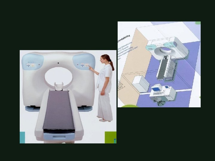
BRAIN IMAGING CT MRI Mamdouh Mahfouz MD Professor

BRAIN IMAGING CT & MRI Mamdouh Mahfouz MD Professor of Radiology Cairo University
![Patient Preparation Patient position Technique Scanogram [frontal, lateral] Scan intervals](http://slidetodoc.com/presentation_image/7d4463ede6b59c94efb3dfddc030a12b/image-2.jpg "Patient Preparation Patient position Technique Scanogram [frontal, lateral] Scan intervals")
Patient Preparation Patient position Technique Scanogram [frontal, lateral] Scan intervals
![Patient Preparation Fasting 4 - 6 hours • Contrast material [ Urographin, Telebrix, …]](http://slidetodoc.com/presentation_image/7d4463ede6b59c94efb3dfddc030a12b/image-3.jpg "Patient Preparation Fasting 4 - 6 hours • Contrast material [ Urographin, Telebrix, …]")
Patient Preparation Fasting 4 - 6 hours • Contrast material [ Urographin, Telebrix, …] 1 -2 ml/kg • Anesthesia Children, Uncooperative patients

Contrast material administration NO Traumatic cases, CVS YES Cold cases [headache, epilepsy, signs of increased ICT, …]


Vallecula

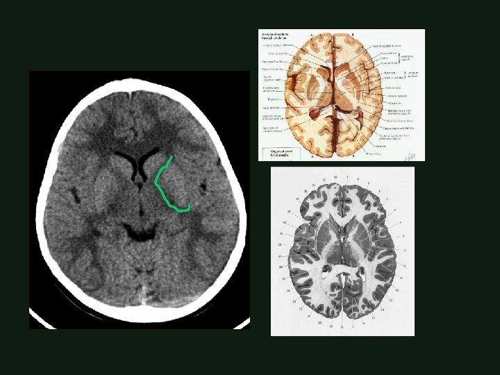
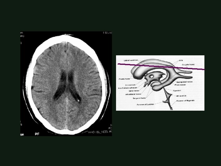
• Ventricular anatomy

Quadrigeminal Cistern

Vellum interpositum Retro-thalamic Cistern Sup. cerebellar Cistern Quadrigeminal Cistern

F T T O F F F T P O O F O P P P

Corona Radiata

Corona Radiata

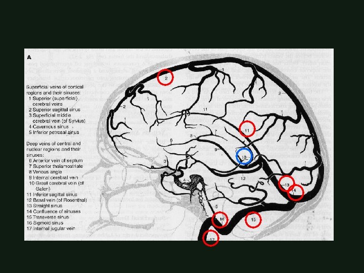
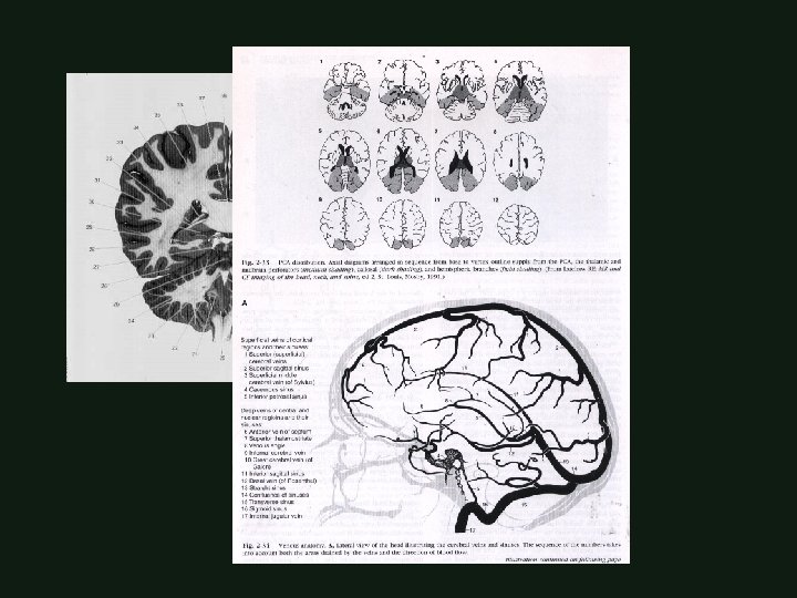
CT Vascular Anatomy
 coils • Imaging are created by")
BRAIN MRI Strong magnetic field and Radiofrequency (RF) coils • Imaging are created by the motion of hydrogen protons in response to the applied radiofrequency • Multiplanar imaging [ axial, sagittal, coronal ] • Any MR examination should include T 1 and T 2 Weighted images

CLOSED MAGNET

CLOSED MAGNET

OPEN MAGNET

OPEN MAGNET

EXTERMITY MAGNET

MR advantages § Multiplanar imaging § Tissue characterization § No bone artifacts § Shows blood vessels without contrast

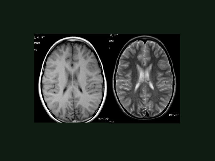
BRAIN MRI • • • T 1 WIs (TR< 800 msec T 2 WIs (TR> 1000 msec PD WIs (TR> 1000 msec • • • T 1 WIs [CSF BLACK ] T 2 WIs [CSF BRIGHT ] FLAIR WIs [ CSF BLACK ] TE 20 msec) TE> 80 msec) TE= 20 msec)

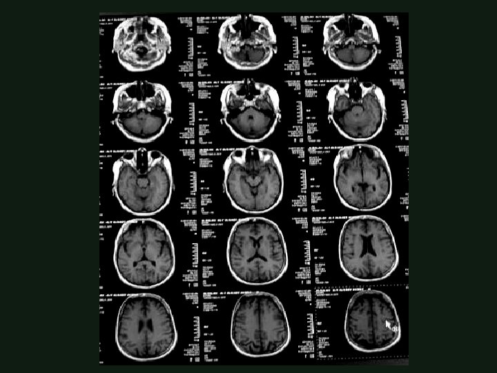
BRAIN MRI PROTOCOL

T 1 T 2 FLAIR

Signal intensity Low signal lesion = hypointense = dark High signal lesion = hyperintense = bright Intermediate signal = isointense = Gray

How to interpret MR Images ? ! Identify T 1 weighted images (CSF low signal) and T 2 Weighted images (CSF high signal) Assess the signal intensity of the structure or lesion in both T 1 and T 2 weighted images Follow the well known common signal behavior
 Calcifications ( physiological, pathological) Flowing")
Cortical bone Mature fibrous tissue ( ligaments and tendons) Calcifications ( physiological, pathological) Flowing blood in the vessels ( fast moving protons) (signal void) Air in the sinuses, lungs, …( minimal hydrogen protons) Others……. .
![T 1[High signal] T 2[High signal] Subacute blood [met Hb] Others…. T 1[High signal]](http://slidetodoc.com/presentation_image/7d4463ede6b59c94efb3dfddc030a12b/image-40.jpg "T 1[High signal] T 2[High signal] Subacute blood [met Hb] Others…. T 1[High signal]")
T 1[High signal] T 2[High signal] Subacute blood [met Hb] Others…. T 1[High signal] T 2[Low signal] Fat ( subcutaneous fat, dermoid cyst, …) Others….
![T 1[Low signal] T 2[High signal] Any structure or lesion not listed before •](http://slidetodoc.com/presentation_image/7d4463ede6b59c94efb3dfddc030a12b/image-41.jpg "T 1[Low signal] T 2[High signal] Any structure or lesion not listed before •")
T 1[Low signal] T 2[High signal] Any structure or lesion not listed before • Fluids ( CSF, urine, pleural effusion, ascites. , …) • Edema and infarctions • Most of tumors • Contrast injection [ Gd- DTPA] ++

Gadolinium – DTPA 0. 1 – 0. 2 mmol/kg body weight Only T 1 weighted images are obtained after Gd- DTPA injection Differentiate SOLs Assess activity of some lesions like MS Assess post operative tumour recurrence

Gyrus rectus Optic tract , mamillary body, cerebral peduncle, substantia negra

Amegdala , hippocampus, superior vermis

Hippocampal region

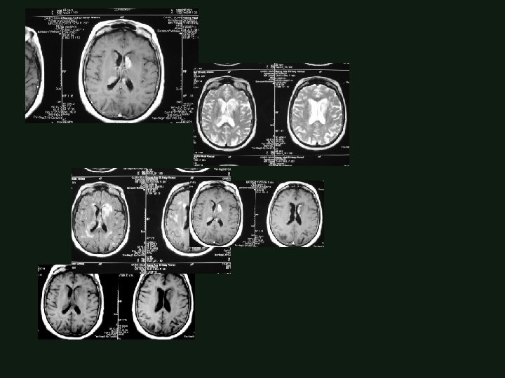
Mesial temporal sclerosis

Choroidal fissure , Hippocampal tail , Vein of Galen

Verchaw – Robben’s spaces Seen in T 1 WIs Not seen in FLAIR No clinical correlation Anatomic sites
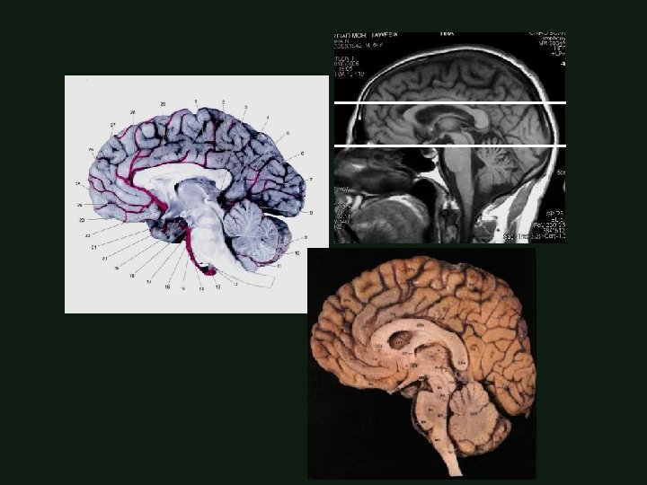
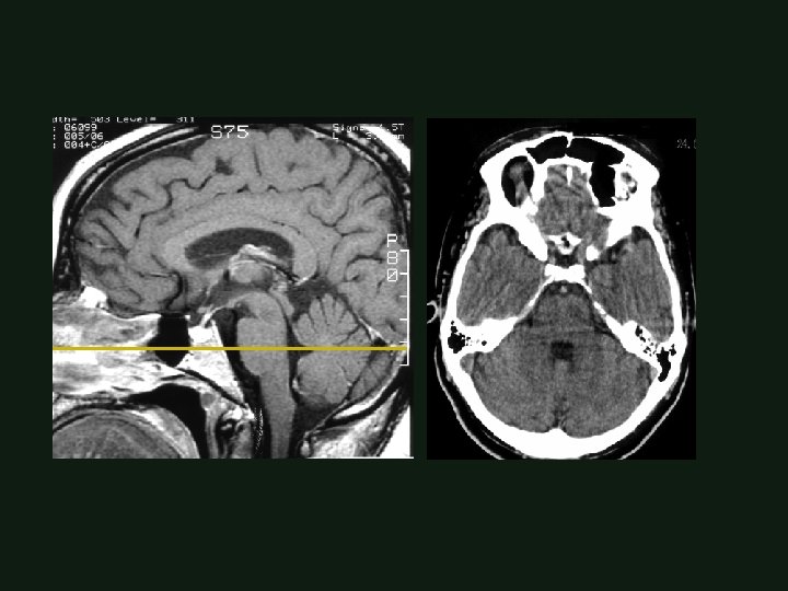
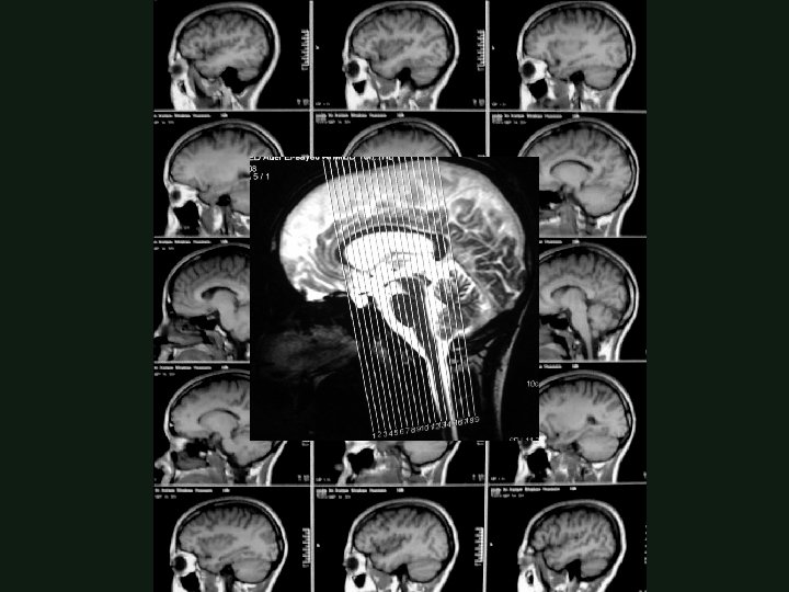
![Value of sagittal images Anatomic localization [Lobes] Corpus callosum delineation Cranio cervical junction Evaluation](http://slidetodoc.com/presentation_image/7d4463ede6b59c94efb3dfddc030a12b/image-53.jpg "Value of sagittal images Anatomic localization [Lobes] Corpus callosum delineation Cranio cervical junction Evaluation")
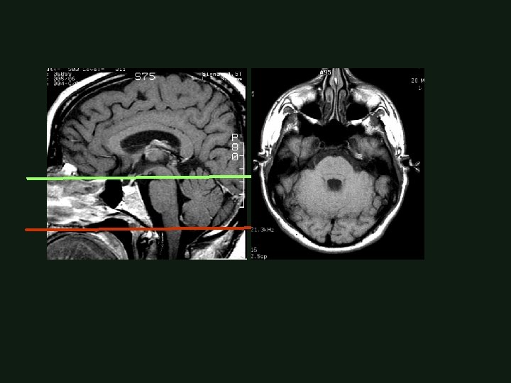
Value of sagittal images Anatomic localization [Lobes] Corpus callosum delineation Cranio cervical junction Evaluation of the venous sinuses Pituitary gland

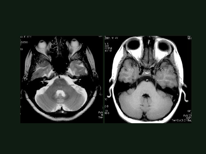
Value of coronal images Pituitary gland, chiasm, hypothalamus Hippocampal region Skull base and posterior fossa Trigeminal nerve Vascular anatomy

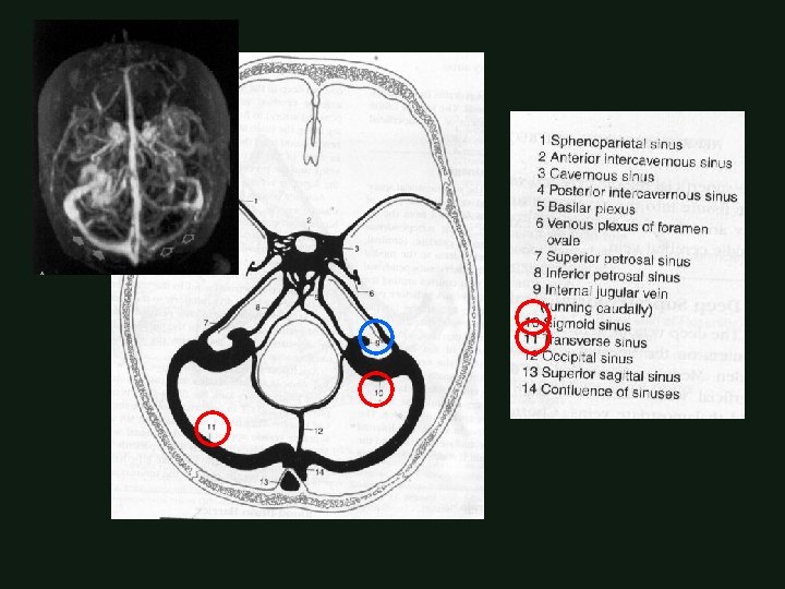
MRI Vascular Anatomy


- Slides: 65