MRI 3 MRI o Magnetic resonance imaging MRI

 is a technique of visualizing thin anatomical")


 Localized: Localized thyroid ophthalmopathy, granuloma, abscess, local pseudotumor, optic")
 Good ultrasonic transmission: (1) Rounded, cystic : mucocele, dermoid,")


- Slides: 26
MRI
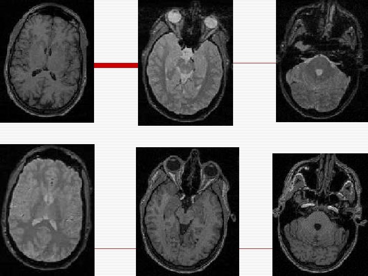
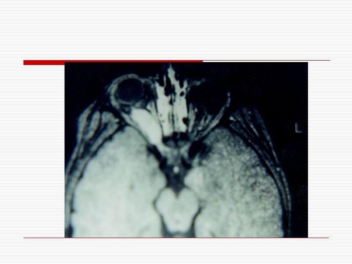
3. MRI o Magnetic resonance imaging (MRI) is a technique of visualizing thin anatomical sections by using a radio frequency pulse applied in a strong magnetic field to excite protons and then collect the energy emitted by these protons to formulate an image
o The four basic tissue parameters used in MRI include: 1. 2. 3. 4. Hydrogen density Bulk motion of the hydrogen nuclei (flow) Spin lattice relaxation time (T 1 ) Spin-spin relaxation time (T 2)
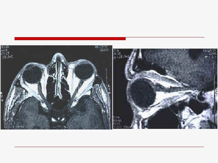
o. In short: A. In a T 1 accentuated image, orbital fat is hyperintense (bright) and vitreous is hypointense (dark) whereas, B. In a T 2 accentuated image, orbital fat is hypointense and vitreous is hyperintense (Bright).
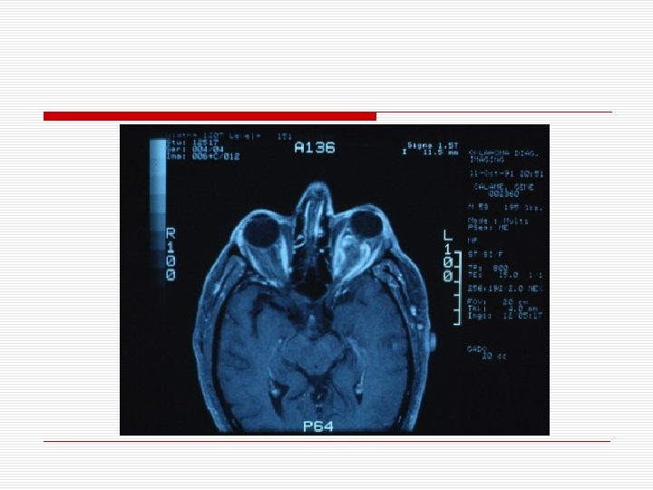
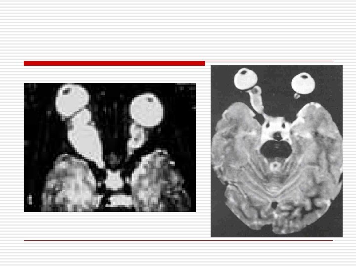
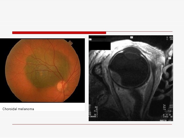
o Orbital MRI is superior over CT in diagnosis of patients with orbital bleeding, vascular tumors, metastatic tumors, melanomas and other soft tissue diseases. o While orbital CT is superior over MRI in diagnosis of diseases, tumors & trauma related to the orbital bone
4. US o US is complementary to CT in diagnosis of orbital lesions. o It is of particular value as an initial screening procedure
o Both conventional A-scan and B-scan techniques of ultrasonography have been employed.
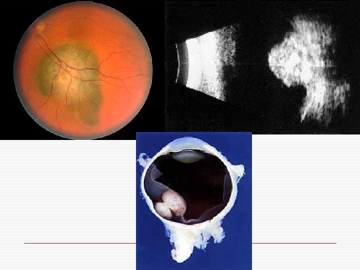
o o o The normal A scan is characterized by a linear echo or spike at surfaces where there is a change in acoustic impedance such as the junctions between the aqueous and the lens, the lens and the vitreous, and the vitreous and the posterior eye wall. Because the compact soft tissues of the orbit do not contain significant acoustic interfaces, the ultrasound echo gradually decreases in amplitude as the sound passes from the anterior to the mid-portions of the orbit. Clear cysts and homogeneous solid tumors in the orbit tend to produce low amplitude echoes within the normal orbital pattern, whereas heterogeneous cysts and tumors tend to produce higher amplitude echoes within the normal orbital pattern.
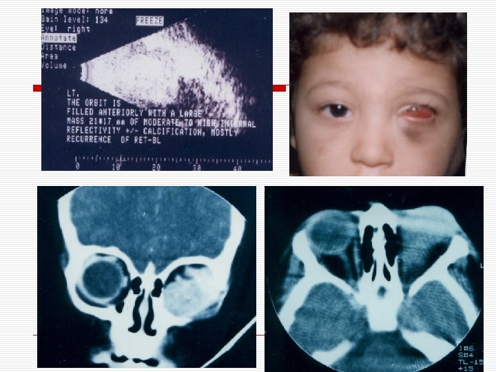
o o o B-scan ultrasonography provides a two-dimensional display of echoes rather than the linear echoes seen in A scans. As a result, B scans of the globe can depict a cross-sectional plane, showing the intraocular and orbital structures in two dimensions. The B scan of the orbit is depicted as a triangular area of high reflectivity (acoustic solidity) that tends to diminish more posteriorly in the orbit. Direct anteroposterior views often show the optic nerve, which appears as a homogeneous linear dark shadow replacing the lighter normal orbital soft tissue patterns. Rather characteristic alterations of the normal orbital pattern can be seen in scans of tumors, cysts, and inflammations.
o Coleman and others* have outlined the following scheme for ultrasonographic differential diagnosis of orbital lesions which is based on the degree of localization, and the shape, regularity, and acoustic transmission characteristics of the lesion. * LIoyd GAS: The impact of CT scanning and ultrasonography on orbital diagnosis. Clin Radiol 28: 583, 1977. * Dallw RL: Reliability of orbital diagnostic tests- ultrasonography, computed tomography, and radiography. Ophthalmol 85: 1218, 1978.
o 1. Inflammatory process: (a) Localized: Localized thyroid ophthalmopathy, granuloma, abscess, local pseudotumor, optic neuritis (b) Diffuse: Diffuse cellulitis, diffuse pseudotumor
o 2. Mass lesion: (a) Good ultrasonic transmission: (1) Rounded, cystic : mucocele, dermoid, cavernous hemangioma (2) Irregular, angiomatous : diffuse hemagioma, lymphangioma (b) Poor ultrasonic transmission: (1) Rounded, solid : meningioma, glioma, neurofibroma, most lacrimal gland tumors (2) Irregular, infiltrative: Iymphoma, metastatic carcinoma, pseudotumor
o 3. Foreign bodies
5. Biopsy techniques o Several biopsy techniques are available depending on the suspected diagnosis and the size and extent of the orbital lesion: 1. Fine Needle Aspiration Biopsy 2. Incisional biopsy 3. Excisional Biopsy
6. Pathology o The most essential aspect of diagnosing an orbital tumor is the microscopic study of the excised tissue. o In recent years a number of advances have been made in the microscopic diagnosis of orbital tumors and related lesions
● Types o o o Routine Histopathology Cytology Histochemistry Immuno-histochemistry Electron Microscopy
TREATMENT OF PROPTOSIS
III. Treatment o Depends upon the cause of proptosis e. g. dysthyroid orbitopathy, inflammatory pseudotumor, orbital tumor …etc.