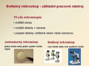
TYPES OF MICROSCOPES 1 2 3 Mikroskop Cahaya


- Slides: 75

TYPES OF MICROSCOPES 1. 2. 3. Mikroskop Cahaya (Visible Light Microscope) Fluorescence Microscope (Mikroskop Fluoresen) Electron Microscope

Visible Light Microscope 1. 2. 3. 4. 5. Brightfield Microscopy Darkfield Microscopy DIC (Difference Interference Contrast) Phase Contrast Microscopy Polarisasi Microscopy

Fluorescence Microscope 1. 2. Epi-fluorescence Confocal Laser Scanning Microscope


BRIGHT FIELD MICROSCOPY • Most commonly used microscopy imaging technique is bright field microscopy, where light is either passed through or reflected off a specimen • Biologists and histologists have used counter staining for over one hundred years; and this helps to differentiate the various tissues and organelles that can be found in a variety of subjects that would otherwise be rendered invisible Drawbacks: The cells are usually killed and therefore cannot be studied whilst moving around their natural habitat When to use bright field microscopy • Viewing stained or naturally pigmented specimens such as stained prepared slides of tissue sections • Used when there is enough contrast in the subject matter or artificial staining techniques are employed.

BRIGHT FIELD Main uses: • Viewing stained specimens • Pathological exams • Blood tests • Water inspections • Liquid crystal board inspections


Brightfield Requirements Condenser Objectives

Blue Filter n Illumination being emitted by Halogen Lamp tends to be on the Yellow Hue. Colour of image are being influence by it. Thus, it tend to be yellowish. n A Blue filter is required to compensate or to correct the colour of this error.

DARK FIELD MICROSCOPY: • A special condenser lens is used to illuminate the specimen diagonally, then observe light scattering off it. • The field of view is darker than bright field microscopy because illumination light does not enter the objective lens. • Oblique illumination is used to increase the visibility of specimens • Useful in revealing very fine detail especially bacteria Drawbacks: • Dark field is only black and white and is missing the information from the shading ability of phase contrast • For serious dark field work, one needs to use a dedicated darkfield condenser.

DARK FIELD All of us are quite familiar with the appearance and visibility of stars on a dark night, this despite their enormous distances from the Earth. Stars can be readily observed at night primarily because of the stark contrast between their faint light and the black sky. Main uses: • Microbiological imaging • Blood tests • Detecting microscopic scratches or irregularities

Main use DARKFIELD: Microbiological imaging Amoeba Proteus Bright Field Dark Field

Darkfield Requirements Darkfield Condenser


PHASE CONTRAST MICROSCOPY: • Optical phenomena of diffraction and interference are used to add light/dark contrast to a transparent specimen for imaging. • There is no need to stain the specimen as in brightfield microscopy, so live specimens can be used. • Enhances contrasts of transparent and colorless objects by influencing the optical path of light Drawbacks: A disadvantage of this method is the appearance of light halos around some objects ("halo-effect"). When to use Phase Contrast microscopy • Phase contrast is preferable to bright field microscopy when high magnifications (400 x, 1000 x) are needed and the specimen is colorless or the details so fine that color does not show up well. • Cilia and flagella, for example, are nearly invisible in bright field but show up in sharp contrast in phase contrast

PHASE CONTRAST For the procedure itself a special condenser with a ring-shaped mask and an additional "phase-ring" that is fixed within the back focal plane of the objective is needed Main uses: • Imaging cultured cells • Imaging blood or living cells


Phase Contrast Requirements Phase Contrast Turret Condenser Green Interference Filter Centering Device Phase Contrast Objective


DIFFERENTIAL INTERFERENCE CONTRAST MICROSCOPY (D. I. C) • Transforms minute differences in refraction indexes of light passing through an unstained specimen, or optical path differences from the specimen surface shape, into a monochromatic shadow-cast image enabling observation. • 3 D-pseudo effect that it gives and also, unlike phase contrast there are no halos around the subject Drawbacks: • DIC utilizes optical path differences within the specimen (i. e. : product of refractive index and geometric path length) to generate contrast the three-dimensional appearance may not represent reality • Birefringent specimens such as those found in crystals may not be suitable because of their effect upon polarized light. Similarly, specimen carriers, such as culture vessels, Petri dishes, etc. , made of plastic may not be suitable When to use DIC? As with phase contrast microscopy, DIC microscopy may be used with living specimens. However, it is better suited to thicker specimens.

DIC The DIC set-up consists of: A Polarizer, a Wollaston prism the Object, Wave train, Objective, Wollaston prism, Analyzer, and Eyepiece. Main uses: • Imaging fibrous structure of nerve • Imaging mitotic spindles • Imaging cellular nucleic structures or other thick unstained specimens

Example of DIC image The stereoscopic effect unique to DIC is observed in this example. A B C Retardation A>B>C Nematode

Example of DIC image Contrast has directivity. Volvox

Example of DIC image The specimen was observed using both fluorescence and DIC microscopy. Salivary gland of drosophila

DIC Requirements Universal DIC Condenser Polarizer DIC Elements Analyzer DIC Slider

DIC vs Phase Contrast DIC Phase Contrast

DIC PHASE CONTRAST Higher revolving power Lower revolving power (40 X: 0. 95 / 60 X: 1. 30 -1. 42) (40 X: 0. 75 / 60 X: 1. 25) Thicker specimen Only for thinner specimen Good with Fluorescence Not good with Fluorescence Glass only Plastic and Glass

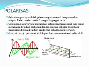
POLARIZING MICROSCOPY • This technique uses the phenomenon of polarization to add contrast and color to specimen images. • Designed to observe and photograph specimens that are visible primarily due to their optically anisotropic character. • When this beam passes through certain specimens the plane of the waves is "rotated". In some cases the extent of rotation varies with wave length, or "colour" (birefringence) • Second filter, referred to as the analyser, prior to viewing • When a birefringent specimen is viewed under these conditions, the rotated light can pass through the analyser Drawbacks: Proper alignment of the various optical and mechanical components is a critical step that must be conducted prior to undertaking quantitative analysis between crossed polarizers alone, or in combination with retardation plates and compensators When to use Polarizing microscopy Polarized light is a contrast-enhancing technique that improves the quality of the image obtained with birefringent materials when compared to other techniques


The basic configuration of polarized optical microscope. Copyright: Nikon Corporation. A schematic

representation of the polarization of light waves. Copyright: Nikon Corporation.

A schematic representation of a Nicol polarzing prism. Copyright: Nikon Corporation.

Microscope must be equipped with both a polarizer, positioned in the light path somewhere before the specimen, and an analyzer (a second polarizer), placed in the optical pathway between the objective rear aperture and the observation tubes or camera port. POLARIZING Incident light is polarized, passes through the sample and crossed polar analyzer to an image of a brightly colored (interference colored) image of the pigment crystallite. Main uses: • Analysis of optical properties of rocks, ores • Polarization analysis of fine structures within living organisms and cytoskeletons • Gout testing


Simple Polarizing Requirements Polarizer Analyzer

Polarizing Requirements For Measurement of various retardation For identification of orthoscopic & conoscopic materials

Relief Contrast A contrast technique made possible by using a special aperture located in the objective and condenser. ICSI procedure

Relief Contrast Requirements • Relief Contrast Condenser RC Modulator for 10 x, 20 x, 40 x,

FLUORESCENCE MICROSCOPY • Specimen is excited with a specific wavelength of light, then fluorescent emissions are observed. Drawbacks • Photo bleaching can significantly cause measurement error When to use Fluorescence microscopy • Used to study specimens, which can be made to fluoresce. • Certain material emits energy detectable as visible light when irradiated with the light of a specific wavelength. The sample can either be fluorescing in its natural form like chlorophyll and some minerals, or treated with fluorescing chemicals.

FLUORESCENCE WHAT IS FLUORESCENCE?


What is Fluorescence? Light of a short wavelength generates light of a longer wavelength. Jablonski diagram Illustrating the processes involved in the creation of an excited electronic singlet state by optical absorption and subsequent emission of fluorescence. Upon absorbing the excitation light, usually of short wavelengths, electrons may be raised to a higher energy and vibrational excited state excited electrons lose some energy & return to the lowest excited singlet state with simultaneous emission of fluorescent light

Fluorescent Organic Dyes Inorganic Fluorophors High quantum efficiency. Poorer quantum efficiency. More limited selection. Many colors to choose from. Conjugated to anti-bodies and proteins. Many sensitive to p. H, temperature, solvents, etc. Photo-bleaching a problem. Even poor thermal stability once in solution. . Highly stable. Less sensitive to changes in temperature. When in host, not sensitive to p. H, moisture, or solvents. Makes great standards!

FLUORESCENCE • Sample you want to study is itself the light source. Main uses: • Imaging and quantification • Assaying antigens in antigen/antibody reactions • Imaging and quantification of intracellular DNA • Analysis of chromosomal abnormalities Principle of Fluorescence 1. Energy is absorbed by the atom which becomes excited. 2. The electron jumps to a higher energy level. 3. Soon, the electron drops back to the ground state, emitting a photon (or a packet of light) - the atom is fluorescing

Properties of Fluorochrome Excitation (nm) Emission (nm) Color DAPI 365 420 Blue Fluorescein 495 525 Green Hoechts 33258 360 470 Blue R-phycocyanin 555, 618 634 Red B-phycoerythrin 545, 565 575 Orange, red R-phycoerythrin 480, 545, 565 578 Orange, red Rhodamine 552 570 Red Texas red 596 620 Red


Aktin-mitokondria

Aktin-palloidin

Endotel-mitokondria

Mitokondria-aktin-nukleus

Lisosom-mitotracker


IMAGE COMPONENTS


 Mikroskop cahaya
 Three types of microscope
 Types of electron microscopes
 When focusing a specimen you should always start with the
 When focusing a specimen you should always start with the
 T.trimpe 2006 http sciencespot.net
 What is purpose of microscope
 Forensic science microscopes
 Which organelle breaks down organelles that are no
 Uses of a compound microscope
 Electron microscopes main idea
 Skaningowy mikroskop tunelowy
 ışık mikroskobu bölümleri
 Farmastik
 Statif mikroskop
 Mitosephasen mikroskop
 Mikroskop metalograficzny
 Mikroskop princip
 Mikroskop djelovi
 Bagian-bagian mikroskop
 Hidra mitoz bölünme geçirir mi
 Hans and zacharias janssen
 Interferencijski mikroskop
 Iris zaslon mikroskop
 Bieg promieni w mikroskopie optycznym
 Mikroskop in mikroskopiranje
 Iris zaslon mikroskop
 Perawatan mikroskop
 Clsm mikroskop
 Iodamoeba butschlii
 Billur kumu mikroskop
 Veliki vijak mikroskop
 Mikroskop popis částí
 Mikroskop optyczny budowa
 Spm mikroskop
 Svjetlosni mikroskop
 Neutronenmikroskop
 Mikroskop 1660
 Mikroskop 1660
 Opticki instrumenti
 Mikroskop
 Stavba mikroskopu
 Spreadsheet adalah program
 Spekular mikroskop
 Rumus perbesaran bayangan
 Kopetensi dasar
 Stanini
 Mikroskop 3000x
 Fungsi sekrup pada mikroskop
 Diagnosis topis vertigo
 Terserapnya sebagian arah getar cahaya disebut dengan
 Salah satu cahaya alamiah adalah
 Cahaya utama sebagai penerang pokok utama disebut
 Fotometri
 Sinar istimewa lensa cembung
 Cahaya dapat dipantulkan
 Konsep dasar pencahayaan adalah
 Isyarat cahaya
 Diaskop adalah
 Gambar cahaya menembus benda bening
 Difraksi celah tunggal dan ganda
 Ukuran warna yang sangat murni dan cemerlang disebut …
 Cahaya merupakan gelombang longitudinal
 Ppt sifat cahaya
 Rumus snellius
 Pemantulan cahaya pada cermin cekung
 Optika geometris
 Panjang gelombang cahaya tampak mulai dari 4000
 Pt cahaya pertiwi indonesia
 Sebuah ketel listrik dihubungkan ke baterai 12 volt
 Warna cahaya dan warna pigmen
 Cahaya baru elektronik
 Contoh soal dispersi cahaya
 Pengertian visual art
 Penerangan
 Dualisme cahaya adalah
