Super Cells Types of Microscopes Compound light microscopes


- Slides: 14
Super Cells!
Types of Microscopes • Compound light microscopes use a series of lenses to refract light.
Types of Microscopes • Stereoscopic Dissecting- uses binocular vision to view larger specimen
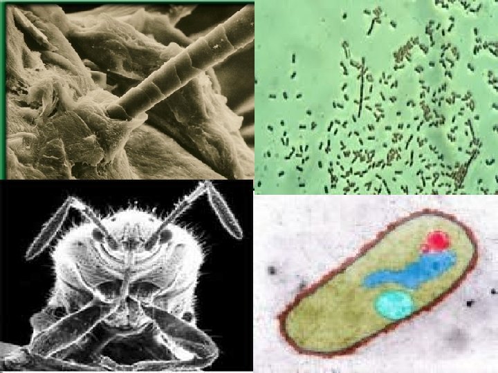
Types of Microscopes • Electron Microscope- uses a beam of electrons to view extremely small objects like atoms; there are 2 types: – Scanning- scans the 3 D surface of the object – Transmission- sees within a thin slice of the object
Introduction to the Microscope §Care §Parts §Focusing
• • • Always carry with 2 hands Only use lens paper for cleaning Do not force knobs Always store covered Keep objects clear of desk and cords
Eyepiece Body Tube Revolving Nosepiece Objective Lens Stage Clips Diaphragm Light Arm Stage Coarse Focus Fine Focus Base
• Place the Slide on the Microscope • Use Stage Clips • Click Nosepiece to the scanning (shortest) setting • Look into the Eyepiece • Use the Coarse Focus
• • Follow steps to focus using low power Click the nosepiece to the longest objective Do NOT use the Coarse Focusing Knob Use the Fine Focus Knob to bring the slide What can you find on your slide?
For each observation • START OVER with step one for each NEW observation!
Magnification • Your microscope has 3 magnifications: Scanning, Low and High. Each objective will have written the magnification. In addition to this, the ocular lens (eyepiece) has a magnification. The total magnification is the ocular x objective • Eyepiece=10 x • Scanning=4 x Low=10 x High=40 x
Drawing Specimens 1. Make detailed drawing of exactly what you see! Make sure the specimen is drawn to scale. 2. LABEL what you are observing AND the TOTAL magnification. 3. LABEL any structures to the side and connect to a line.
Example