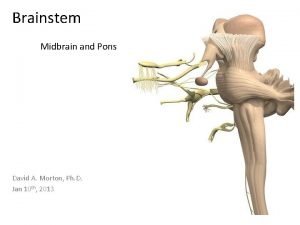
The Pons The Pons Nuclei involved with respiration


- Slides: 125

The Pons • The Pons – Nuclei involved with respiration • Apneustic center and pneumotaxic center: – modify respiratory rhythmicity center activity – Nuclei and tracts • Process and relay information to and from cerebellum • Ascending, descending, and transverse tracts: – transverse fibers (axons): » link nuclei of pons with opposite cerebellar hemisphere Copyright © 2010 Pearson Education, Inc.

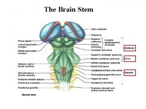
The Pons Figure 13– 6 b The Medulla Oblongata and Pons. Copyright © 2010 Pearson Education, Inc.

The Pons Figure 13– 6 c The Medulla Oblongata and Pons. Copyright © 2010 Pearson Education, Inc.

The Pons Copyright © 2010 Pearson Education, Inc.

The Pons Copyright © 2010 Pearson Education, Inc.

The Cerebellum • Functions of the Cerebellum – Adjusts postural muscles – Fine-tunes conscious and subconscious movements Copyright © 2010 Pearson Education, Inc.

The Cerebellum • Structures of the Cerebellum – Folia • Surface of cerebellum • Highly folded neural cortex – Anterior and posterior lobes • Separated by primary fissure – Cerebellar hemispheres: • Separated at midline by vermis – Vermis • Narrow band of cortex – Flocculonodular lobe • Below fourth ventricle Copyright © 2010 Pearson Education, Inc.

The Cerebellum • Structures of the Cerebellum – Purkinje cells • Large, branched cells • Found in cerebellar cortex • Receive input from up to 200, 000 synapses – Arbor vitae • Highly branched, internal white matter of cerebellum • Cerebellar nuclei: embedded in arbor vitae: – relay information to Purkinje cells Copyright © 2010 Pearson Education, Inc.

The Cerebellum • Structures of the Cerebellum – The peduncles • Tracts link cerebellum with brain stem, cerebrum, and spinal cord: – superior cerebellar peduncles – middle cerebellar peduncles – inferior cerebellar peduncles Copyright © 2010 Pearson Education, Inc.

The Cerebellum • Disorders of the Cerebellum – Ataxia • Damage from trauma or stroke • Intoxication (temporary impairment) • Disturbs muscle coordination Copyright © 2010 Pearson Education, Inc.

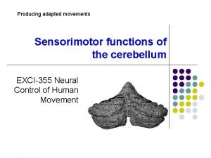
The Cerebellum Figure 13– 7 a The Cerebellum. Copyright © 2010 Pearson Education, Inc.

The Cerebellum Figure 13– 7 b The Cerebellum. Copyright © 2010 Pearson Education, Inc.

The Cerebellum Copyright © 2010 Pearson Education, Inc.

The Mesencephalon • Structures of the Mesencephalon – Tectum • Two pairs of sensory nuclei (corpora quadrigemina): – superior colliculus (visual) – inferior colliculus (auditory) – Tegmentum • Red nucleus (many blood vessels) • Substantia nigra (pigmented gray matter) Copyright © 2010 Pearson Education, Inc.

The Mesencephalon • Structures of the Mesencephalon – Cerebral peduncles • Nerve fiber bundles on ventrolateral surfaces • Contain: – descending fibers to cerebellum – motor command fibers Copyright © 2010 Pearson Education, Inc.

The Mesencephalon Figure 13– 8 a The Mesencephalon. Copyright © 2010 Pearson Education, Inc.

The Mesencephalon Figure 13– 8 b The Mesencephalon. Copyright © 2010 Pearson Education, Inc.

The Mesencephalon Copyright © 2010 Pearson Education, Inc.

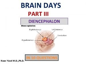
The Diencephalon • Integrates sensory information and motor commands • Thalamus, epithalamus, and hypothalamus – The pineal gland • Found in posterior epithalamus • Secretes hormone melatonin Copyright © 2010 Pearson Education, Inc.

The Diencephalon • The Thalamus – Filters ascending sensory information for primary sensory cortex – Relays information between basal nuclei and cerebral cortex – The third ventricle • Separates left thalamus and right thalamus • Interthalamic adhesion (or intermediate mass): – projection of gray matter – extends into ventricle from each side Copyright © 2010 Pearson Education, Inc.

The Diencephalon • The Thalamus – Thalamic nuclei • Are rounded masses that form thalamus • Relay sensory information to basal nuclei and cerebral cortex Copyright © 2010 Pearson Education, Inc.

The Diencephalon • Five Groups of Thalamic Nuclei – Anterior group • Anterior nuclei • Part of limbic system (emotions) – Medial group • Provides awareness of emotional states – Ventral group • Relays sensory information Copyright © 2010 Pearson Education, Inc.

The Diencephalon • Five Groups of Thalamic Nuclei – Posterior group • Pulvinar nucleus (sensory) • Lateral geniculate nucleus (visual) • Medial geniculate nucleus (auditory) – Lateral group • Affects emotional states • Integrates sensory information Copyright © 2010 Pearson Education, Inc.

The Diencephalon Figure 13– 9 The Thalamus. Copyright © 2010 Pearson Education, Inc.

The Diencephalon Figure 13– 9 a The Thalamus. Copyright © 2010 Pearson Education, Inc.

The Diencephalon Figure 13– 9 b The Thalamus. Copyright © 2010 Pearson Education, Inc.

The Diencephalon Copyright © 2010 Pearson Education, Inc.

The Diencephalon • The Hypothalamus – Mamillary bodies • Process olfactory and other sensory information • Control reflex eating movements – Infundibulum • A narrow stalk • Connects hypothalamus to pituitary gland – Tuberal area • Located between the infundibulum and mamillary bodies • Helps control pituitary gland function Copyright © 2010 Pearson Education, Inc.

The Diencephalon Figure 13– 10 a The Hypothalamus in Sagittal Section. Copyright © 2010 Pearson Education, Inc.

The Diencephalon Figure 13– 10 b The Hypothalamus in Sagittal Section. Copyright © 2010 Pearson Education, Inc.

The Diencephalon • Eight Functions of the Hypothalamus – Provides subconscious control of skeletal muscle – Controls autonomic function – Coordinates activities of nervous and endocrine systems – Secretes hormones • Antidiuretic hormone (ADH) by supraoptic nucleus • Oxytocin (OT; OXT) by paraventricular nucleus Copyright © 2010 Pearson Education, Inc.

The Diencephalon • Eight Functions of the Hypothalamus – Produces emotions and behavioral drives • The feeding center (hunger) • The thirst center (thirst) – Coordinates voluntary and autonomic functions – Regulates body temperature • Preoptic area of hypothalamus – Controls circadian rhythms (day–night cycles) • Suprachiasmatic nucleus Copyright © 2010 Pearson Education, Inc.

The Diencephalon Copyright © 2010 Pearson Education, Inc.

The Limbic System • The Limbic System – Is a functional grouping that • Establishes emotional states • Links conscious functions of cerebral cortex with autonomic functions of brain stem • Facilitates memory storage and retrieval Copyright © 2010 Pearson Education, Inc.

The Limbic System • Components of the Limbic System – Amygdaloid body • Acts as interface between the limbic system, the cerebrum, and various sensory systems – Limbic lobe of cerebral hemisphere • Cingulate gyrus • Dentate gyrus • Parahippocampal gyrus • Hippocampus Copyright © 2010 Pearson Education, Inc.

The Limbic System • Components of the Limbic System – Fornix • Tract of white matter • Connects hippocampus with hypothalamus – Anterior nucleus of the thalamus • Relays information from mamillary body to cingulate gyrus – Reticular formation • Stimulation or inhibition affects emotions (rage, fear, pain, sexual arousal, pleasure) Copyright © 2010 Pearson Education, Inc.

The Limbic System Figure 13– 11 a The Limbic System. Copyright © 2010 Pearson Education, Inc.

The Limbic System Figure 13– 11 b The Limbic System. Copyright © 2010 Pearson Education, Inc.

The Limbic System Copyright © 2010 Pearson Education, Inc.

The Cerebrum • The Cerebrum – Is the largest part of the brain – Controls all conscious thoughts and intellectual functions – Processes somatic sensory and motor information Copyright © 2010 Pearson Education, Inc.

The Cerebrum • Gray matter – In cerebral cortex and basal nuclei • White matter – Deep to basal cortex – Around basal nuclei Copyright © 2010 Pearson Education, Inc.

The Cerebrum • Structures of the Cerebrum – Gyri of neural cortex • Increase surface area (number of cortical neurons) – Insula (island) of cortex • Lies medial to lateral sulcus – Longitudinal fissure • Separates cerebral hemispheres – Lobes • Divisions of hemispheres Copyright © 2010 Pearson Education, Inc.

The Cerebrum • Structures of the Cerebrum – Central sulcus divides • Anterior frontal lobe from posterior parietal lobe – Lateral sulcus divides • Frontal lobe from temporal lobe – Parieto-occipital sulcus divides • Parietal lobe from occipital lobe Copyright © 2010 Pearson Education, Inc.

The Cerebrum Figure 13– 12 a The Brain in Lateral View. Copyright © 2010 Pearson Education, Inc.

The Cerebrum Figure 13– 12 b The Brain in Lateral View. Copyright © 2010 Pearson Education, Inc.

The Cerebrum Figure 13– 12 c The Brain in Lateral View. Copyright © 2010 Pearson Education, Inc.

The Cerebrum • Three Functional Principles of the Cerebrum – Each cerebral hemisphere receives sensory information from, and sends motor commands to, the opposite side of the body – The two hemispheres have different functions, although their structures are alike – Correspondence between a specific function and a specific region of cerebral cortex is not precise Copyright © 2010 Pearson Education, Inc.

The Cerebrum • White Matter of the Cerebrum – Association fibers – Commissural fibers – Projection fibers Copyright © 2010 Pearson Education, Inc.

The Cerebrum • White Matter of the Cerebrum – Association fibers • Connections within one hemisphere: – arcuate fibers: » are short fibers » connect one gyrus to another – longitudinal fasciculi: » are longer bundles » connect frontal lobe to other lobes in same hemisphere Copyright © 2010 Pearson Education, Inc.

The Cerebrum • White Matter of the Cerebrum – Commissural fibers • Bands of fibers connecting two hemispheres: – corpus callosum – anterior commissure Copyright © 2010 Pearson Education, Inc.

The Cerebrum • White Matter of the Cerebrum – Projection fibers • Pass through diencephalon • Link cerebral cortex with: – diencephalon, brain stem, cerebellum, and spinal cord • Internal capsule: – all ascending and descending projection fibers Copyright © 2010 Pearson Education, Inc.

The Cerebrum Figure 13– 13 a Fibers of the White Matter of the Cerebrum. Copyright © 2010 Pearson Education, Inc.

The Cerebrum Figure 13– 13 b Fibers of the White Matter of the Cerebrum. Copyright © 2010 Pearson Education, Inc.

The Cerebrum • The Basal Nuclei – Also called cerebral nuclei – Are masses of gray matter – Are embedded in white matter of cerebrum – Direct subconscious activities Copyright © 2010 Pearson Education, Inc.

The Cerebrum • Structures of Basal Nuclei – Caudate nucleus • Curving, slender tail – Lentiform nucleus • Globus pallidus • Putamen Copyright © 2010 Pearson Education, Inc.

The Cerebrum Figure 13– 14 a The Basal Nuclei. Copyright © 2010 Pearson Education, Inc.

The Cerebrum Figure 13– 14 b The Basal Nuclei. Copyright © 2010 Pearson Education, Inc.

The Cerebrum Figure 13– 14 c The Basal Nuclei. Copyright © 2010 Pearson Education, Inc.

The Cerebrum • Functions of Basal Nuclei – Involved with • The subconscious control of skeletal muscle tone • The coordination of learned movement patterns (walking, lifting) Copyright © 2010 Pearson Education, Inc.

The Cerebrum • Motor and Sensory Areas of the Cortex – Central sulcus separates motor and sensory areas – Motor areas • Precentral gyrus of frontal lobe: – directs voluntary movements • Primary motor cortex: – is the surface of precentral gyrus • Pyramidal cells: – are neurons of primary motor cortex Copyright © 2010 Pearson Education, Inc.

The Cerebrum • Motor and Sensory Areas of the Cortex – Sensory areas • Postcentral gyrus of parietal lobe: – receives somatic sensory information (touch, pressure, pain, vibration, taste, and temperature) • Primary sensory cortex: – surface of postcentral gyrus Copyright © 2010 Pearson Education, Inc.

The Cerebrum • Special Sensory Cortexes – Visual cortex • Information from sight receptors – Auditory cortex • Information from sound receptors – Olfactory cortex • Information from odor receptors – Gustatory cortex • Information from taste receptors Copyright © 2010 Pearson Education, Inc.

The Cerebrum Figure 13– 15 a Motor and Sensory Regions of the Cerebral Cortex. Copyright © 2010 Pearson Education, Inc.

The Cerebrum • Association Areas – Sensory association areas • Monitor and interpret arriving information at sensory areas of cortex – Somatic motor association area (premotor cortex) • Coordinates motor responses (learned movements) Copyright © 2010 Pearson Education, Inc.

The Cerebrum • Sensory Association Areas – Somatic sensory association area • Interprets input to primary sensory cortex (e. g. , recognizes and responds to touch) – Visual association area • Interprets activity in visual cortex – Auditory association area • Monitors auditory cortex Copyright © 2010 Pearson Education, Inc.

The Cerebrum • Integrative Centers – Are located in lobes and cortical areas of both cerebral hemispheres – Receive information from association areas – Direct complex motor or analytical activities Copyright © 2010 Pearson Education, Inc.

The Cerebrum • General Interpretive Area – Also called Wernicke area – Present in only one hemisphere – Receives information from all sensory association areas – Coordinates access to complex visual and auditory memories Copyright © 2010 Pearson Education, Inc.

The Cerebrum • Other Integrative Areas – Speech center • Is associated with general interpretive area • Coordinates all vocalization functions – Prefrontal cortex of frontal lobe • Integrates information from sensory association areas • Performs abstract intellectual activities (e. g. , predicting consequences of actions) Copyright © 2010 Pearson Education, Inc.

The Cerebrum Figure 13– 15 b Motor and Sensory Regions of the Cerebral Cortex. Copyright © 2010 Pearson Education, Inc.

The Cerebrum • Interpretive Areas of Cortex – Brodmann areas • Patterns of cellular organization in cerebral cortex Copyright © 2010 Pearson Education, Inc.

The Cerebrum Figure 13– 15 c Motor and Sensory Regions of the Cerebral Cortex. Copyright © 2010 Pearson Education, Inc.

The Cerebrum Copyright © 2010 Pearson Education, Inc.

The Cerebrum • Hemispheric Lateralization – Functional differences between left and right hemispheres – Each cerebral hemisphere performs certain functions that are not ordinarily performed by the opposite hemisphere Copyright © 2010 Pearson Education, Inc.

The Cerebrum • The Left Hemisphere – In most people, left brain (dominant hemisphere) controls • Reading, writing, and math • Decision making • Speech and language • The Right Hemisphere – Right cerebral hemisphere relates to • Senses (touch, smell, sight, taste, feel) • Recognition (faces, voice inflections) Copyright © 2010 Pearson Education, Inc.

The Cerebrum Figure 13– 16 Hemispheric Lateralization. Copyright © 2010 Pearson Education, Inc.

The Cerebrum • Monitoring Brain Activity – Brain activity is assessed by an electroencephalogram (EEG) • Electrodes are placed on the skull • Patterns of electrical activity (brain waves) are printed out Copyright © 2010 Pearson Education, Inc.

The Cerebrum • Four Categories of Brain Waves – Alpha waves • Found in healthy, awake adults at rest with eyes closed – Beta waves • Higher frequency • Found in adults concentrating or mentally stressed – Theta waves • Found in children • Found in intensely frustrated adults • May indicate brain disorder in adults – Delta waves • During sleep • Found in awake adults with brain damage Copyright © 2010 Pearson Education, Inc.

The Cerebrum Figure 13– 17 a-d Brain Waves. Copyright © 2010 Pearson Education, Inc.

The Cerebrum • Synchronization – A pacemaker mechanism • Synchronizes electrical activity between hemispheres – Brain damage can cause desynchronization • Seizure – Is a temporary cerebral disorder – Changes the electroencephalogram – Symptoms depend on regions affected Copyright © 2010 Pearson Education, Inc.

Cranial Nerves • 12 pairs connected to brain • Four Classifications of Cranial Nerves – Sensory nerves: carry somatic sensory information, including touch, pressure, vibration, temperature, and pain – Special sensory nerves: carry sensations such as smell, sight, hearing, balance – Motor nerves: axons of somatic motor neurons – Mixed nerves: mixture of motor and sensory fibers Copyright © 2010 Pearson Education, Inc.

Cranial Nerves • Cranial nerves are classified by primary functions • May also have important secondary functions – Distributing autonomic fibers to peripheral ganglia • The 12 cranial nerve groups are identified by – Primary function – Origin – Pathway – Destination Copyright © 2010 Pearson Education, Inc.

Cranial Nerves Figure 13– 18 Origins of the Cranial Nerves. Copyright © 2010 Pearson Education, Inc.

Cranial Nerves • Olfactory Nerves (I) – Primary function • Special sensory (smell) – Origin • Receptors of olfactory epithelium – Pathway • Olfactory foramina in cribriform plate of ethmoid – Destination • Olfactory bulbs Copyright © 2010 Pearson Education, Inc.

Cranial Nerves • Olfactory Nerve Structures – Olfactory bulbs • Located on either side of crista galli – Olfactory tracts • Axons of postsynaptic neurons • Leading to cerebrum Copyright © 2010 Pearson Education, Inc.

Cranial Nerves Figure 13– 19 The Olfactory Nerve. Copyright © 2010 Pearson Education, Inc.

Cranial Nerves • Optic Nerves (II) – Primary function • Special sensory (vision) – Origin • Retina of eye – Pathway • Optic canals of sphenoid – Destination • Diencephalon via optic chiasm Copyright © 2010 Pearson Education, Inc.

Cranial Nerves • Optic Nerve Structures – Optic chiasm • Where sensory fibers converge • And cross to opposite side of brain – Optic tracts • Reorganized axons • Leading to lateral geniculate nuclei Copyright © 2010 Pearson Education, Inc.

Cranial Nerves Figure 13– 20 The Optic Nerve. Copyright © 2010 Pearson Education, Inc.

Cranial Nerves • Oculomotor Nerves (III) – Primary function • Motor (eye movements) – Origin • Mesencephalon – Pathway • Superior orbital fissures of sphenoid – Destination • Somatic motor: – superior, inferior, and medial rectus muscles – inferior oblique muscle – levator palpebrae superioris muscle • Visceral motor: – intrinsic eye muscles Copyright © 2010 Pearson Education, Inc.

Cranial Nerves • Oculomotor Nerve Structures – Oculomotor nerve • Controls four of six eye-movement muscles • Delivers autonomic fibers to ciliary ganglion: – ciliary ganglion: controls intrinsic muscles of iris and lens Copyright © 2010 Pearson Education, Inc.

Cranial Nerves • The Trochlear Nerves (IV) – Primary function • Motor (eye movements) – Origin • Mesencephalon – Pathway • Superior orbital fissure of sphenoid – Destination • Superior oblique muscle Copyright © 2010 Pearson Education, Inc.

Cranial Nerves • The Abducens Nerves (VI) – Primary function • Motor (eye movements) – Origin • Pons – Pathway • Superior orbital fissures of sphenoid – Destination • Lateral rectus muscle Copyright © 2010 Pearson Education, Inc.

Cranial Nerves Figure 13– 21 Cranial Nerves Controlling the Extra-Ocular Muscles. Copyright © 2010 Pearson Education, Inc.

Cranial Nerves • The Trigeminal Nerves (V) – Primary function • Mixed (sensory and motor) to face – Origin • Ophthalmic branch (sensory): – orbital structures – nasal cavity – skin of forehead, upper eyelid, and eyebrow – part of nose Copyright © 2010 Pearson Education, Inc.

Cranial Nerves • The Trigeminal Nerves (V) – Origin • Maxillary branch (sensory): – – lower eyelid upper lip, gums, and teeth cheek and nose palate and part of pharynx • Mandibular branch (sensory): – lower gums, teeth, and lips – palate and part of tongue • Mandibular branch (motor): – motor nuclei of pons Copyright © 2010 Pearson Education, Inc.

Cranial Nerves • The Trigeminal Nerves (V) – Pathway • Ophthalmic branch: – superior orbital fissure • Maxillary branch: – foramen rotundum • Mandibular branch: – foramen ovale Copyright © 2010 Pearson Education, Inc.

Cranial Nerves • The Trigeminal Nerves (V) – Destination • Sensory nerves: – sensory nuclei in pons • Motor nerves of mandibular branch: – muscles of mastication Copyright © 2010 Pearson Education, Inc.

Cranial Nerves • Trigeminal Nerve Structures – Trigeminal nerves • Largest cranial nerves • With three major branches – Semilunar ganglion • Contains cell bodies of sensory neurons Copyright © 2010 Pearson Education, Inc.

Cranial Nerves Figure 13– 22 The Trigeminal Nerve. Copyright © 2010 Pearson Education, Inc.

Cranial Nerves • The Facial Nerves (VII) – Primary function • Mixed (sensory and motor) to face – Origin • Sensory: – taste receptors on anterior 2/3 of tongue • Motor: – motor nuclei of pons – Pathway • Internal acoustic meatus to facial canals (stylomastoid foramina) Copyright © 2010 Pearson Education, Inc.

Cranial Nerves • The Facial Nerves (VII) – Destination • Sensory: – sensory nuclei of pons • Somatic motor: – muscles of facial expression • Visceral motor: – tear and nasal mucous glands – submandibular and sublingual salivary glands Copyright © 2010 Pearson Education, Inc.

Cranial Nerves • Facial Nerve Structures – Facial nerve branches • Temporal • Zygomatic • Buccal • Mandibular • Cervical branches Copyright © 2010 Pearson Education, Inc.

Cranial Nerves • Facial Nerve Structures – Geniculate ganglia • Hold cell bodies of sensory neurons – Pterygopalatine ganglia • Postganglionic fibers innervate glands (lacrimal, nasal cavity, and pharynx) – Submandibular ganglia • Innervate salivary glands Copyright © 2010 Pearson Education, Inc.

Cranial Nerves Figure 13– 23 a The Facial Nerve. Copyright © 2010 Pearson Education, Inc.

Cranial Nerves Figure 13– 23 b The Facial Nerve. Copyright © 2010 Pearson Education, Inc.

Cranial Nerves • The Vestibulocochlear Nerves (VIII) – Primary function: special sensory • Vestibular branch: – balance and equilibrium • Cochlear branch: – hearing – Origin • Receptors of inner ear – Pathway • Internal acoustic meatus of temporal bones – Destination • Vestibular and cochlear nuclei of pons and medulla oblongata Copyright © 2010 Pearson Education, Inc.

Cranial Nerves • Vestibulocochlear Nerve Structures – Vestibular branch • Originates at receptors of vestibule (balance) • Connects to vestibular nuclei of pons and medulla oblongata – Cochlear branch • Originates at sensors of cochlea (hearing) • Connects with cochlear nuclei of pons and medulla oblongata Copyright © 2010 Pearson Education, Inc.

Cranial Nerves Figure 13– 24 The Vestibulocochlear Nerve. Copyright © 2010 Pearson Education, Inc.

Cranial Nerves • The Glossopharyngeal Nerves (IX) – Primary function • Mixed (sensory and motor) to head and neck – Origins • Sensory: – posterior 1/3 of tongue – part of pharynx and palate – carotid arteries • Motor: – motor nuclei of medulla oblongata Copyright © 2010 Pearson Education, Inc.

Cranial Nerves • The Glossopharyngeal Nerves (IX) – Pathway • Jugular foramina between occipital and temporal bones – Destination • Sensory: – sensory nuclei of medulla oblongata • Somatic motor: – nerves involved in swallowing • Visceral motor: – parotid salivary gland Copyright © 2010 Pearson Education, Inc.

Cranial Nerves • Glossopharyngeal Nerve Structures – Superior and inferior ganglion • Sensory neurons of tongue and pharynx – Otic ganglion • Synapse visceral motor fibers Copyright © 2010 Pearson Education, Inc.

Cranial Nerves Figure 13– 25 The Glossopharyngeal Nerve. Copyright © 2010 Pearson Education, Inc.

Cranial Nerves • The Vagus Nerves (X) – Primary function • Mixed (sensory and motor) • Widely distributed in thorax and abdomen – Origins • Sensory: – – part of pharynx auricle and external acoustic meatus diaphragm visceral organs of thoracic and abdominopelvic cavities • Motor: – motor nuclei in medulla oblongata Copyright © 2010 Pearson Education, Inc.

Cranial Nerves • The Vagus Nerves (X) – Pathway • Jugular foramina • Between occipital and temporal bones – Destination • Sensory: – sensory nuclei and autonomic centers of medulla oblongata • Visceral motor: – muscles of the palate and pharynx – muscles of the digestive, respiratory, and cardiovascular systems in thoracic and abdominal cavities Copyright © 2010 Pearson Education, Inc.

Cranial Nerves • Vagus Nerve Structures – Vagus nerves • Branch and radiate extensively – Superior (jugular) ganglion and inferior (nodose) ganglion • Hold sensory neurons Copyright © 2010 Pearson Education, Inc.

Cranial Nerves Figure 13– 26 The Vagus Nerve. Copyright © 2010 Pearson Education, Inc.

Cranial Nerves Figure 13– 26 The Vagus Nerve. Copyright © 2010 Pearson Education, Inc.

Cranial Nerves • The Accessory Nerves (XI) – Primary function • Motor to muscles of neck and upper back – Origin • Motor nuclei of spinal cord and medulla oblongata – Pathway • Jugular foramina between occipital and temporal bones – Destination • Internal branch: – voluntary muscles of palate, pharynx, and larynx • External branch: – sternocleidomastoid and trapezius muscles Copyright © 2010 Pearson Education, Inc.

Cranial Nerves • Accessory Nerve Structures – Spinal root • Motor fibers that originate in anterior gray horns of first five cervical segments of spinal cord – Cranial root • Motor fibers that originate in medulla oblongata – Internal branch • Joins the vagus nerve – External branch • Controls muscles of neck and back Copyright © 2010 Pearson Education, Inc.

Cranial Nerves • The Hypoglossal Nerves (XII) – Primary function • Motor (tongue movements) – Origin • Motor nuclei of medulla oblongata – Pathway • Hypoglossal canals of occipital bone – Destination • Muscles of tongue Copyright © 2010 Pearson Education, Inc.

Cranial Nerves Figure 13– 27 The Accessory and Hypoglossal Nerves. Copyright © 2010 Pearson Education, Inc.

Cranial Nerves Copyright © 2010 Pearson Education, Inc.

Cranial Nerves Copyright © 2010 Pearson Education, Inc.

Cranial Reflexes • Cranial Reflexes – Monosynaptic and polysynaptic reflex arcs – Involve sensory and motor fibers of cranial nerves – Clinically useful to check cranial nerve or brain damage Copyright © 2010 Pearson Education, Inc.

Cranial Reflexes Copyright © 2010 Pearson Education, Inc.
 Muscles of inspiration
 Internal respiration vs external respiration
 External and internal respiration
 Pons function
 Pons
Pons Cerebellar cortex
 Pons function in psychology
 Superior medullary velum
 Cerebellum ct anatomy
 Myelencephalon
 What is the brainstem
 Internal structure of pons
 Nicolas pons vignon
 Glossopharyngeal nerve examination
 Pons
Pons Gerard pons voyages
 Mesencephalon
 Pons sublicius iter paene
 Mnemonic for reticular formation
 Pons
Pons Function of the pons
 Caudal pons
 Pons
Pons Pons function
 Mesencephalon pons medulla oblongata
 Site:slidetodoc.com
 Vttt tract
 Basilar artery course
 Neo malthusians definition ap human geography
 Vascularizatia cerebelului
 Vmn body lift
 Oblongata
 Mirror nuclei
 Septal nuclei
 Nutmeg liver
 Nuclei of thalamus
 The distinct threadlike structures that contain the genetic
 Urban realms model
 Active galactic nuclei
 How unstable atoms gain stability
 The multiple nuclei model
 Nucleii cohleari
 Active galactic nuclei
 Bid rent theory example
 Nuclei proxy
 Atn
Atn Robbins
 Nuclei
 Harris y ullman
 Multiple nuclei model
 Multiple nuclei model ap human geography
 Simple2do
 Pulvinar thalami
 Cerebral cortex same as cerebrum
 Nuclei della base
 Nuclei
 Thalamus hand
 Comment démarrer un nuclei
 Pontine nuclei
 Concentric zones
 Multiple nuclei model
 Nuclei della base
 Nuclei narrativi promessi sposi
 Shell model of nucleus
 Inferior cerebellar peduncle
 Brain diencephalon
 Via acustica schema
 Reticular formation
 Limbic system
 Urban realms model example
 High nc ratio
 Three models of urban structure
 Nuclei di cure primarie
 Somatoefferent
 Fibrae arcuata externa anteriores
 Nuclei
 Multiple nuclei model
 Harris ullman model
 Multiple nuclei model
 Vestibulocochlear nerve.
 Aree encefalo
 Atomic nuclei
 African city model
 Dottoressa slon
 Cerebriform nuclei
 Mesencephalon
 Suburban sprawl ap human geography
 Active galactic nuclei
 Multiple nuclei model example
 Strengths of the multiple nuclei model
 Nmr active and inactive nuclei
 M
M What continents were involved in the columbian exchange
 Simplex method
 Who was involved in dday
 First favorite novel of rizal
 No taxation without representation
 Justice involved supportive housing
 What is output design
 Chapter 8 thinking language and intelligence
 Measuring the actual performance
 Patient admission process in hospital
 How are p orbitals involved in double bonds
 A purposeful refusal to become generally involved
 Professional and ethical issues during internship
 Which organs are involved in respiratory system
 Opportunity analysis canvas example
 What is public issue management mean
 Mount st helens plates involved
 Channel design and management decisions
 Enzymes involved in central dogma
 Enzyme involved
 Slidetodoc.com
 What countries were involved in wwi
 With erp implementations why would an auditorget involved
 American researcher who involved in getting heart rate
 1s22s2sp63s23p4
 How many countries were involved in wwi
 Hercules 7th labor
 The principle enzyme involved in dna replication is called
 What are the 7 steps of portfolio process
 Locational break-even analysis
 Enzyme involved in dna replication
 Problem identification research
 The principle involved in the image formation by lenses
 Who was involved in dday
