PHYSIOLOGY EXTERNAL AND INTERNAL RESPIRATION EXTERNAL RESPIRATION Respiration


 responsible for holding the lungs to the thoracic wall is/are A. The")


 l At the end of a quiet inspiration, take in")
 l Stop at the end of a normal exhalation, then")
 l Cannot be measured direction l Even if you blow out")


 l Asthma, emphysema, chronic bronchitis")


 essential to hemoglobin saturation and the release")

- Slides: 83
PHYSIOLOGY EXTERNAL AND INTERNAL RESPIRATION
EXTERNAL RESPIRATION
Respiration l Movement of gases between the environment and the body’s cells – The exchange of air between the atmosphere and the lungs l l Known as ventilation or breathing Inspiration and Expiration – The exchanges of O 2 and CO 2 between the lungs and the blood – Transport of O 2 and CO 2 by the blood – The exchange of gases between blood and the cells
RESPIRATION
Anatomy of the Respiratory System l Nasal Concha – Air eddies Air is cleaned l Warmed l Humidified l l Tonsils and Adenoids – Lymph nodes that filter the air – Located in the nose, back of the throat, below the tongue
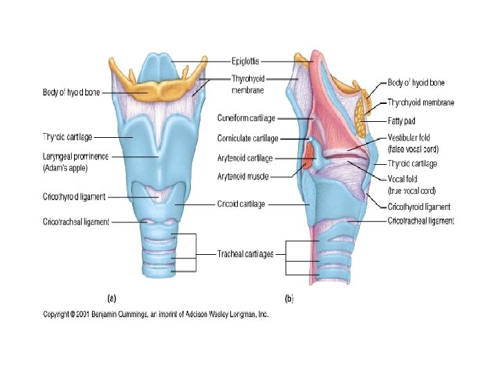
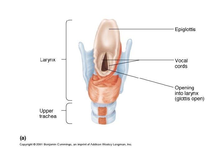
Larynx l Contains Vocal Cords – Connective tissue bands that tighten to create sound when air moves past them l Thyroid Cartilage – Sensitive to Testosterone levels
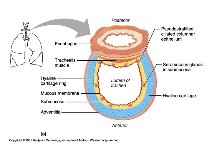
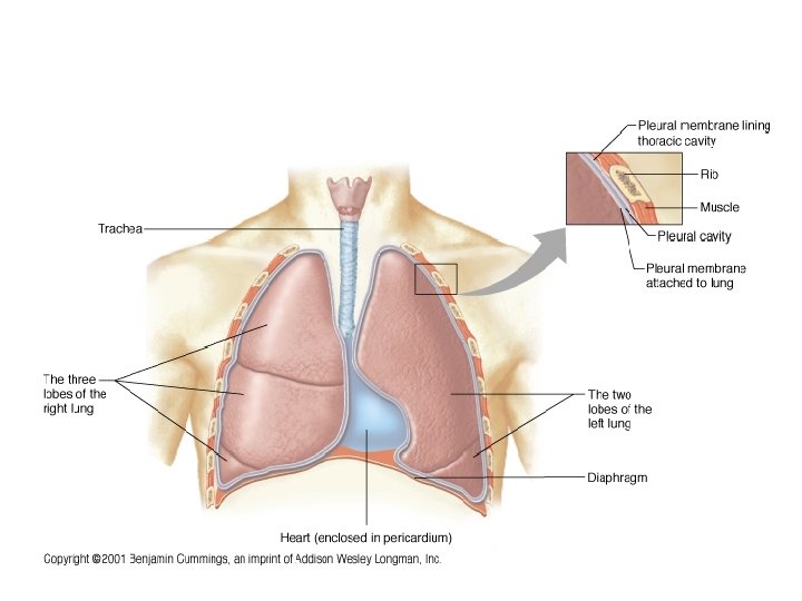
Trachea l Conducts Air – Lined with pseudostratified ciliated columnar epithelium – Cilia can be paralyzed by cigarette smoke l Surrounded by C-shaped Cartilagenous rings and the trachealis muscle – Esophagus is dorsal to the trachea l Approximately 4 inches long
The nose serves all the following functions except A. As a passageway for air movement B. As the initiator of the cough reflex C. Warming and humidifying the air D. Cleansing the air
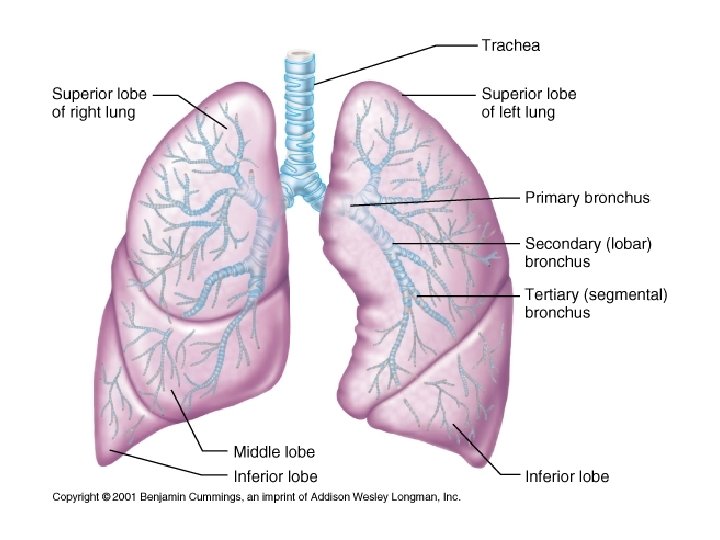
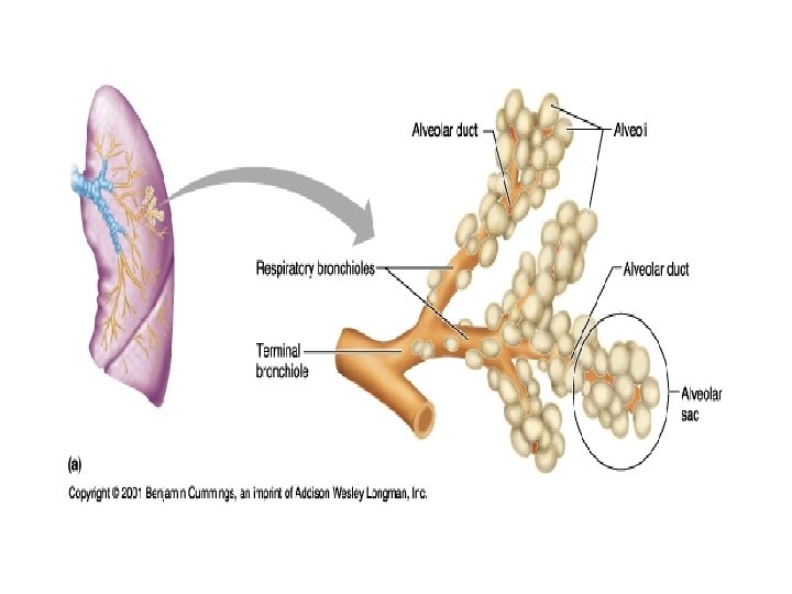
Conducting System or Respiratory Tree l Primary Bronchi – Surrounded by O-shaped cartilagenous rings – Bifurcates to Secondary Bronchi in the lungs – Respiratory Bronchioles Surrounded by smooth muscles l Diameter of the airways becomes progressively smaller from the trachea to the bronchioles l The total cross-sectional area increases with each division of the airways l
Pleural Membranes l Visceral Pleura – Attached directly to the lungs l Parietal Pleura – Attaches to the visceral pleura – Also attaches to the thoracic cavity l Serous Fluid – Separates the two pleura and lubricates in order to decrease friction – Consistency of egg whites – Pleurisy occurs when the fluid decreases l The Function of the Pleural Membranes is to hold the lungs open
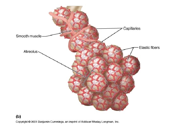
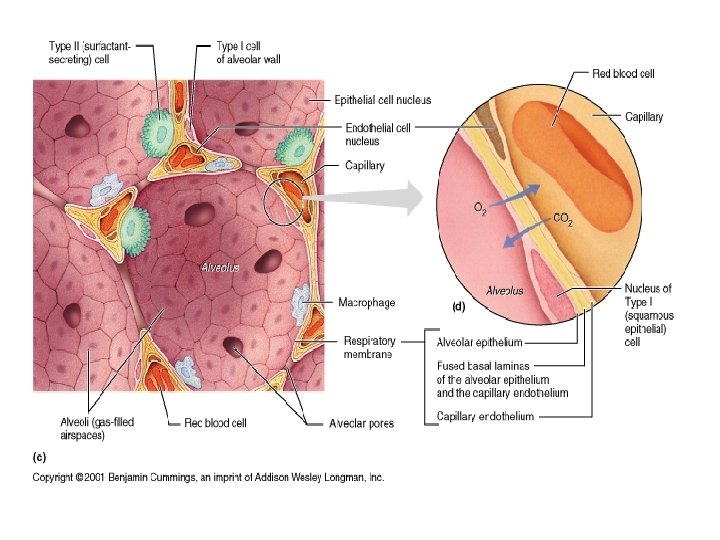
Alveoli l Clustered at the ends of the terminal bronchioles l Makes up the bulk of lung tissue l Primary function is the exchange of gases between themselves and the blood l Surrounded by elastic fibers – Creates Elastic Recoil
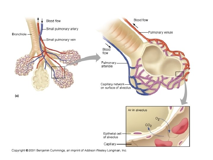
Capillaries l The alveoli are closely associated with an extensive network of capillaries – Blood vessels cover 80 -90% of the alveolar surface forming a continuous “sheet” of blood in close contact with the air-filled alveoli
Respiratory Membrane l Consists of – The Wall of the Alveoli – The Respiratory Space This is a fluid filled space l Pneumonia may cause the space to fill with more fluid than normal l – This decreases the ability to exchange gases – The Wall of the Capillary
Surfactant helps to prevent the alveoli from collapsing by A. Humidifying the air before it enters B. Warming the air before it enters C. Interfering with the cohesiveness of water molecules, thereby reducing the surface tension of alveolar fluid D. Protecting the surface of alveoli from dehydration and other environment variations
The respiratory membrane is a combination of A. Respiratory bronchioles and alveolar ducts B. Alveolar walls, alveolar space and capillary walls C. Atria and alveolar sacs D. None of the above
Gas Laws l At sea level normal atmospheric pressure is 760 mm. Hg – On top of Mt. Everest Patm = 153 mm. Hg
Dalton’s Law l The total pressure exerted by a mixture of gases is the sum of the pressures exerted by the individual gases – 78% N 2 – 21% O 2 – 1% CO 2 l Partial Pressure of gases – The pressure of a single gas in a mixture
Gas Law The total pressure of a mixture of gases, is the sum of the pressures of the individual gases (Dalton’s Law) l Gases, singly or in a mixture, move from areas of higher pressure to areas of lower pressure l If the volume of a container of gas changes, the pressure of the gas will change in an inverse manner (Boyle’s Law) l
Dalton’s Law l To find the partial pressure of any one gas in a sample of air, multiply the atmospheric Pressure (Patm) by the gas’s relative contribution (%) to Patm. – Partial pressure of an atmospheric gas = l Patm X % of gas in atmosphere – Partial pressure of oxygen = 760 mm. Hg X 21% l PO 2 = 760 X 0. 21 = 160 mm. Hg
Gases Move from High Pressure to Low Pressure l Air flow occurs whenever there is a pressure gradient
Boyle’s Law l The pressure exerted by a gas or mixture of gases in a sealed container is created by the collisions of moving gas molecules with the walls of the container and with each other. – P 1 V 1 = P 2 V 2 l An increase in volume will create a decrease in pressure and a decrease in volume will create an increase in pressure
Boyle’s Law l Changes in the volume of the chest cavity during ventilation cause pressure gradients that create air flow – When the chest volume increases, the alveolar pressure falls, and air flows into the respiratory system l When the chest volume decreases, the alveolar pressure rises, and air flows out into the atmosphere
Alveoli Composed of a single layer of epithelium called Type I cells l Type II alveolar cells l – Secretes surfactant – Surfactant decreases the surface tension of the water within the alveoli – Coats the inside of the alveoli – Cortisol causes the maturation of the type II cells in the fetal stage of development l Dust Cells – Phagocytes
Law of La. Place l The pressure inside a bubble formed by a fluid film is a function of two factors – Surface tension of the fluid (T) – Radius of the bubble (r) l P = 2 T/r Surfactant decreases the surface tension of water in the alveoli l Newborn Respiratory Distress Syndrome (RDS) l
Air Flow l Flow = changes in P / R – P = Pressure – R = Resistance to Flow l Air flow in response to a pressure gradient l The flow decreases as the resistance to flow increases
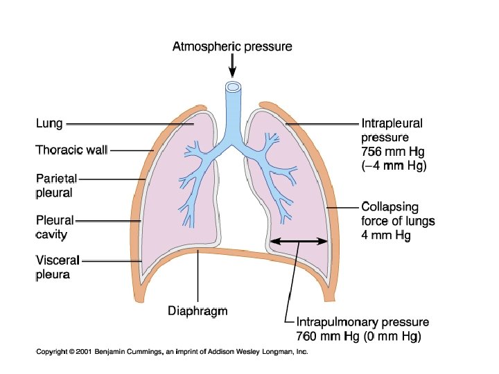
Pressure in the System l Alveolar Pressure – Pressure in the air spaces of the lungs l Intrapleural Pressure – Pressure in the pleural fluid l Intrapulmonary Pressure – Pressure within the lungs as a whole l Atmospheric Pressure – Pressure in the atmosphere due to a column of air up to the stratosphere
The pleurae are vital to the integrity of the lungs because they A. Contain cilia that protect the lungs B. control the volume of the lungs C. Secrete a lubricating serous fluid, allowing the lungs to glide over the thoracic wall during breathing D. Maintain the proper temperature of the lungs during sleep
The factor(s) responsible for holding the lungs to the thoracic wall is/are A. The smooth muscles of the lung B. The diaphragm and the intercostals muscles C. The visceral pleurae and the changing volume of the lungs D. Surface tension from pleural fluid, positive pressure, and atmospheric pressure on the thorax
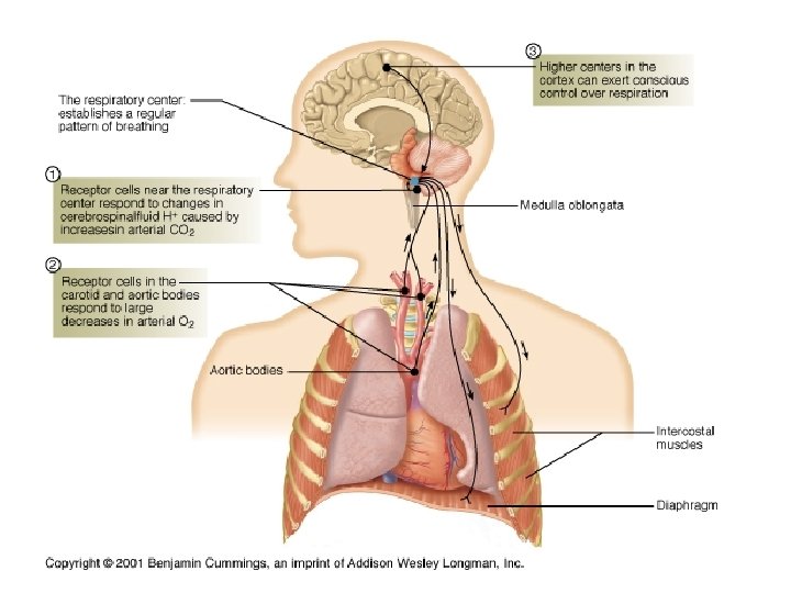
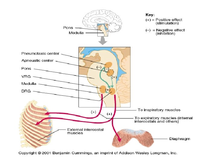
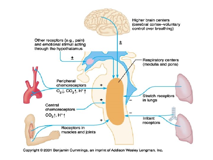
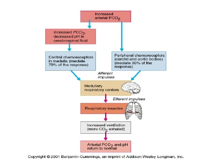
Inspiration l Time 0. – In the brief pause between breathes, alveolar pressure is equal to atmospheric pressure – When pressures are equal, there is no air flow l Time 0 -2 sec – Oxygen levels fall and Carbon Dioxide levels rise – Peripheral Chemoreceptors are stimulated l Located in the carotid arteries l Sensitive to oxygen levels – Central Chemoreceptors are stimulated l Located in the Medulla Oblongata of the brain l Sensitive ot carbon dioxide levels
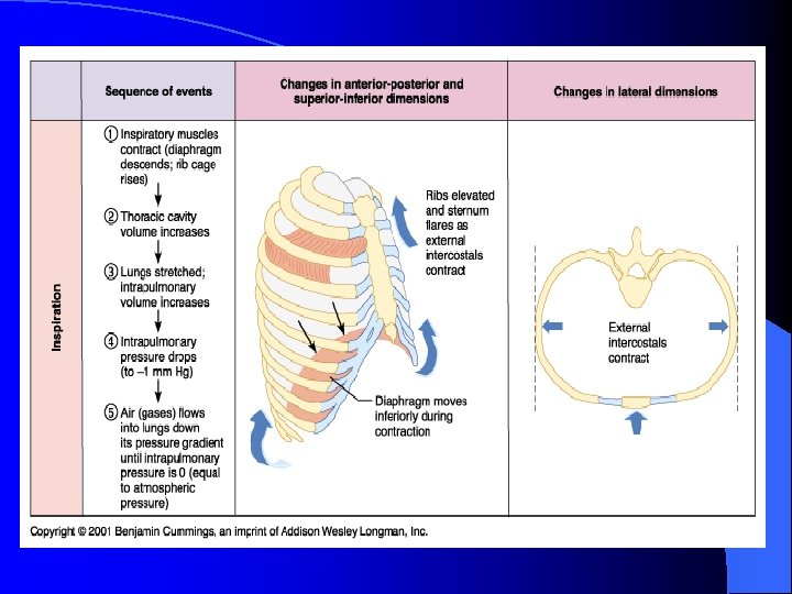
Inspiration l Chemoreceptors stimulate the Medulla Oblongata l The MO stimulates the Phrenic Nerve l The Phrenic Nerve stimulates the respiratory muscles of the thoracic cage and the diaphragm l The muscles contract and the thoracic volume increases
Inspiration l When thoracic volume increases then alveolar pressure fall approximately 4 mm. Hg below atmospheric pressure l Air flows from high pressure to low pressure until the pressures reach equilibrium
Exhalation l Time 2 -4 sec: – As lung and thoracic volumes decrease air pressure in the lungs increases until the pressures equal equilibrium – Stretch receptors in the lung tissue are stimulated – Stretch receptors send information to the MO and this stops the phrenic nerve stimulation – Respiratory muscles relax l Time 4 sec: – Elastic Recoil occurs – Alveolar pressure is now higher than atmospheric pressure due to a decrease in lung volume – Air leaves the lungs until pressures reach equilibrium
Intrapleural Pressure Changes During Ventilation l The lungs are “stuck” to the thoracic cage by the cohesive forces exerted by the fluid between the two pleural membranes l If the thoracic cage moves, the lungs move with it
Intrapleural Pressure l The pressure between the pleural membranes is normally subatmospheric l The combination of the outward pull of the thoracic cage and in inward recoil of the elastic lungs creates a subatmospheric intrapleural pressure of about -3 mm. Hg
What happens to subatmospheric intrapleural pressure if an opening is made between the sealed pleural cavity and the atmosphere?
A knifing? Air in the pleural cavity breaks the fluid bond holding the lung to the chest wall l The chest wall expands outward while the elastic lung collapses to an unstretched state l – Like a deflated balloon – Pneumothorax l Results in a collapsed lung that is unable to function normally – Correction of a Pneumothorax l l Removing as much air from the pleural cavity as possible with a suction pum Sealing the hole
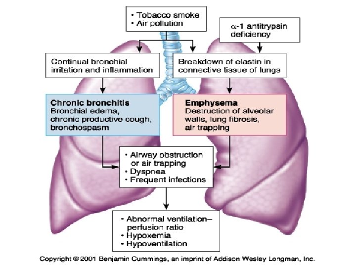
Emphysema l Loss of elastic fibers for elastic recoil during expiration – Elastin is destroyed by elastase l An enzyme released by immune cells l Have more difficulty exhaling than inhaling
Select the incorrect statement about external respiration A. Carbon dioxide is exchanged in the alveoli. B. Cells produce nitrogen by their metabolism. C. Diffusion accounts for the transport of gases. D. Oxygen is exchanged in the alveoli. E. The pulmonary capillaries are involved.
The intra-alveolar pressure A. Is the pressure within the air sacs of the lung. B. Never equilibrates with atmospheric pressure. C. Is always less than intrapleural pressure. D. All of the above are correct.
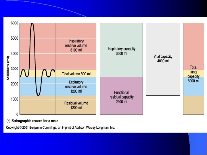
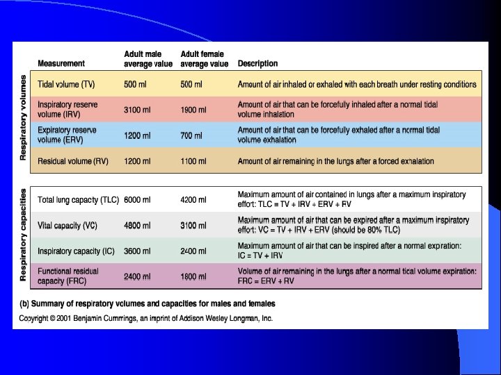
Ventilation l Lung Volumes – Use a spirometer to measure pulmonary function l Tidal Volume l Inspiratory Reserve Volume l Expiratory Reserve Volume l Residual Volume
Tidal Volume l Breathing Quietly – The volume of air that moves during a single inspiration or expiration (VT). l Average tidal volume during quiet breathing is about 350 – 500 ml of air
Inspiratory Reserve Volume (IRV) l At the end of a quiet inspiration, take in as much additional air as you possibly can – About 3000 ml in a 70 kg male
Expiratory Reserve Volume (ERV) l Stop at the end of a normal exhalation, then exhale as much air as you possibly can l This is a forceful exhalation – Average is about 1100 ml
Residual Volume (RV) l Cannot be measured direction l Even if you blow out as much air as you can, air still remains in the lungs and the airways – About 1200 ml
Lung Capacities l The sum of two or more lung volumes – Vital capacity (VC) The sum of the inspiratory reserve volume, expiratory reserve volume and tidal volume l This represents the maximum amount of air that can be voluntarily moved into or out of the respiratory system with one breath l
Total Lung Capacity l Total Lung Capacity – Vital capacity + residual volume l Functional Residual Capacity – Expiratory reserve volume + residual volume
Airway Resistance l Bronchioles are collapsible tubes l Bronchoconstriction – Increases resistance to air flow and decreases the amount of fresh air that reaches the alveoli l Histamine – Bronchoconstrictor l Bronchodilators – Decreases resistance to air flow
PARASYMPATHIC INPUT l PNS is the primary neural control of bronchioles and causes bronchoconstriction l Smooth muscle in the bronchioles have β-2 receptors that respond to epinephrine and norepinephrine – Stimulation of β-2 receptors relaxes airway smooth muscle and results in bronchodilation l Used to treat asthma or allergies
Chronic Obstructive Pulmonary Disease (COPD) l Asthma, emphysema, chronic bronchitis
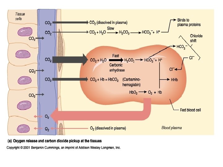
INTERNAL RESPIRATION
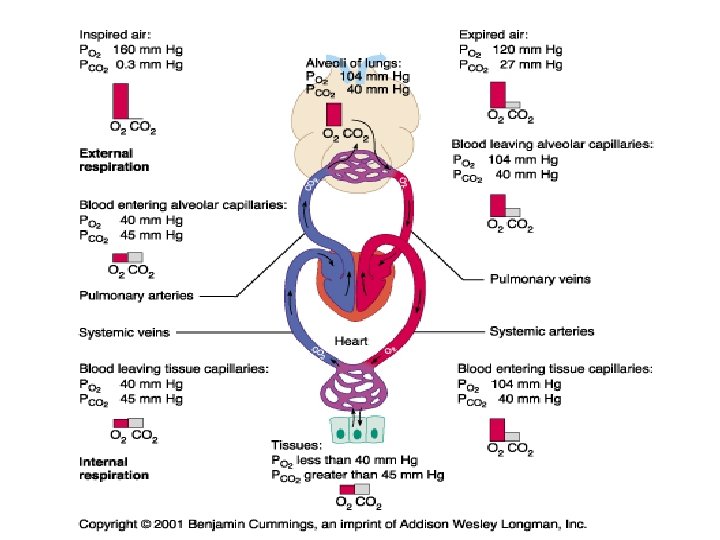
Pressure Gradients l Arterial Pressures – PO 2 = 100 mm. Hg – PCO 2 = 40 mm. Hg l Alveolar Pressures – PO 2 = 100 mm. Hg – PCO 2 = 40 mm. Hg l Venous Pressures – PO 2 = 40 mm. Hg – PCO 2 = 45 mm. Hg l Tissue Pressures – PO 2 = 40 mm. Hg – PCO 2 = 45 mm. Hg
Hemoglobin l Oxygen is transported two – Dissolved in the plasma – Bound to hemoglobin ways in the blood l Mean Corpuscular Hemoglobin – Counting the RBC’s and quantifying the amount of hemoglobin per RBC l Hemoglobin Concentration or %
Hemoglobin l One hemoglobin molecule binds up to four oxygen molecules – Percent oxygen saturation l Globin – Proteins in the hemoglobin Alpha, beta, gamma and delta types l Adults have 2 alpha and 2 bets l Fetal Hb l – Two gamma and two alpha
Affects of Oxygen-Hemoglobin Binding l Any factor that changes the conformation of the hemoglobin protein can affects its ability to bind oxygen l Changes in plasma p. H, PCO 2, 2, 3 DPG and temperature alters oxygen-binding affinity of hemoglobin – 2, 3 DPG = 2, 3 Diphosphoglycerate
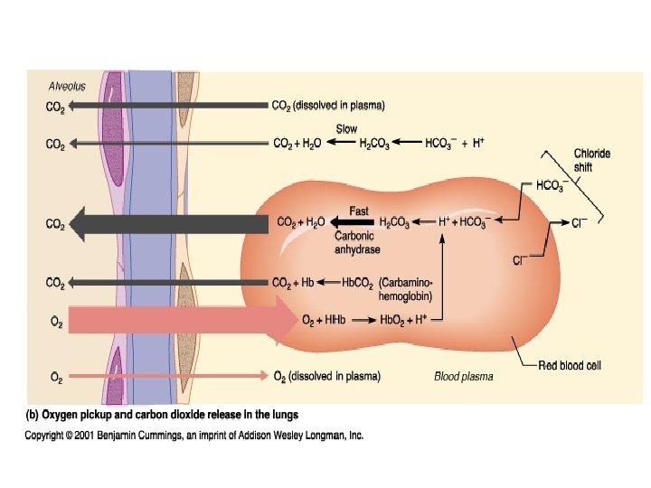
Carbon Dioxide Transportation l 7% of the CO 2 is carried by venous blood dissolved in the blood l 93% diffuses into red blood cells, – 70% is converted to bicarbonate ions HCO 3– 23% binds to hemoglobin as carbaminohemoglobin
Hydrogen Ion Transportation l Hemoglobin binds hydrogen ions – Prevents large shifts in the body’s p. H – Too many hydrogen ions in the blood may cause respiratory acidosis l This is caused by an increase in CO 2 levels in the blood
l H 2 O + CO 2 = H 2 CO 3 = H+ + HCO 3 - l. Carbonic Anyhydrase
Death from carbon monoxide poisoning would be due to A. The body’s destruction of hemoglobin B. The strong affinity of carbon monoxide to hemoglobin C. Formation of cellular toxins D. Inability of form hemoglobin
The entire sequence of events involved in the exchange of O 2 and CO 2 between the body and the external environment is known as A. Internal respiration B. External respiration C. Ventilation D. Breathing E. All of the above are correct
The majority of the carbon dioxide in the blood is found in which chemical form? A. Carboxyhemoglobin B. Bicarbonate C. Dissolved carbon dioxide gas D. Carbonic anhydrase
Which of the following factors is (are) essential to hemoglobin saturation and the release of oxygen from the heme? A. Hydrogen concentration B. Partial pressure of carbon dioxide C. The amount of 2, 3 DPG in the blood D. All of the above
What happens to plasma p. H during hyperventilation? How does this change in p. H affect oxygen binding at the lungs when PO 2 is decreased? How does it affect unloading of oxygen at the cells?