
NONHODGKINS LYMPHOMA DR SANJANA BHAGWAT MODERATOR DR GANAPULE
NON-HODGKINS LYMPHOMA DR. SANJANA BHAGWAT MODERATOR : DR. GANAPULE
Definition �Lymphomas are malignant tumours of lymphoid tissue which give rise to solid tissue masses. �Heterogenous group of disorders.
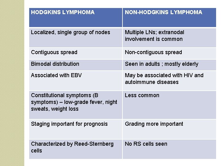
HODGKINS LYMPHOMA NON-HODGKINS LYMPHOMA Localized, single group of nodes Multiple LNs; extranodal involvement is common Contiguous spread Non-contiguous spread Bimodal distribution Seen in adults ; mostly elderly Associated with EBV May be associated with HIV and autoimmune diseases Constitutional symptoms (B symptoms) – low-grade fever, night sweats, weight loss Less common Staging important for prognosis Grading more important Characterized by Reed-Sternberg cells No RS cells seen
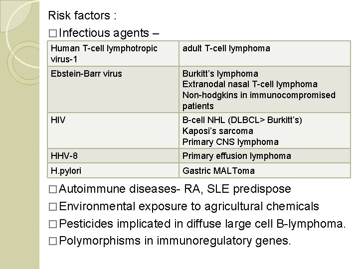
Risk factors : � Infectious agents – Human T-cell lymphotropic virus-1 adult T-cell lymphoma Ebstein-Barr virus Burkitt’s lymphoma Extranodal nasal T-cell lymphoma Non-hodgkins in immunocompromised patients HIV B-cell NHL (DLBCL> Burkitt’s) Kaposi’s sarcoma Primary CNS lymphoma HHV-8 Primary effusion lymphoma H. pylori Gastric MALToma � Immunodeficiency increases the risk � Autoimmune diseases- RA, SLE predispose � Environmental exposure to agricultural chemicals � Pesticides implicated in diffuse large cell B-lymphoma. � Polymorphisms in immunoregulatory genes.
Classification Earlier the REAL classification was used which had some deficiencies Currently the WHO classification is followed

WHO classification Precursor B-cell neoplasms Precursor T-cell neoplasm Mature B-cell Neoplasms mature T-cell and NK-cell lymphomas
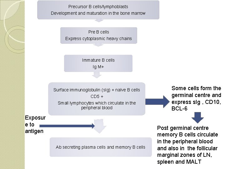
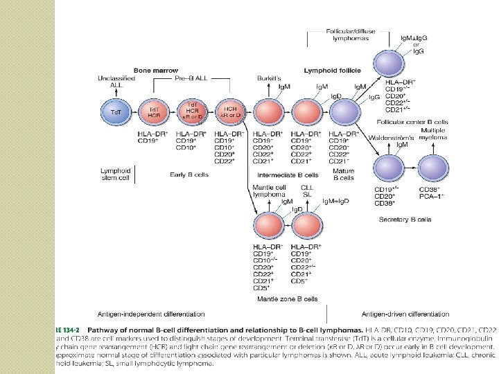
Precursor B cells/lymphoblasts Development and maturation in the bone marrow Pre B cells Express cytoplasmic heavy chains Immature B cells Ig M+ Surface immunoglobulin (s. Ig) + naïve B cells CD 5 + Small lymphocytes which circulate in the peripheral blood Exposur e to antigen Ab secreting plasma cells and memory B cells Some cells form the germinal centre and express s. Ig , CD 10, BCL-6 Post germinal centre memory B cells circulate in the peripheral blood and also in the follicular marginal zones of LN, spleen and MALT
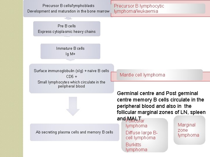
Precursor B cells/lymphoblasts Development and maturation in the bone marrow Precursor B lymphocytic lymphoma/leukaemia Pre B cells Express cytoplasmic heavy chains Immature B cells Ig M+ Surface immunoglobulin (s. Ig) + naïve B cells CD 5 + Small lymphocytes which circulate in the peripheral blood Mantle cell lymphoma Germinal centre and Post germinal centre memory B cells circulate in the peripheral blood and also in the follicular marginal zones of LN, spleen and MALT Follicular lymphoma Ab secreting plasma cells and memory B cells Diffuse large Bcell lymphoma Burkitts lymphoma Marginal zone lymphoma
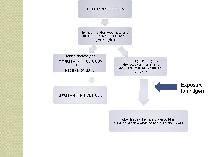
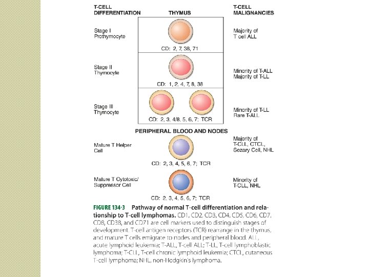
Precursor in bone marrow Thymus – undergoes maturation into various types of naïve tlymphocytes Cortical thymocytes Immature – Td. T, c. CD 3, CD 5 CD 7 Negative for CD 4, 8 Mature – express CD 4, CD 8 Medullary thymocytes phenotypically similar to peripheral mature T-cells and NK-cells Exposure to antigen After leaving thymus undergo blast transformation – effector and memory T cells
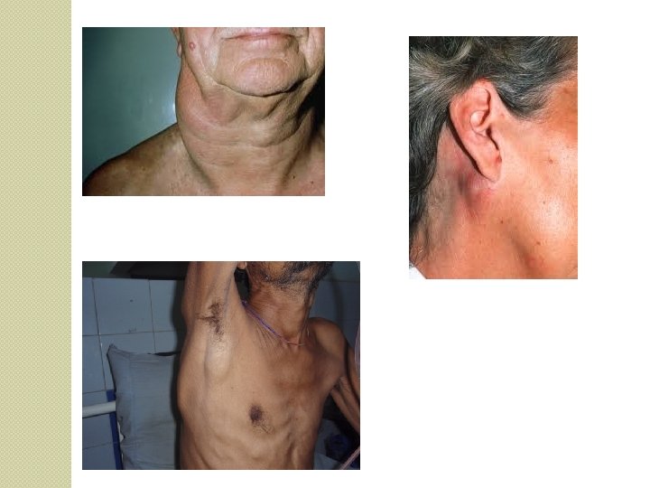
Clinical features �More frequent in elderly �Most subtypes show a male preponderance �Generally present with painless, generalised lymphadenopathy especially in the cervical or supraclavicular region. �Widely disseminated at presentation �Primary extranodal involvement is more common.
�Generalised lymphadenopathy – Enlargement of three or more non-contiguous lymph node groups. �Significant lymphadenopathy – Ø >1 cm in cervical and axillary region Ø > 1. 5 -2 cm in inguinal region Ø Any supraclavicular or scalene lymphadenopathy is always abnormal.
� Nasopharynx or tonsilar involvement : soreness or pain in throat, Ø nasal obstruction or bleeding, Ø lump in throat or dysphagia. Ø � Waldeyer’s ring involvement : Ø Dysphagia Ø Compression of trachea
�GI mc extranodal site – stomach, mesenteric LNs, peritoneum, liver, small bowel, oesophagus. Ø GI bleed, Ø abdominal pain, Ø vomiting, dyspepsia, nausea, Ø change of bowel habits Ø weight loss. Ø Hepatosplenomegaly � Compression syndrome Ø Gut obstruction Ø Ascites Ø SVC obstruction
� Skin involvement – nodules on head or trunk � CNS- Ø headache, Ø lethargy, Ø hemiparesis Ø seizures. Ø May infiltrate meninges. Ø � Blurring of vision, loss of visual acuity – ocular lymphoma.
� Kidney or testicular involvement – Ø urinary tract obstruction or testicular mass. � Metabolic complications – Hyperuricemia Ø hypercalcemia Ø
�Systemic features less common than in Hodgkins. �Pruritis uncommon �Fever indicates secondary infection. �If present indicate advanced disease and apoorer prognosis. �Autoimmune disorders more common.
� Blood picture – normal hematological parameters early in the disease. Ø Advanced – Hb falls, may be thrombocytopenia and neutropenia. Ø Marrow involvement in 30 -70% Ø
Diagnosis �Rule out other causes of LN enlargement. Bacterial Viral parasitic Others
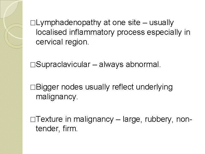
�Lymphadenopathy at one site – usually localised inflammatory process especially in cervical region. �Supraclavicular – always abnormal. �Bigger nodes usually reflect underlying malignancy. �Texture in malignancy – large, rubbery, non- tender, firm.
Ø Lymph node biopsy Ø Fine-needle aspiration cytology Ø Immunophenotyping Ø Immunohistochemistry Ø Flow cytometry Ø Genetics – PCR or southern blotting. Ø If liver or BM involvement suspected – trephine biopsy of BM or liver biopsy. Ø Occassionally, diagnostic laparotomy.

�Serum albumin is used as a prognostic factor in DLBCL. A new prognostic index A- IPI (albumin adjusted) has also been proposed. �Recent studies have also shown that vitamin D 3 deficiency is a negative prognostic factor in aggressive lymphomas. �Current studies are focused on supplementation of vitamin D 3 in deficient patients during treatment and to see if there is any benefit.
Subtyping of NHL �Lineage markers �Lymphoma specific markers �Morphology �Genetics
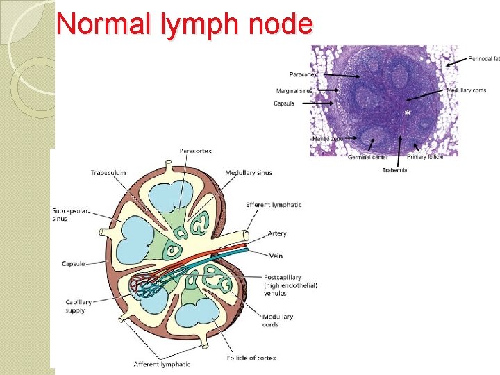
Normal lymph node
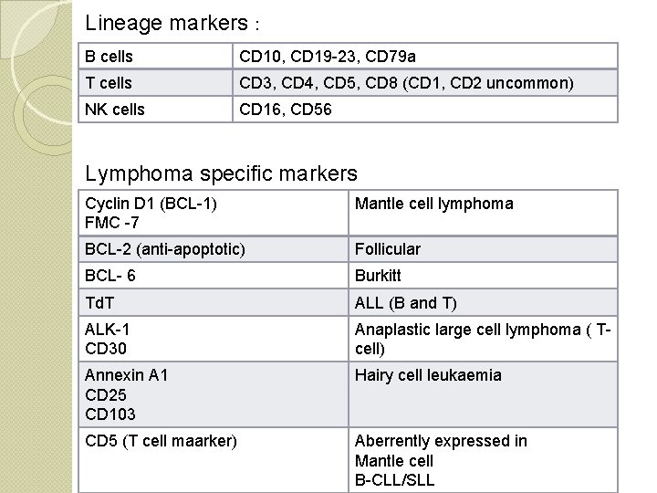
Lineage markers : B cells CD 10, CD 19 -23, CD 79 a T cells CD 3, CD 4, CD 5, CD 8 (CD 1, CD 2 uncommon) NK cells CD 16, CD 56 Lymphoma specific markers Cyclin D 1 (BCL-1) FMC -7 Mantle cell lymphoma BCL-2 (anti-apoptotic) Follicular BCL- 6 Burkitt Td. T ALL (B and T) ALK-1 CD 30 Anaplastic large cell lymphoma ( Tcell) Annexin A 1 CD 25 CD 103 Hairy cell leukaemia CD 5 (T cell maarker) Aberrently expressed in Mantle cell B-CLL/SLL
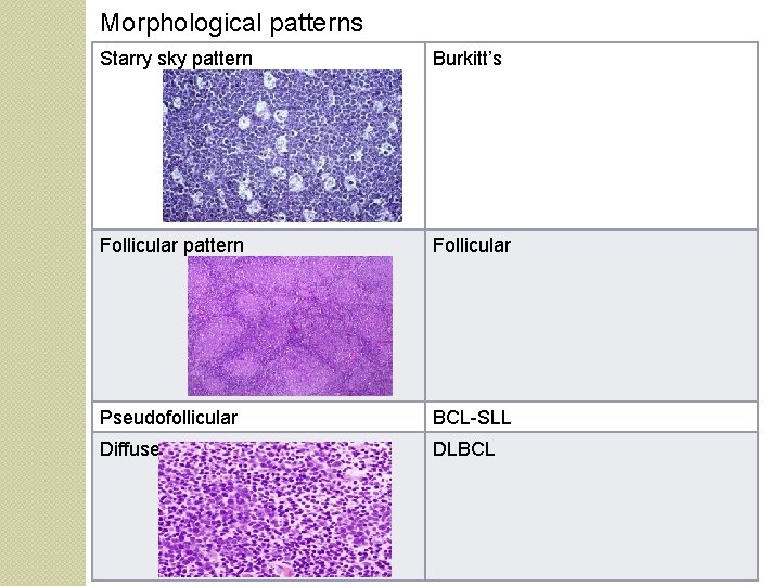
Morphological patterns Starry sky pattern Burkitt’s Follicular pattern Follicular Pseudofollicular BCL-SLL Diffuse DLBCL
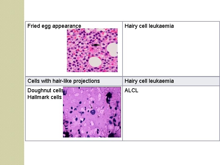
Fried egg appearance Hairy cell leukaemia Cells with hair-like projections Hairy cell leukaemia Doughnut cells Hallmark cells ALCL
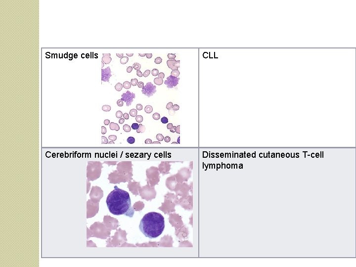
Smudge cells CLL Cerebriform nuclei / sezary cells Disseminated cutaneous T-cell lymphoma
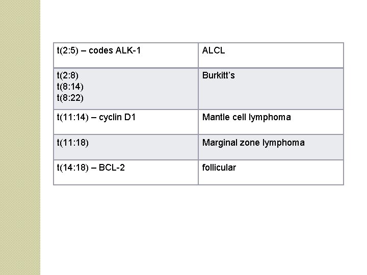
t(2: 5) – codes ALK-1 ALCL t(2: 8) t(8: 14) t(8: 22) Burkitt’s t(11: 14) – cyclin D 1 Mantle cell lymphoma t(11: 18) Marginal zone lymphoma t(14: 18) – BCL-2 follicular
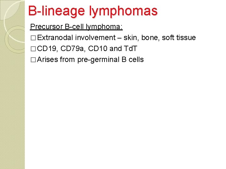
B-lineage lymphomas Precursor B-cell lymphoma: � Extranodal involvement – skin, bone, soft tissue � CD 19, CD 79 a, CD 10 and Td. T � Arises from pre-germinal B cells
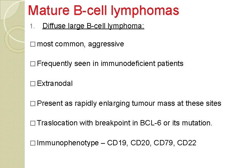
Mature B-cell lymphomas 1. Diffuse large B-cell lymphoma: � most common, aggressive � Frequently seen in immunodeficient patients � Extranodal � Present as rapidly enlarging tumour mass at these sites � Traslocation with breakpoint in BCL-6 or its mutation. � Immunophenotype – CD 19, CD 20, CD 79, CD 22
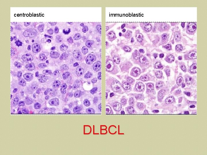
centroblastic immunoblastic DLBCL
2. Follicular lymphoma : � Middle-aged adults � From germinal centre B-cells. � Nodal involvement is common but extranodal may be seen. � Bone marrow – 85% � B symptoms common � Indolent course � Moleucular characteristic – t(14, 18) – overexpression of BCL-2 which prevents apoptosis – 90% of follicular lymphomas
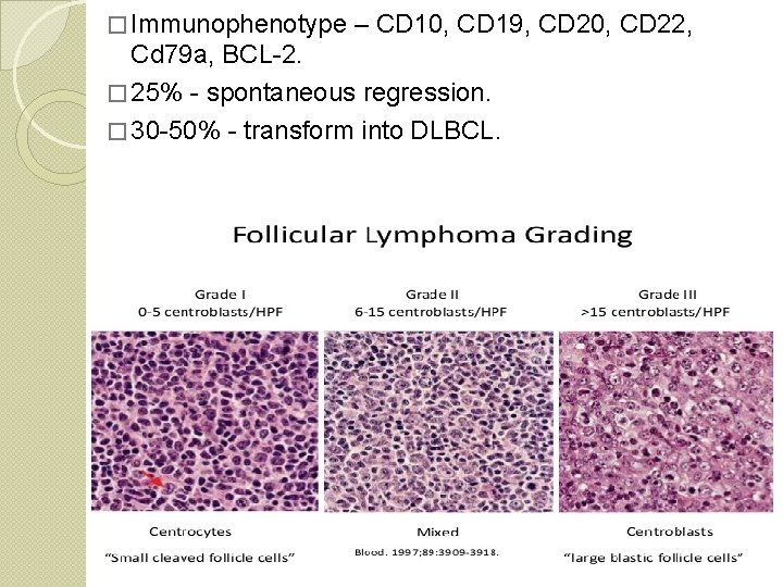
� Immunophenotype – CD 10, CD 19, CD 20, CD 22, Cd 79 a, BCL-2. � 25% - spontaneous regression. � 30 -50% - transform into DLBCL.
Mantle cell lymphoma – � From naïve B-cells, pre-germinal. � t(11: 14) 3. Extranodal marginal zone lymphoma of MALT (MALToma) � Arises in extranodal tissues involved in chronic inflammation � Arises from marginal zone B-cells. � Indolent and remains localized for years. � Prognosis is good � May transform into DLBCL 4.
� Burkitts – � From mature B-cells. � Three variants � Associated with EBV � Also in HIV-infected patients � CNS, jaw and other facial bones commonly involved � Aggressive tumour � Responds to chemotherapy
T-lineage lymphomas � Precursor T-cell lymphomas – � Highly aggressive � Patients present with a mass in anterior mediastinum along with supradiaphragmatic lymphadenopathy and pleural effusion. � Can involve CNS � Diffuse pattern � Disseminated disease and B symptoms are common
Mature T-cell lymphoma 1. Mycosis fungoides/ cutaneous T-ell lymphoma � Indolent � Presents with eczematous skin lesions � Median survival is 1 year from development of systemic disease. � Lymphopenia � Increase in suppressor: helper T-cell ratio. � Histology – pautrier’s micro-abscesses. � Circulating tumour cells – Sezary syndrome
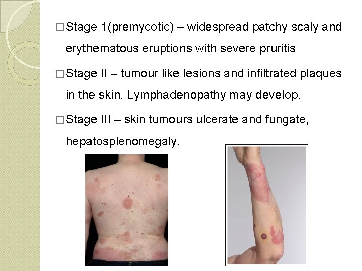
� Stage 1(premycotic) – widespread patchy scaly and erythematous eruptions with severe pruritis � Stage II – tumour like lesions and infiltrated plaques in the skin. Lymphadenopathy may develop. � Stage III – skin tumours ulcerate and fungate, hepatosplenomegaly.
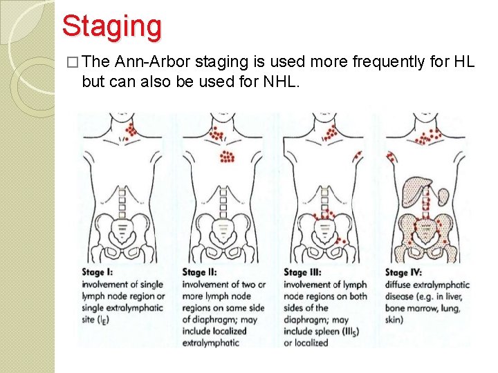
Staging � The Ann-Arbor staging is used more frequently for HL but can also be used for NHL.
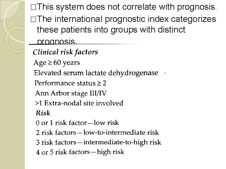
�This system does not correlate with prognosis. �The international prognostic index categorizes these patients into groups with distinct prognosis.
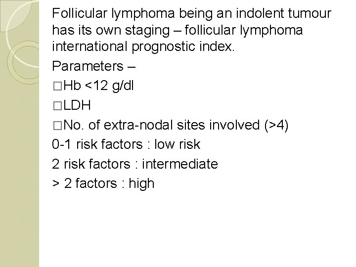
Follicular lymphoma being an indolent tumour has its own staging – follicular lymphoma international prognostic index. Parameters – �Hb <12 g/dl �LDH �No. of extra-nodal sites involved (>4) 0 -1 risk factors : low risk 2 risk factors : intermediate > 2 factors : high
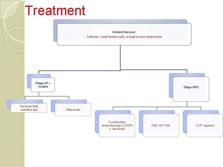
Treatment Indolent tumours: follicular, small lymphocytic, marginal zone lymphomas Stage I/II – curable Involved field radiotherapy Stage III/IV Rituximab Combination chemotherapy (CHOP) + rituximab FND OR FAD CVP regimen

�DLBCL- Most popular – CHOP+ Rituximab Stage I/II (non-bulky) – 3 -4 cycles Bulky II/IV – 6 -8 cycles with/without subsequent field radiotherapy. 30 -40% - refractory or relapse – salvage therapy Autologous bone marrow transplant superior to salvage therapy in patients whose lymphomas are chemo-sensitive after relapse.
�Follicular lymphoma – Ø Responds well to chemotherapy and radiotherapy. Ø Asymptomatic – wait and watch. Ø Single agent - Chlorambucil/cyclophosphamide if treatment is required. Or CVP or CHOP Ø Localised – field radiotherapy Ø Remission - Addition of interferon alpha Ø Relapse – Rituximab Or bedamustine with rituximab
�Chronic lymphoid leukaemia (CLL)– Ø Cholarambucil alone or in combination. Ø Fludarabine based regimens are preferred Ø Anti-CD 52 antibody – alemtuzimab but causes severe immunosuppression. Ø Young patients – bone marrow transplant considered but not very encouraging. Ø 2 newer antibodies – Ofatumumab and Obinutuzumab Ø Others- Ibrutinib and idelalisib
�Marginal zone lymphoma – is localized hence responds to local therapy- radiation or surgery.
�Mantle cell lymphoma – Ø Intermediate behaviour hence does not respond to conventional chemotherapy. Ø Hyper C-VAD (cyclophosphamide, vincristine, adriamycin and dexamethasone) regimen in combination with rituximab. Plus high dose methotrexate and cytarabine. Ø Bendamustine + rituximab

THANK YOU
- Slides: 53