
Manuscript Explicit Constructivism a missing link in ineffective




- Slides: 54
Manuscript: Explicit Constructivism: a missing link in ineffective lectures? Author: E. S. Prakash Supplement 1: Typical Lecture Note - Copyrighted images have been removed and replaced with a URL link to those images. This is an open access article distributed under the terms of the Creative Commons Attribution License http: //creativecommons. org/licenses/by-nc-sa/3. 0/
Chemical and neural regulation of respiration E. S. Prakash, School of Medicine, AIMST University, Malaysia E-mail: dresprakash@gmail. com
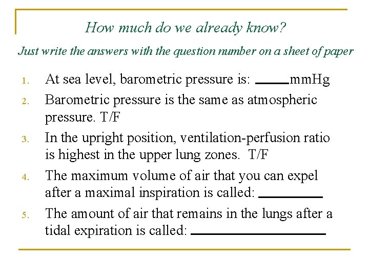
How much do we already know? Just write the answers with the question number on a sheet of paper 1. 2. 3. 4. 5. At sea level, barometric pressure is: mm. Hg Barometric pressure is the same as atmospheric pressure. T/F In the upright position, ventilation-perfusion ratio is highest in the upper lung zones. T/F The maximum volume of air that you can expel after a maximal inspiration is called: The amount of air that remains in the lungs after a tidal expiration is called:
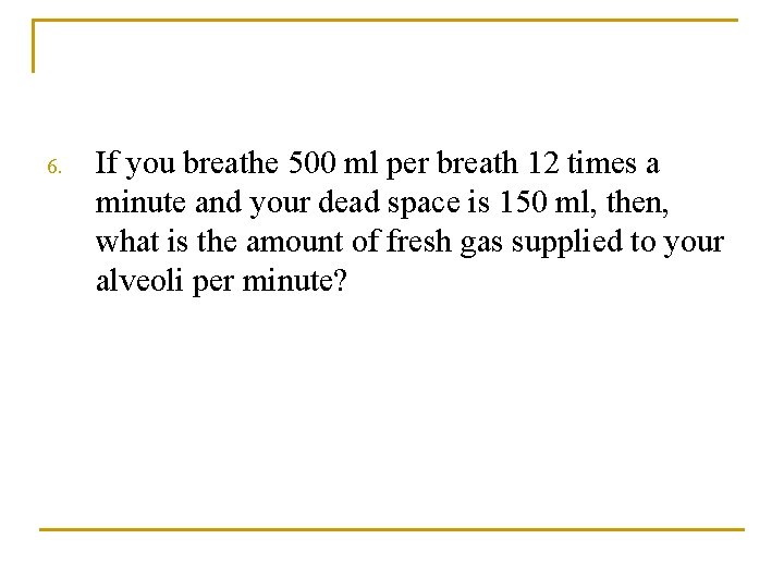
6. If you breathe 500 ml per breath 12 times a minute and your dead space is 150 ml, then, what is the amount of fresh gas supplied to your alveoli per minute?
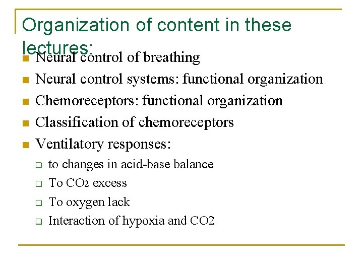
Organization of content in these lectures: n Neural control of breathing n n Neural control systems: functional organization Chemoreceptors: functional organization Classification of chemoreceptors Ventilatory responses: q q to changes in acid-base balance To CO 2 excess To oxygen lack Interaction of hypoxia and CO 2
Content outline continued: n Nonchemical influences on respiration q q q n n Responses mediated by airway receptors Responses mediated by receptors in the lung parenchyma Coughing & sneezing Regulation of respiration during sleep Abnormal respiratory patterns
Neural control of breathing There are 2 systems: n One for voluntary control n One for spontaneous breathing
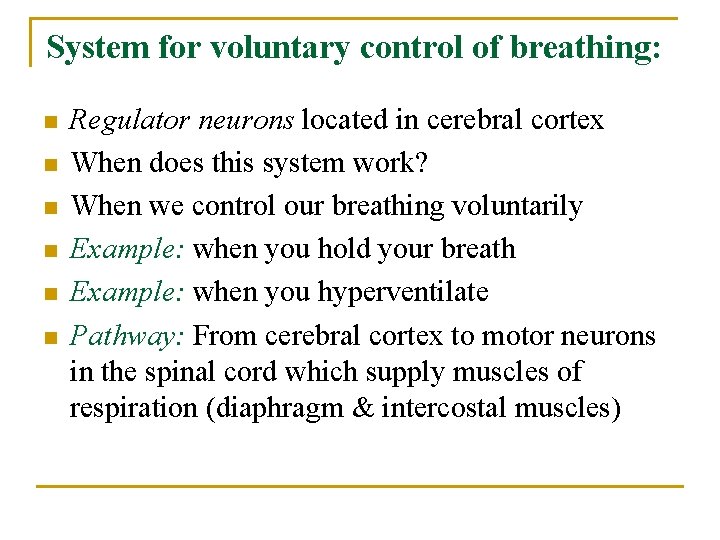
System for voluntary control of breathing: n n n Regulator neurons located in cerebral cortex When does this system work? When we control our breathing voluntarily Example: when you hold your breath Example: when you hyperventilate Pathway: From cerebral cortex to motor neurons in the spinal cord which supply muscles of respiration (diaphragm & intercostal muscles)
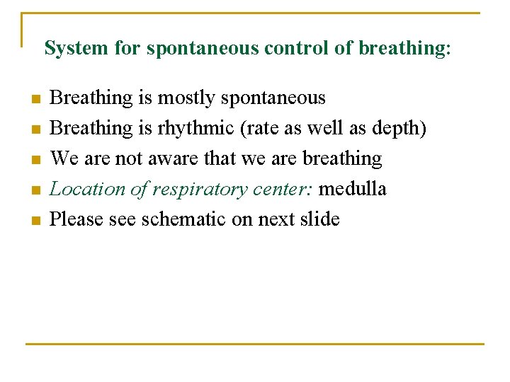
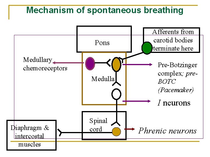
System for spontaneous control of breathing: n n n Breathing is mostly spontaneous Breathing is rhythmic (rate as well as depth) We are not aware that we are breathing Location of respiratory center: medulla Please see schematic on next slide
Mechanism of spontaneous breathing Pons Medullary chemoreceptors Medulla Afferents from carotid bodies terminate here Pre-Botzinger complex; pre. BOTC (Pacemaker) I neurons Diaphragm & intercostal muscles Spinal cord Phrenic neurons
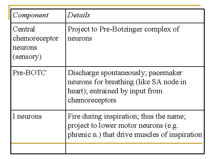
Component Details Central chemoreceptor neurons (sensory) Project to Pre-Botzinger complex of neurons Pre-BOTC Discharge spontaneously; pacemaker neurons for breathing (like SA node in heart); entrained by input from chemoreceptors I neurons Fire during inspiration; thus the name; project to lower motor neurons (e. g. phrenic n. ) that drive muscles of inspiration
Feedback to the respiratory center from lungs: n During inspiration, q lungs expand, and lung parenchyma is stretched; q stretch receptors are present here; q these are activated and convey information to the brain via sensory branches of vagus nerve
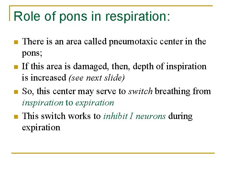
Role of pons in respiration: n n There is an area called pneumotaxic center in the pons; If this area is damaged, then, depth of inspiration is increased (see next slide) So, this center may serve to switch breathing from inspiration to expiration This switch works to inhibit I neurons during expiration
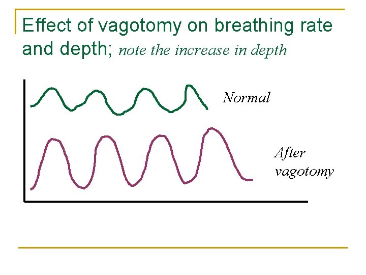
Effect of vagotomy on breathing rate and depth; note the increase in depth Normal After vagotomy
Effect of damage to the pneumotaxic center in vagotomized animals: normal After vagotomy apneusis
Functional organization of chemoreceptors: n A rise in PCO 2, a fall in p. H or PO 2 of arterial blood increases respiratory neuron activity in the medulla. n Stimulus: a change in blood chemistry … Sensed by: receptors called chemoreceptors Response: change in minute ventilation n n
Functional organization of chemoreceptors (contd. ) Location of chemoreceptors: n Central chemoreceptors (in medulla); also called medullary chemoreceptors n Arterial chemoreceptors (in carotid & aortic bodies); sometimes called peripheral chemoreceptors
Functional organization of chemoreceptors (contd. ) n Innervation of peripheral (systemic arterial chemoreceptors); n Figure at Link: http: //www. medicine. mcgill. ca/physio/resp-web/Figures/Figtt 20. jpg n Note carotid body is supplied by branch of IX nerve and aortic bodies are supplied by branch of X nerve.
Some facts about systemic arterial chemoreceptors: n n n There are 2 types of cells in the carotid body; Type I glomus cells contain oxygen sensitive K channels (these are the chemoreceptors) Type II cells are supporting cells They have a very high blood flow In carotid bodies, blood flow rate: 2000 ml/100 g tissue/min For example, the brain gets 50 ml/100 g/min
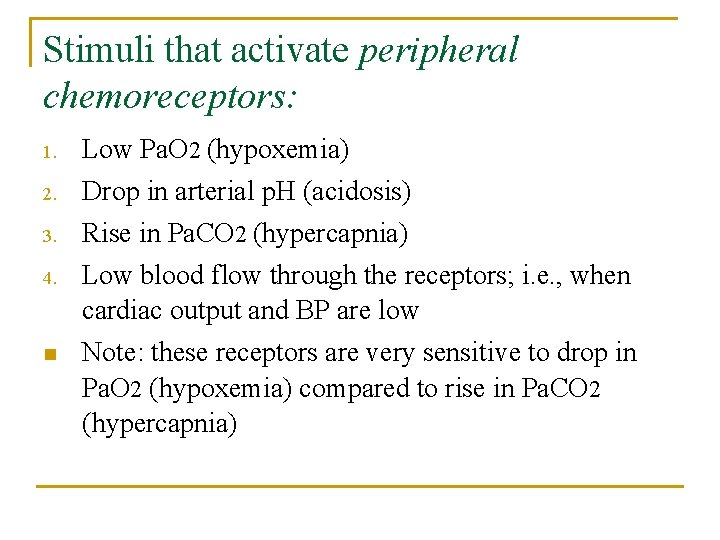
Stimuli that activate peripheral chemoreceptors: 1. Low Pa. O 2 (hypoxemia) 2. Drop in arterial p. H (acidosis) 3. Rise in Pa. CO 2 (hypercapnia) 4. Low blood flow through the receptors; i. e. , when cardiac output and BP are low Note: these receptors are very sensitive to drop in Pa. O 2 (hypoxemia) compared to rise in Pa. CO 2 (hypercapnia) n
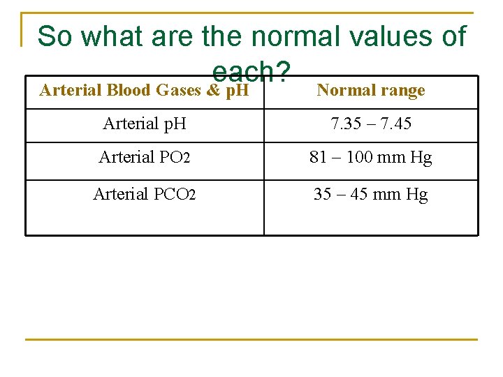
So what are the normal values of each? Arterial Blood Gases & p. H Normal range Arterial p. H 7. 35 – 7. 45 Arterial PO 2 81 – 100 mm Hg Arterial PCO 2 35 – 45 mm Hg
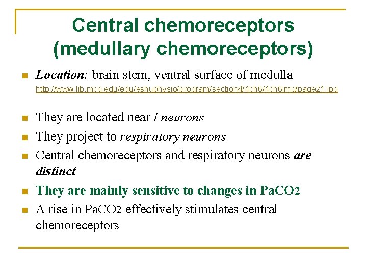
Central chemoreceptors (medullary chemoreceptors) n Location: brain stem, ventral surface of medulla http: //www. lib. mcg. edu/eshuphysio/program/section 4/4 ch 6 img/page 21. jpg n n n They are located near I neurons They project to respiratory neurons Central chemoreceptors and respiratory neurons are distinct They are mainly sensitive to changes in Pa. CO 2 A rise in Pa. CO 2 effectively stimulates central chemoreceptors
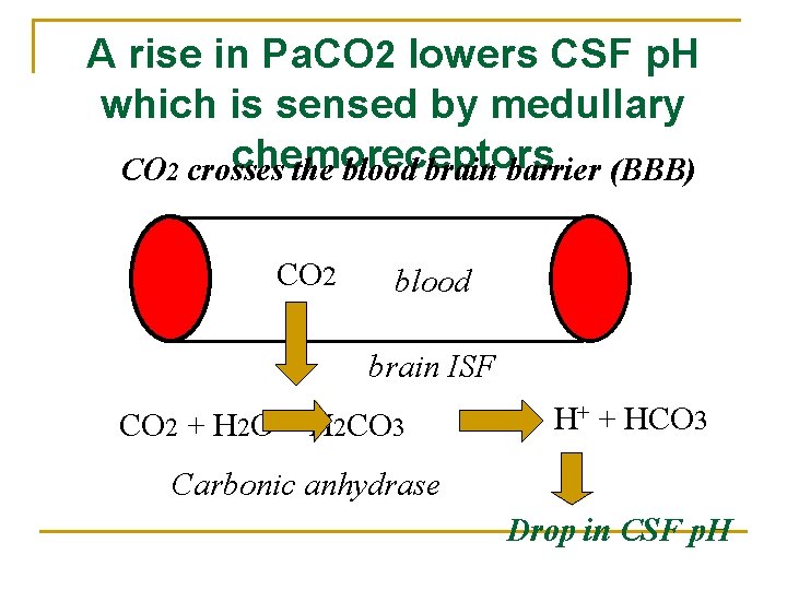
A rise in Pa. CO 2 lowers CSF p. H which is sensed by medullary chemoreceptors CO 2 crosses the blood brain barrier (BBB) CO 2 blood brain ISF CO 2 + H 2 O H 2 CO 3 H+ + HCO 3 Carbonic anhydrase Drop in CSF p. H
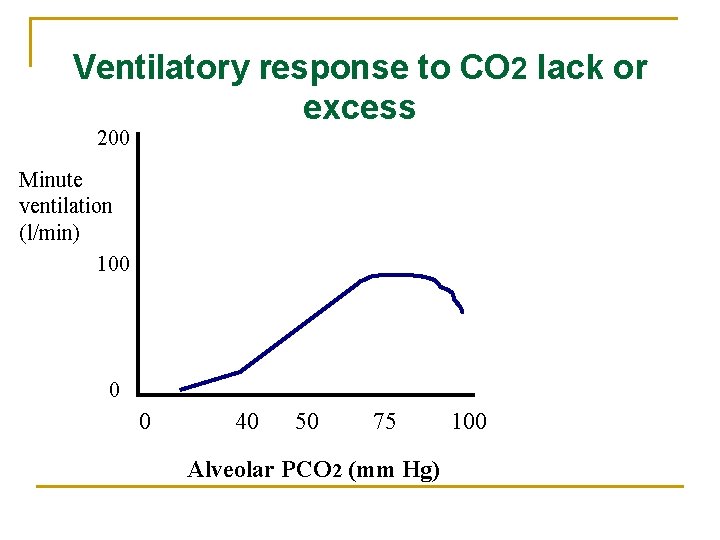
n n n Central chemoreceptor neurons monitor the H+ ion concentration of brain ISF; Greater the Pa. CO 2, > the minute ventilation; If you lower Pa. CO 2, minute ventilation is lowered
Effect of addition of metabolic acid (e. g. , lactic acid, on ventilation) n n Example: lactic acidosis (metabolic acidosis) Arterial p. H is low (< 7. 35); Breathing is rapid and deep (Kussmaul’s respiration) and CO 2 is blown off This response is mediated by carotid bodies (peripheral chemoreceptors) and is lost if they are removed.
Effect of a rise in blood p. H on minute ventilation n n Example: metabolic alkalosis due to vomiting; i. e. , loss of HCl; Arterial p. H is high (> 7. 45) Respiration is slowed; i. e. , decrease in minute ventilation) As a result Pa. CO 2 gradually rises
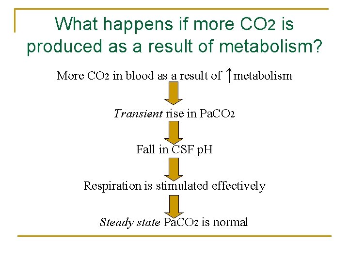
What happens if more CO 2 is produced as a result of metabolism? More CO 2 in blood as a result of ↑metabolism Transient rise in Pa. CO 2 Fall in CSF p. H Respiration is stimulated effectively Steady state Pa. CO 2 is normal
Ventilatory response to CO 2 lack or excess 200 Minute ventilation (l/min) 100 0 0 40 50 75 Alveolar PCO 2 (mm Hg) 100
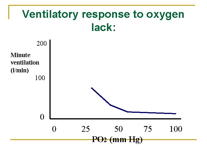
Ventilatory response to oxygen lack: 200 Minute ventilation (l/min) 100 0 0 25 50 75 PO 2 (mm Hg) 100
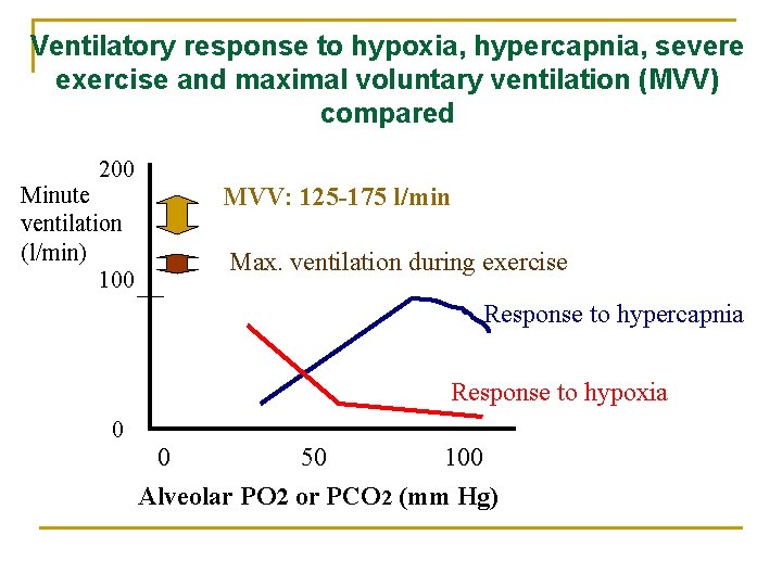
Ventilatory response to hypoxia, hypercapnia, severe exercise and maximal voluntary ventilation (MVV) compared 200 Minute ventilation (l/min) 100 MVV: 125 -175 l/min Max. ventilation during exercise Response to hypercapnia Response to hypoxia 0 0 50 100 Alveolar PO 2 or PCO 2 (mm Hg)
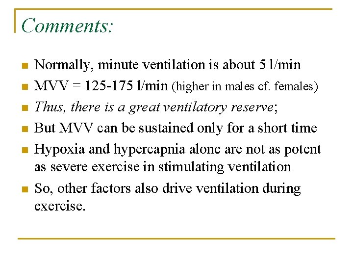
Comments: n n n Normally, minute ventilation is about 5 l/min MVV = 125 -175 l/min (higher in males cf. females) Thus, there is a great ventilatory reserve; But MVV can be sustained only for a short time Hypoxia and hypercapnia alone are not as potent as severe exercise in stimulating ventilation So, other factors also drive ventilation during exercise.
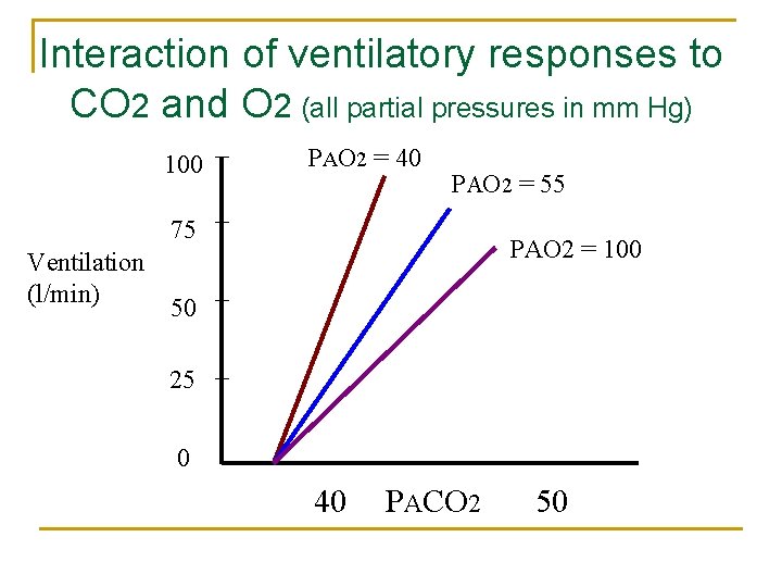
Interaction of ventilatory responses to CO 2 and O 2 (all partial pressures in mm Hg) 100 PAO 2 = 40 PAO 2 = 55 75 Ventilation (l/min) PAO 2 = 100 50 25 0 40 PACO 2 50
Conclusion: n Conclusion: Hypoxia makes an individual more sensitive to CO 2 excess
Ventilation at high altitudes: n n n n Barometric (atmospheric) pressure is lower; When Pa. O 2 is < 60 mm Hg, min. ventilation ↑ What happens to Pa. CO 2? It is lowered as a result of hyperventilation What happens to p. H of arterial blood? p. H increases slightly say from 7. 4 to 7. 45 Arterial blood gases: Hypoxemia (low Pa. O 2); hypocapnia (Pa. CO 2 < 35 mm Hg); respiratory alkalosis (p. H > 7. 45 because of hypocapnia)
Some working definitions for you: n n n Normocapnia: Pa. CO 2 between 35 and 45 mm Hg Hypocapnia: Pa. CO 2 < 35 mm Hg Hypercapnia: Pa. CO 2 > 45 mm Hg Hypoxemia: Pa. O 2 < 80 mm Hg Note: significant activation of carotid bodies occurs only when Pa. O 2 < 60 mm Hg
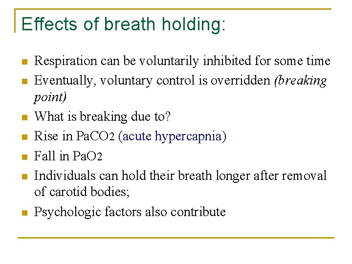
Effects of breath holding: n n n n Respiration can be voluntarily inhibited for some time Eventually, voluntary control is overridden (breaking point) What is breaking due to? Rise in Pa. CO 2 (acute hypercapnia) Fall in Pa. O 2 Individuals can hold their breath longer after removal of carotid bodies; Psychologic factors also contribute
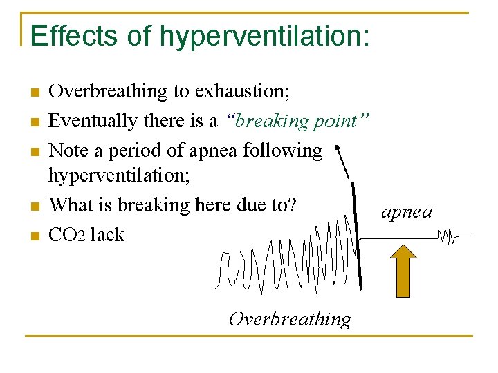
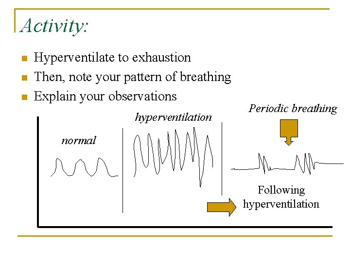
Effects of hyperventilation: n n n Overbreathing to exhaustion; Eventually there is a “breaking point” Note a period of apnea following hyperventilation; What is breaking here due to? apnea CO 2 lack Overbreathing
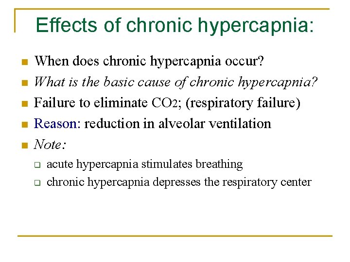
Effects of chronic hypercapnia: n n n When does chronic hypercapnia occur? What is the basic cause of chronic hypercapnia? Failure to eliminate CO 2; (respiratory failure) Reason: reduction in alveolar ventilation Note: q q acute hypercapnia stimulates breathing chronic hypercapnia depresses the respiratory center
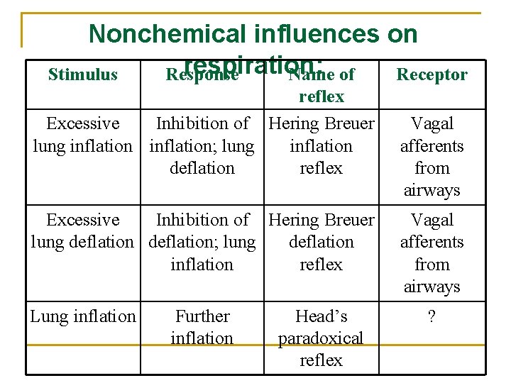
Nonchemical influences on respiration: Stimulus Response Name of Receptor reflex Excessive Inhibition of Hering Breuer lung inflation; lung inflation deflation reflex Excessive Inhibition of Hering Breuer lung deflation; lung deflation inflation reflex Lung inflation Further inflation Head’s paradoxical reflex Vagal afferents from airways ?
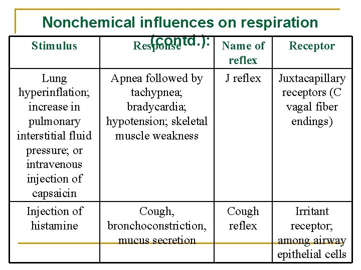
Nonchemical influences on respiration (contd. ): Name of Stimulus Response Receptor Lung hyperinflation; increase in pulmonary interstitial fluid pressure; or intravenous injection of capsaicin Injection of histamine Apnea followed by tachypnea; bradycardia; hypotension; skeletal muscle weakness Cough, bronchoconstriction, mucus secretion reflex J reflex Cough reflex Juxtacapillary receptors (C vagal fiber endings) Irritant receptor; among airway epithelial cells
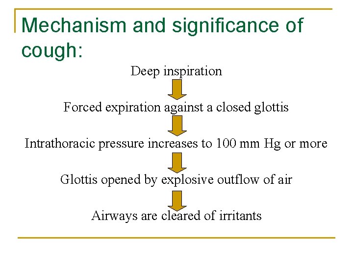
Mechanism and significance of cough: Deep inspiration Forced expiration against a closed glottis Intrathoracic pressure increases to 100 mm Hg or more Glottis opened by explosive outflow of air Airways are cleared of irritants
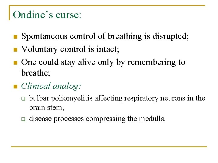
Ondine’s curse: n n Spontaneous control of breathing is disrupted; Voluntary control is intact; One could stay alive only by remembering to breathe; Clinical analog: q q bulbar poliomyelitis affecting respiratory neurons in the brain stem; disease processes compressing the medulla
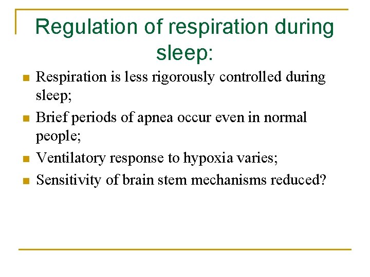
Regulation of respiration during sleep: n n Respiration is less rigorously controlled during sleep; Brief periods of apnea occur even in normal people; Ventilatory response to hypoxia varies; Sensitivity of brain stem mechanisms reduced?
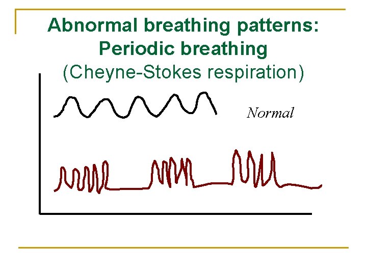
Abnormal breathing patterns: Periodic breathing (Cheyne-Stokes respiration) Normal
Cheyne-Stokes respiration: Periods of apnea punctuated by periods of hyperpnea It occurs in: n n q q 1. 2. congestive heart failure brain stem disease affecting respiratory centers Mechanisms postulated to explain this: Prolonged lung-to-brain circulation time Changes in sensitivity of medullary respiratory neurons
Activity: n n n Hyperventilate to exhaustion Then, note your pattern of breathing Explain your observations hyperventilation Periodic breathing normal Following hyperventilation
Outline of the explanation: n n n n Hyperventilation eliminates CO 2; Apnea is due to lack of CO 2 During apnea, Pa. O 2 falls & stimulates breathing Few breaths eliminate hypoxia Now there is no stimulus for breathing So there is apnea again Normal breathing resumes only when Pa. CO 2 is 40 mm Hg Conclusion: normal breathing pattern is entrained by Pa. CO 2 not Pa. O 2
Some items for self-study: n n n How is breathing regulated during exercise? What is the mechanism of hiccups? What is the mechanism of yawning? What is the mechanism of sneezing? What happens when you sigh?
POST-TEST
You should also be able to answer these questions: 1. 2. 3. Describe with the help of schematic diagram, the neural mechanism of spontaneous breathing Describe with the help of schematic diagram, the neural mechanism of voluntary control of respiration Describe with the help of schematic diagram, the role of systemic arterial chemoreceptors in the regulation of alveolar ventilation
4. 5. 6. 7. Describe with the help of schematic diagrams the functional organization and functions of medullary chemoreceptors. How does CO 2 stimulate breathing? What is the relationship between Pa. CO 2 and minute ventilation? Describe the mechanism responsible for periodic breathing following voluntary hyperventilation
8. 9. 10. Explain the factors that affect breath holding time. Briefly explain the effect of damage to the pneumotaxic center on the pattern of breathing Briefly explain the effect of vagotomy on the pattern of breathing in experimental animals.
11. 12. 13. 14. 15. What is the difference between the effect of acute hypercapnia and chronic hypercapnia on minute ventilation? What is Kussmaul’s respiration? When does it occur? What is the mechanism involved? What is periodic breathing? When does it occur? What is Cheyne-Stokes respiration? What are the Hering Breuer reflexes? What is Head’s paradoxical reflex?
Required Reading: n Chapter 36. Regulation of respiration. Ganong WF. Review of Medical Physiology, Mc Graw Hill Co, 2005