Pulmonary infections Pneumonia Pneumonia can be very broadly
 Pneumonia can be very broadly defined as any infection in the")
Pulmonary infections (Pneumonia) Pneumonia can be very broadly defined as any infection in the lung

Pulmonary infections • Respiratory tract infections are more frequent than infections of any other organ and account for the largest number of workdays lost in the general population, why? – The epithelium of the lung is exposed to liters of contaminated air – Nasopharyngeal flora are aspirated during sleep – Underlying lung diseases render the lung parenchyma vulnerable to virulent organism.

Pulmonary infections • Upper respiratory tract infection are common, caused mainly by viruses (common cold, pharyngitis) • Infection of the lung by virus, mycoplasma, bacteria and fungi account for enormous amount of morbidity and mortality.

Pathogenesis of pneumonia Each day, the respiratory tract is exposed to more than 10, 000 liters of air containing hazardous dust, Chemicals and microorganisms. - Particle > 10 mm deposited in nose. - Particle 3 -10 mm impacted in trachea and bronchi. - Particle 1 -3 mm (bacteria) deposited in terminal airways and alveoli. - Smaller particles < 1 mm may remain suspended in air. - Normal lung is free from bacteria.

Pathogenesis of pneumonia • Pneumonia can result whenever: – defense mechanisms are impaired – the resistance of the host in general is lowered.

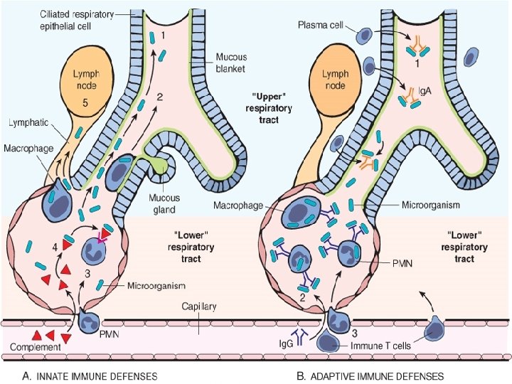
Pulmonary host defenses • Upper airways: – Nasopharynx: Nasal hair, turbinates, mucociliary apparatus, Ig. A secretion – Oropharynx Saliva, sloughing of epithelium, local complement production, interference from resident flora
: Cough, epiglottic")
Pulmonary host defenses • Upper airways: • Conducting airways (trachea and bronchi): Cough, epiglottic reflexes, sharp angled branches of the airways, mucociliary apparatus, Immunoglobulin (Ig. M, Ig. G, and Ig. A) secretion

Pulmonary host defenses • Lower respiratory tract: Alveolar lining fluid ( surfactant, immunoglobulin, complement and fibronectin), Cytokines (IL-1, TNF), alveolar macrophages, polymorphonuclear leukocyte, cell mediated immunity

Pathogenesis of pneumonia Impaired defense mechanisms: • Loss or suppression of the cough reflex, as a result of coma, anesthesia, neuromuscular disorders, drugs, or chest pain. • Injury to the mucociliary apparatus, by either impairment of ciliary function or destruction of ciliated epithelium e. g. cigarette smoke, inhalation of hot or corrosive gases, viral diseases, or genetic disturbances • Interference with the phagocytic or bactericidal action of alveolar macrophages by alcohol, tobacco smoke, anoxia, or oxygen intoxication • Pulmonary congestion and edema • Accumulation of secretions e. g. cystic fibrosis and bronchial obstruction • Defect in innate immunity Include neutrophil, complement, humoral and cell mediated immune defects
 and")
Pathogenesis of pneumonia Ø Defects in innate immunity (including neutrophil and complement defects) and humoral immunodeficiency lead to an increased incidence of infections with pyogenic bacteria. Ø Cell-mediated immune defects lead to increased infections with intracellular microbes such as mycobacteria , herpesviruses and Pneumocystis jiroveci. Ø Several exogenous aspects of lifestyle interfere with host immune defense mechanisms and facilitate infections. § Examples: • cigarette smoke compromises mucociliary clearance and pulmonary macrophage activity • alcohol not only impairs cough and epiglottic reflexes, thereby increasing the risk of aspiration, but also interferes with neutrophil mobilization and chemotaxis.

Pathogenesis of pneumonia • General factors that affect resistance: – – – chronic diseases immunologic deficiency treatment with immunosuppressive agents leukopenia unusually virulent infections.

Pathogenesis of pneumonia • One type of pneumonia sometimes predisposes to another, especially in debilitated patients. • Portal of entry for most pneumonias is the respiratory tract, hematogenous spread from one organ to other organs can occur. • Many patients with chronic diseases acquire terminal pneumonias while hospitalized (nosocomial infection).

Pathogenesis of pneumonia • Pneumonia can be acute or chronic • The histologic spectrum may vary from fibrinopurulent alveolar exudate to mononuclear interstitial infiltrates to granulomatous inflammation

Bacterial pneumonia • Bacterial invasion of lung parenchyma • Classification may be made according to causative agent or gross anatomic distribution of the disease.

Anatomic distribution of pneumonia • Bronchopneumonia: -Represent an extension from preexisting bronchitis or bronchiolitis. -Extremely common tends to occur in two extremes of life. • Lobar pneumonia: - Acute bacterial infection of a large portion of a lobe or entire lobe. -Classic lobar pneumonia is now infrequent.

Bronchopneumonia – most common agents are: Ø Streptococcus pneumonea, Ø Haemophilus Influenza, ØPseudomonas Aeroginosa Øcoliform bacteria. Lobar pneumonia - 90 -95% are caused by pneumococci (type 1, 3, 7 & 2) - Rare agents: K. pneumoniae staphylococci - streptococci H. influenzae - Pseudomonas and Proteus

• Overlap of the two patterns often occur. • Identification of clinical pattern is more important.

The pneumonia syndromes • Community-Acquired Acute Pneumonia • Community-Acquired Atypical Pneumonia • Nosocomial Pneumonia • Aspiration Pneumonia • Chronic Pneumonia • Pneumonia in the Immunocompromised Host

The pneumonia syndromes • Community-Acquired Acute Pneumonia • Community-Acquired Atypical Pneumonia • Nosocomial Pneumonia • Aspiration Pneumonia • Chronic Pneumonia • Pneumonia in the Immunocompromised Host

Etiology of pneumonia Community-Acquired Acute Pneumonia – – Bacterial Can follows viral URT infection Sudden onset of high fever, chills, pleuritic chest pain and productive cough, may be with hemoptysis Streptococcus pneumoniae is the most common cause of Community-Acquired Acute Pneumonia

Etiology of pneumonia Community-Acquired Acute Pneumonia – Frequently affected pt. are those with: 1. Underlying chronic disease e. g. DM, COPD, and congestive heart failure 2. Congenital or acquired immune deficiency 3. Decreased or absent splenic function – Other causative organisms are: • Haemophilus influenzae, Moraxella catarrhalis, Staphylococcus aureus, Legionella pneumophila, Enterobacteriaceae (Klebsiella pneumoniae) and Pseudomonas spp.

Staphylococcus aureus • S. aureus is an important cause of secondary bacterial pneumonia in children and healthy adults after viral respiratory illnesses (e. g. , measles in children and influenza in both children and adults). • It is associated with a high incidence of complications, such as lung abscess and empyema. • Right-sided staphylococcal endocarditis is a serious complication of intravenous drug abuse. • It is also an important cause of nosocomial pneumonia

Haemophilus influenzae • encapsulated and unencapsulated forms are important causes of community-acquired pneumonias. • The former can cause a particularly lifethreatening form of pneumonia in children, often following a respiratory viral infection. • Adults at risk are those with chronic pulmonary diseases such as chronic bronchitis, cystic fibrosis, and bronchiectasis ( the most common bacterial cause of acute exacerbation of COPD)

Pseudomonas aeruginosa § it is associated with infections in cystic fibrosis, § P. aeruginosa is most commonly seen in nosocomial Infection and in persons who are neutropenic, usually secondary to chemotherapy, extensive burns and in those requiring mechanical ventilation. § P. aeruginosa has a propensity to invade blood vessels at the site of infection with consequent extrapulmonary spread

Morphology of pneumonia Community-Acquired Acute Pneumonia • Lobar or bronchopneumonia may occur. • The lower lobes or the right middle lobe are most frequently involved. • Widespread fibrinosuppurative consolidation.
")
Community-Acquired Acute Pneumonia Stages of pneumonia I. III. IV. Congestion Red hepatization ( solidification) Gray hepatization Resolution

Community-Acquired Acute Pneumonia Morphology of pneumonia Congestion – vascular congestion can be seen with proteinaceous fluid, scattered neutrophils and many bacteria in the alveoli. Red hepatization – alveolar spaces are packed with neutrophils, red cells, and fibrin, pleura fibrinous or fibrinopurulent exudate

Community-Acquired Acute Pneumonia Stages of pneumonia Gray hepatization – fibrinous exudate persists within the alveoli. lung is dry, gray and firm

Community-Acquired Acute Pneumonia Stages of pneumonia Resolution – exudates within the alveoli are enzymatically digested.

Clinical features • Abrupt onset of – high fever – shaking chills – cough productive of mucopurulent sputum occasional patients may have hemoptysis. • When fibrinosuppurative pleuritis is present, it is accompanied by pleuritic pain and pleural friction rub
. • Empyema. • Organization of alveolar exudate")
Complications of pneumonia • Tissue destruction (abscess). • Empyema. • Organization of alveolar exudate – solid fibrinous tissue. • Bacteremic dissemination may lead to meningitis, arthritis or infective endocarditis.

Community-Acquired Acute Pneumonia Dx & Rx • Examination of Gram-stained sputum smear is helpful in diagnosis • Blood culture is more specific (only +ve in 20% to 30% of pt. ) • Pneumococcal pneumonia respond to penicillin Rx
 is the most common cause of")
• Acute Pneumonias – S. pneumoniae (pneumococcus) is the most common cause of community-acquired acute pneumonia – Other common causes of acute pneumonias in the community include: ü H. influenzae and Moraxella catarrhalis (both associated with acute ü ü exacerbations of COPD) S. aureus (usually secondary to viral respiratory infections), K. pneumoniae (observed in chronic alcoholics), P. aeruginosa (seen in individuals with cystic fibrosis, in burn patients and in neutropenics), L. pneumophila, seen particularly in individuals who have undergone organ

The pneumonia syndromes • Community-Acquired Acute Pneumonia • Community-Acquired Atypical Pneumonia • • Nosocomial Pneumonia Aspiration Pneumonia Chronic Pneumonia in the Immunocompromised Host

Community-Acquired Atypical Pneumonia: Primary atypical pneumonia – Pt. Usually present with flulike symptoms with pharyngitis evolved into laryngitis, trachiobronchitis and pneumonia with little sputum and no lung consolidation – Mycoplasma pneumoniae, Chlamydia spp. (C. pneumoniae, C. psittaci, C. trachomatis) – Coxiella burnetti (Q fever) – Viruses: respiratory syncytial virus, parainfluenza virus (children); influenza A and B (adults); adenovirus and SARS virus – Mycoplasma pneumoniae is associated with production of Ig. M antibody ( this react with red cells having I antigen leading to hemagglutination of cooled blood)

Community-Acquired Atypical Pneumonia Primary atypical pneumonia • Circumstances that favor extension to lower respiratory tract: – malnutrition – Alcoholism – underlying debilitating disease.

Community-Acquired Atypical Pneumonia: Primary atypical pneumonia Acute febrile respiratory disease characterized by patchy inflammatory infiltration by lymphocyte and plasma cells largely confined to the alveolar septa and pulmonary interstitium- (Interstitial pneumonitis).

Community-Acquired Atypical Pneumonia Gross: Primary atypical pneumonia • Pneumonic involvement may be patchy, or involve whole lobes bilaterally or unilaterally. • Affected areas are red-blue congested. Micro: • Predominant interstitial inflammatory reaction. • Alveolar septa are widened and edematous with mononuclear inflammatory infiltrate (and neutrophils in acute cases only). • Intra-alveolar proteinaceous material with pink hyaline membrane lining the alveolar walls (diffuse alveolar damage).

Community-Acquired Atypical Pneumonia Primary atypical pneumonia Clinical course: • Extremely variable course. • URTI life-threatening infection. • Commonly: - bronchopneumonia. - mycoplasma – lobar pneumonia. • Identification of the organism is difficult. • Treatment: antibiotic. • Prognosis in uncomplicated pt. is good

The pneumonia syndromes • Community-Acquired Acute Pneumonia • Community-Acquired Atypical Pneumonia • Nosocomial Pneumonia • Aspiration Pneumonia • Chronic Pneumonia • Pneumonia in the Immunocompromised Host

Nosocomial pneumonia • Nosocomial Pneumonia: – Hospital acquired Pneumonia – Common in pt. with sever underlying conditions e. g. immunosuppression, prolonged antibiotic therapy, intravascular catheter and pt. with mechanical ventlator – Organism include: Gram-negative rods belonging to Enterobacteriaceae (Serratia marcescens, Escherichia coli, Klebsiella spp. ), Pseudomonas spp. and Staphylococcus aureus (usually penicillinresistant)

The pneumonia syndromes • Community-Acquired Acute Pneumonia • Community-Acquired Atypical Pneumonia • Nosocomial Pneumonia • Aspiration Pneumonia • Chronic Pneumonia • Pneumonia in the Immunocompromised Host

Aspiration pneumonia • Aspiration Pneumonia – Occur in debilitated patients or those who aspirated gastric contents – Chemical injury due gastric acid and bacterial infection including: • Anaerobic oral flora (Bacteroides, Prevotella, Fusobacterium, Peptostreptococcus), admixed with aerobic bacteria (Streptococcus pneumoniae, Staphylococcus aureus, Haemophilas influenzae, and Pseudomonas aeruginosa) • A necrotizing pneumonia with fulminant clinical course, common complication (abscess) and frequent cause of death.

The pneumonia syndromes • • Community-Acquired Acute Pneumonia Community-Acquired Atypical Pneumonia Nosocomial Pneumonia Aspiration Pneumonia • Chronic Pneumonia • Pneumonia in the Immunocompromised Host

Chronic pneumonia v is most often a localized lesion in an immunocompetent person, with or without regional lymph node involvement. v There is typically granulomatous inflammation, v may be due to bacteria (e. g. , M. tuberculosis) or fungi (Histoplasma capsulatum, Coccidioides immitis, Blastomyces ) v In the immunocompromised, there is usually systemic dissemination of the causative organism, accompanied by widespread disease. v Tuberculosis is by far the most important entity within the spectrum of chronic pneumonias.

The pneumonia syndromes • • • Community-Acquired Acute Pneumonia Community-Acquired Atypical Pneumonia Nosocomial Pneumonia Aspiration Pneumonia Chronic Pneumonia • Pneumonia in the Immunocompromised Host

Pneumonia in the Immunocompromised Host • • • Cytomegalovirus Pneumocystis jiroveci Mycobacterium avium-intracellulare Invasive aspergillosis Invasive candidiasis "Usual" bacterial, viral, and fungal organisms
, an opportunistic infectious agent")
Pneumocystis Pneumonia • P. jiroveci (formerly known as P. carinii), an opportunistic infectious agent long considered to be a protozoan, is now believed to be more closely related to fungi. • Serologic evidence indicates that virtually all persons are exposed to Pneumocystis during the first few years of life, but in most the infection remains latent. • Reactivation and clinical disease occurs almost exclusively in those who are immunocompromised (AIDS)

Pneumocystis Pneumonia Microscopically, involved areas of the lung demonstrate a characteristic intraalveolar foamy, pink-staining exudate with H&E stains Silver stain demonstrates cup-shaped cyst walls within the exudate

Pneumocystis Pneumonia • Fever, dry cough, and dyspnea occur in 90% to 95% of patients, who typically demonstrate bilateral perihilar and basilar infiltrates. • Hypoxia is frequent; pulmonary function studies show a restrictive lung defect. • The most sensitive and effective methods of diagnosis: – to identify the organism in bronchoalveolar lavage fluids or in a transbronchial biopsy specimen. – immunofluorescence antibody kits and PCR-based assays have also become available for use on clinical specimens

Lung abscess • A localized suppurative process within the pulmonary parenchyma • features: tissue necrosis and marked acute inflammation • Posssile causes: aerobic and anaerobic streptococci, Staphylococcus aureus, and many gram negative organisms • Can follow aspiration ( one abscess of Rt. lung) • occur as complication of pneumonia ( multiple) • Abscess is filled with necrotic suppurative debri

Lung abscess Clinical Features - Prominent cough producing copious amount of foul- smelling purulent sputum - Change in position evoke paroxysm of cough - Fever malaise and clubbing of fingers

• Chest radiograph of a patient who had foul-smelling and bad-tasting sputum, an almost diagnostic feature of anaerobic lung abscess.

Lung abscess
 formation resulting from a bronchopleural fistula • massive")
Lung abscess Complications Pleural involvement (empyema) formation resulting from a bronchopleural fistula • massive hemoptysis, spontaneous rupture into uninvolved lung segments • non-resolution of abscess cavity • Bacteremia could result in brain abscess and meningitis • • with antibiotic therapy 75% of abscess resolve
- Slides: 56