Emergency Management of Congestive Heart Failure in Small


- Slides: 43

Emergency Management of Congestive Heart Failure in Small Animal Practice **Name**, DVM, DACVIM-Cardiology **Date**

Outline • Cardiac Auscultation and History – Useful tools for distinguishing cardiac and respiratory disease • Radiographs • Pathophysiology of Heart Failure • Treatment of Heart Failure

Cardic Auscultation: Tips • • Take your time Adequately restrain Minimize panting, growling, purring Focus on heart sounds first, breath sounds second Develop a repeatable pattern Listen for 3 rd heart sounds (clicks, gallops) Consider the signalment of the patient – Common things happen commonly – Congenital vs. Acquired – Breed-specific defects

Cardiac Auscultation: Quick Review • Determine heart rate and rhythm • Correlate with femoral pulses • Auscult over all heart valves – Cranially - PDAs in big dogs • Normal Sounds – S 1 - closure of MV and TV • Onset of systole • High frequency, PMI left apex • Pulse occurs just after S 1 – S 2 - closure of Ao. V and PV • Onset of diastole • High frequency, PMI left base

Cardiac Auscultation: Quick Review • Transient Heart Sounds – Systolic Click • Dogs - usually associated with DMVD (prolapse of AMVL) • Cats - hyperdynamic function, systolic contact of LV walls – S 3 Gallop (ventricular gallop) • Low frequency • Represents ventricular stiffness (diastolic dysfunction - reduced compliance while filling) • Caused by sudden termination of expansion of LV walls during period of rapid ventricular filling • HCM, DCM, Severe DMVD – S 4 Gallop (atrial gallop) • Low frequency • Represents ventricular stiffness • Caused by atria trying to force blood into an already over-distended ventricle; atria forcing blood into stiff ventricle (atrial contraction - late ventricular diastole) • HCM • Can also hear S 4 with some 3 rd degree AVB

Cardiac Auscultation: Quick Review • Heart Murmurs – Timing • Systolic - b/w S 1 and S 2 (“lub-shh-dub”) • Diastolic - after S 2 (“lub-dub-shh”) • Continuous – PMI (base vs. apex, L vs R) – Intensity (I-VI) – Quality • Regurgitant = plateau shape – MR, TR • Ejection = crescendo-decrescendo – SAS, PS • Machinery (PDA) • Decrescendo (VSD, MR, TR)

Cardiology Auscultation: Quick Review • I - faint, requires concentration/quiet room • II - soft, consistently ausculted over 1 valve area • III - radiates on the same side of the chest • IV - radiates to both sides of the chest • V - loud with palpable precordial thrill • VI - audible with stethoscope off chest wall

Cardiac Auscultation: Summary • Slow down and take your time • It matters - YOU are the gate keeper • Making the correct diagnosis makes a difference in outcome and survival • 30% of Boxers, 1 st sign to owner = SCD • Does the presence of crackles = CHF? • Document…Document – Vital signs and trends matter – Can you explain changes? • If present, when did arrhythmia start?

Dyspneic Patient: Recognizing Breathing Patterns • Upper Airway Disease – Obstructive breathing pattern • Long, slow inspiration – Exception - intrathoracic obstruction cause expiratory distress – Narrowing of airway causes reduced and turbulent airflow – Animal works harder to breathe against obstruction exacerbating edema and inflammation - vicious cycle • Stridor or Stertor • Increased RE +/- abdominal component • Orthopnea – +/- Hyperthermia – Auscultation • Referred upper airway sounds, be sure to listen over trachea • Obstructions can cause non-cardiogenic pulmonary edema (crackles)

Dyspneic Patient: Recognizing Breathing Patterns • Lower Airway Disease – Expiratory distress • Normal inspiration, exaggerated and prolonged expiration • Edema and cellular infiltrates of bronchiole walls lead to thickening and weakening of bronchial walls, excessive secretion, mucus plugs • Narrowing causes acute bronchospasm • As animal inhales, radial traction on the lungs pull the airways open and allows air to enter alveolus • As animal exhales, negative intrathoracic pressure causes airways to collapse, trapping air in alveolus – Alveolus full on next inspiration - reduced gas exchange – Marked expiratory abdominal “push” – Cats - cough (“coughing up hairballs”) – Auscultation • Expiratory wheezes • Loud crackles • No murmur – Exception - presence of cor pulmonale or concurrent cardiac disease

Dyspneic Patient: Recognizing Breathing Patterns • Pulmonary Parenchymal Disease – No specific respiratory pattern – Respirations usually short, rapid, and deep • Can mimic nearly any respiratory pattern – +/- Abdominal component – Dogs - cough – Auscultation • Soft crackles – Sound of collapsed alveoli and lower airways “popping” open at endinspiration (indicate fluid-filled alveoli) – Fluid - water, hemorrhage, pus • +/- Murmur

Dyspneic Patient: Recognizing Breathing Patterns • Pleural Space Disease – Restrictive breathing pattern • Short, shallow, and rapid respirations – Presence of air/fluid/organs/masses prevents expansion of lungs – Intrapleural pressure > Intrapulmonary pressure » Causes increased tidal volume - animals must breathe faster that normal to maintain minute volume – Auscultation • Dull lung +/- heart sounds – Fluid - lungs loudest dorsal, dull ventral – Air - lungs loudest ventral, dull dorsal

CARDIAC RESPIRATORY Cough Soft +/- productive (mucus) Occurs at rest Harsh/honking Occurs w/ activity/excitement Activity +/- Exercise intolerance Normal Weight/BCS +/- Weight loss +/- Obese Lungs Sounds Normal to BV sounds +/- Soft crackles (dyspneic) BV sounds (pleural effusion) Normal to BV sounds +/- Loud crackles (eupneic) +/- Wheezes RR/RE/Pattern Usually short, rapid, deep +/abdominal effort Exaggerated, prolonged expiration +/ - exp abd. push HR/Rhythm Normal to Sinus Tachycardia Possible arrhythmia Normal to sinus bradycardia +/- RSA Murmur Often L apex in dogs +/- Cor pulmonale, Concurrent cardiac disease (R vs L? ) CXR Interstitial to alveolar pattern LAE Bronchial and nodular patterns unlikely CHF Bronchial, Broncho-interstitial pattern Peri-bronchial enhancement

Cardiac vs. Respiratory Dogs • Causes of dyspnea – – – Bronchitis Collapsing airway disorders Pulmonary interstitial disease (IPF) Pulmonary hypertension Pneumonia • Cardiac cough = “hack/gag” – Cardiomegaly and bronchial compression – RR should be normal • Sequence of pulmonary edema: – Perihilar Cd-D (R then L) Cr-V – Resolves in reverse order w/ Tx

Cardiac vs. Respiratory - Cats • Causes of Dyspnea – CHF – Asthma / Inflammatory bronchial disease – Mycoplasma – Pneumonia • CHF Origin Pulmonary Infiltrates (usually) – Cardiomegaly – No bronchial pattern – Can put edema anywhere • Cardiac Origin Pleural Effusion – TP < 4. 5 g/d. L – Mixed cell population with low TCC – No bacteria • Arrhythmias – – – CV disease Hypoxia (global or local) Metabolic disease Drugs / Toxins Infection (sepsis) Autonomic disease (usuall diagnosis of exclusion

History and Presenting Complaint • Early Signs of CHF – Dogs • Exercise intolerance • +/-Coughing – Cats • Hiding, decreased appetite, behavioral change – Both • Increase sleeping/resting RR • Late Signs of CHF – Rapid breathing – Exaggerated chest and abdominal motion to respiration – Extended head/neck – Coughing up pink foam (pulmonary edema)

Chest Radiographs: Quick Review

Chest Radiographs: Quick Review • <3 ICS - Avg Dog – <3. 5 - Small Breeds – < 2. 5 - Deep-Chested • A = 1/3 -1/4 (A+B) • B = 2/3 -3/4 (A+B) • Cats - <70% height of chest; < 2 -3 ICS • Greatest horizontal dimension should be <2/3 chest dimension at that location • Cats - <50% width of chest

Chest Radiographs: Quick Review • Normal dogs: 8. 5 -10. 7 (9. 7 +/- 0. 5) • Boxers: 10. 3 -12. 6 • Labrador Retrievers: 9. 7 -11. 7 • CKCS: 9. 9 -11. 7 • Cats: 6. 7 -8. 1 (mean 7. 5)

Cat in a Box Radiograph Trick

Ok…. so it’s CHF… Now what? ? ?

Congestive Heart Failure • Syndrome, not a disease – Abnormality of cardiac function that results in the failure of the heart to pump blood at a rate commensurate with requirements of metabolizing tissues

CHF - Classification • Forward Failure – Signs result from low CO and inadequate tissue perfusion – Weakness, lethargy, pre-renal azotemia • Backward Failure – Failure of heart to empty blood from the veins - leading to elevated venous and capillary pressures – Pulmonary edema, ascites, pleural effusion • Cardiogenic Shock – Signs of forward and backward failure + systemic hypotension

CHF: Classification NYHA Class I (Mild) Asymptomatic; Heart disease present but no CS Class II (Mild) CS present with strenuous activity; Comfortable at rest Class III (Moderate) CS with routine daily activities and mild exercise; Comfortable at rest Class IV (Severe) CS severe, even at rest; Requires hospitalization ACVIM 2009 Consensus Statement A Dogs are risk for CHF; No apparent structural abnormality; No murmur B-1 Structural heart disease present; Never had signs of CHF; Asymptomatic B-2 Asymptomatic; Hemodynamically significant, remodeling noted on echo C Past or current signs of CHF assoc w/ structural heart disease D End stage heart disease; CHF refractory to standard therapy

CHF - Mechanisms • 5 Mechanisms of CHF – Volume overload • MR, PDA – Pressure overload • SAH, SAS, PS – Myocardial failure • DCM – Diastolic dysfunction • HCM – Arrhythmias • AFib, SVT, VT

CHF - CV System Priorities • Priorities of the CV System – Maintain normal systemic BP – Maintain normal tissue blood flow – Maintain normal systemic and pulmonary capillary pressures • Why does the CV system have priorities? – 3 critical vascular beds in the body (brain, heart, kidneys) have high innate resistance to blood flow • Ie. They need high pressures to force blood through them

CHF - Consequences • CHF results in reduction of cardiac output - triggers cascades of physiologic events to restore BP (CV System 1 st priority) – Sympathetic stimulation of the heart – Vasoconstriction – Redistribution of blood flow • • SNS RAAS Vasopressin Vascular endothelial systems – Na/H 2 O Retention • • Changes in RBF Aldosterone Vasopressin Inhibition of natriuretic hormones

CHF - Systems altered… What’s the harm? • So, if these events are beneficial, why do we try to block them with medications? – No permanent harm if systems return to normal – Chronic activation - physiologic balance shifts toward • • • Vasoconstriction Na retention Mediators of inflammation Mediators of tissue growth Remodeling/fibrosis – Structural and functional damage to heart muscle

CHF - ER Management • Goals – Reduce venous congestion, edema, and effusion formation – Increase CO – Normalize HR and rhythm – Address forward failure signs (hypotension, hypothermia) • O 2 – Pulmonary edema can case life-threatening hypoxemia d/t decreased ability of O 2 to diffuse from alveoli into pulmonary capillaries – Increased inspired O 2 concentration increases pressure gradient of O 2 from alveoli to capillaries – Emergency - 60 -100% Fi. O 2 – Chronic O 2 therapy - 35 -40% Fi. O 2

Methods for Supplemental O 2 Delivery • Blow-by O 2 • O 2 Hood (temporary treatment) – Place head inside plastic bag; O 2 tubing through small hole in front of bag; Back of bag left open for gas to escape; Monitor temp. – Provides 85 -95% O 2 • O 2 Collar (short or long-term treatment) – Cover ventral 50 -75% E-collar with plastic wrap; E-collar should be 1 size larger than normally used; O 2 tubing placed along inside of collar and taped ventrally – O 2 concentration up to 80% can be achieved – Flow rate of 1 L / 10 kg of BW usually provides adequate Fi. O 2 • Nasal Cannula (long-term) – – – Infant - cat and small dogs (2 -5 kg) Pediatric - medium dogs (11 -24 kg) Adult - large dogs (>25 kg) Flow rate 50 -100 m. L/kg, up to 5 -6 L/min Provides at least 40% O 2

Methods for Supplemental O 2 Delivery • Transtracheal Catheter – Patient with upper airway obstruction – Large-bore over-the-needle catheter or commercial tracheal catheter placed between tracheal rings in midcervical region – Humidify O 2 • O 2 Cage – Easy – Disadvantage - time it takes to oxygenate cage (up to 30 min to reach 45% at 15 L/min), inability to evaluate/treat patient, opening doors for evaluation/treatment

Methods for Supplemental O 2 Delivery • Nasal Catheter (long-term) – One of the most effective methods – Red rubber tube placed in ventral nasal meatus and sutured to face • Measure from tip of nose to lateral canthus • 3. 5 -5 Fr - small dogs and cats – 50 m. L/kg/min • 5 -8 Fr - medium dogs • 8 Fr - large dogs – 100 m. L/kg/min – Provides 40 -50% O 2 • If higher flow rates needed, – Place 2 nd nasal catheter – Nasopharyngeal O 2 catheter » Tip ends in proximal pharynx, ie. angle of mandible » Provides 60 -70% O 2 at same flow rates as nasal catheter – Nasotracheal O 2 catheter » Tip ends in proximal tracheal lumen, ie. level of thoracic inlet (elevated head to help facilitate blind passage) » Provides 80 -90% O 2 at 50% of nasal catheter flow rates » Remember to humidify O 2 – *Avoid in dogs w/ severe nasal/pharyngeal disease, thrombocytopenia, bleeding disorders, head trauma (sneezing increases ICP)

REMEMBER…it’s simple physiology…

Goal #1: Reduce Congestion, Edema, Effusions • Reduce vascular volume (preload) – Diuretics - Lasix • • Cats - 1 -3 mg/kg IM/IV q 1 -2 hr initially Dogs - 2 -4 mg/kg IM/IV q 1 -2 hr initially Then reduce to 2 mg/kg TID-QID If RR/RE not improving to <40 -50/min within 2 -3 hours (ie. 2 -3 doses), consider additional therapies or incorrect Dx – Nitroprusside +/- Dobutamine • Morphine – Anxiolytic and may increased pulmonary venous compliance – Cats - 0. 02 -0. 1 mg/kg IV q 1 -4 hr; 0. 2 -0. 5 mg/kg IM or SC q 3 -4 hr – Dogs - 0. 1 -1 mg/kg IV q 1 -4 hr; 0. 2 -2 mg/kg IM or SC q 2 -4 hr • Reduce venous tone (vasodilators - increase venous capacitance) – Pimobendan - 0. 2 -0. 3 mg/kg PO BID – Dobutamine - 1 -10 mcg/kg/min (max. 5 -8 in cats) – Nitroglycerine - 1/4” small dog, 1/2’ large dog

Goal #1: Reduce Congestion, Edema, Effusions • Effusions – If clinically significant, remove – Pericardial effusion • Caution in cats and small dogs…Why?

Goal #2: Improve CO • Decrease Afterload – Arterial vasodilators • Hydralazine – If BP >120 mm. Hg, ok to give PO meds – Monitor BP closely - want to decrease ~15 -20 mm. Hg – Hydralazine - 0. 5 -1 mg/kg PO QD-BID (cats -2. 5 mg PO BID) – Balanced vasodilators • Nitroprusside - 1 -10 mcg/kg/min – Light sensitive – Monitor BP closely - titrate q 5 -15 min – Goal - maintain BP ~100 mm. Hg systolic – Do NOT use >48 hours • Increase Contractility – Pimobendan – Dobutamine • Consider early in Tx plan if DCM suspected • Greatest clinical benefit when left on CRI for 72 hr, then wean off over 8 -12 hours

Pimobendan vs. Dobutamine or Dopamine • Do. Butamine – B 1 > B 2 > A 1 – Synthetic direct beta-1 agonist – Mild beta-2 and alpha-1 • Balance effect on vasculature – Strongest + inotrope available – Less arrhythmogenic and effect on HR • Dop. Amine – Beta; High dose alpha – Precursor to Nor. Ep – Direct and indirect (via NE) effects on alpha and beta-1 rec. – More arrhythmogenic and tachycardia-inducing • Pimobendan – Inodilator • PDEIII inhibtor • Ca sensitizer – Contractility – Vasodilator • Balanced systemic • Pulmonary – Myocardial blood flow – LV filling pressures – *Requires oral dosing and absorption to be effective

Goal #3: Normalize HR and Rhythm • Arrhythmias further compromise cardiac function – Atrial fibrillation – VPCs / Vtach • Stick to medications that do NOT significantly worsen cardiac function – Digoxin - 0. 003 -0. 005 mg/kg of LBW PO BID • Can “load” by giving 2 x dose for 2 doses (ie. 24 hr) – – Diltiazem - 0. 5 -2 mg/kg PO TID Mexilitine - 5 -8 mg/kg PO TID Lidocaine - 40 -80 mcg/kg/min (Sotalol) - 1 -3 mg/kg PO BID • Ideally know systolic function • Beta-blockers are contraindicated in active CHF

Goal #3: Continued • Atrial Fibrillation – Digoxin in acute setting • Primary SE = inappetance – May present inappetant, but you can’t eat if you can’t breathe – +/- Diltiazem • Delay combination if inappetant • If Pt laterally recumbent due to arrhythmia and unable to take oral medications, can give diltiazem IV as CRI – Bolus 0. 1 mg/kg SLOW (over 3 minutes) – CRI 1 -5 mcg/kg/min – Do NOT expect conversion to NSR – Goal is rate control • < 160 bpm in stressful setting ultimately • Start drugs low, then increase based on rate response every 24 hours

Goal #3: Continued • Frequent VPCs or Vtach – Reasons to Treat • Sustained VTach = >160 bpm for 30 seconds • R-on-T phenon (risk factor for VFib) • Mutlifocal VPCs, Couplets, Triplets – Lidocaine • Bolus - 2 mg/kg IV slow (1 -2 min) – Repeat up to 4 times • CRI - 40 -80 mcg/kg/min – Mexilitine • 5 -8 mg/kg PO TID • Primary SE - GI - Recommend giving w/ food • Start Mexitil prior to discontinuing Lidocaine – Give 1 st dose of Mexitil, wait 4 -6 hours, then wean off Lidocaine over 4 -6 hours

Goal #4: Forward Failure, Cardiogenic Shock • Hypotension – Systolic BP < 90 mm. Hg – Consider dobutamine CRI • CRI total volume should be <25% maintenance fluid rates (ie. <15 m. L/kg/d) • What about hypotensive, dehydrated cats in active CHF? – IVF contraindicated – NE tube fluids better tolerated

CHF and Cats • Hypotensive + Hypothermic + Dehydrated = Poor prognosis for short term survival – Need NE tube for caloric and rehydration needs • Cats that do not respond to standard therapy have a poor prognosis • Cats are more sensitive to CRIs in general – Nitroprusside can be useful

Comments / Questions Contact Information: info@cvcavets. com www. cvcavets. com
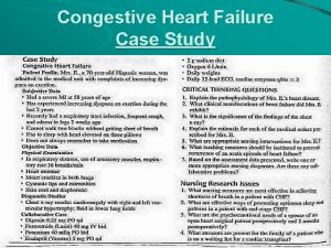
 Congestive heart failure zones for management
 Nursing assessment for congestive heart failure
 Nursing assessment for congestive heart failure
 Congestive heart failure symtoms
 Non conducted pac ecg
 Failure to sense pacemaker
 Ductile fracture
 Forrester classification heart failure
 Cardiomegay
 Heart failure definition
 Causes of cardiomegaly in child
 Heart failure complications
 Chapter 24 heart failure drugs
 Heart failure
 Lmnop heart failure
 Keith rn heart failure case study
 Donkey analogy heart failure
 Heart failure cells are seen in lungs
 Heart failure forward vs backward
 Vetsulin dosage chart for dogs
 Diabetes and heart failure
 Compensatory mechanisms of heart failure
 Cardiac landmarks
 Acute vs chronic heart failure
 Dr nienkemper
 Right border of the relative cardiac dullness is formed by
 Sheep heart diagram
 Heart 2 heart
 What is sems
 Emergency management of maxillofacial trauma
 Hennepin county emergency management
 Emergency management
 Walmart emergency response system
 Ares clint
 Raptor reunification
 Emergency management institute
 Emergency management institute
 Emergency management institute
 Ucf public administration masters
 Tulane university emergency management
 Raptor emergency management
 Emergency management
 Canadian emergency management college
 Ims incident management


