Patras University Hospital Patras University Hospital Patras University

- Slides: 94
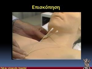
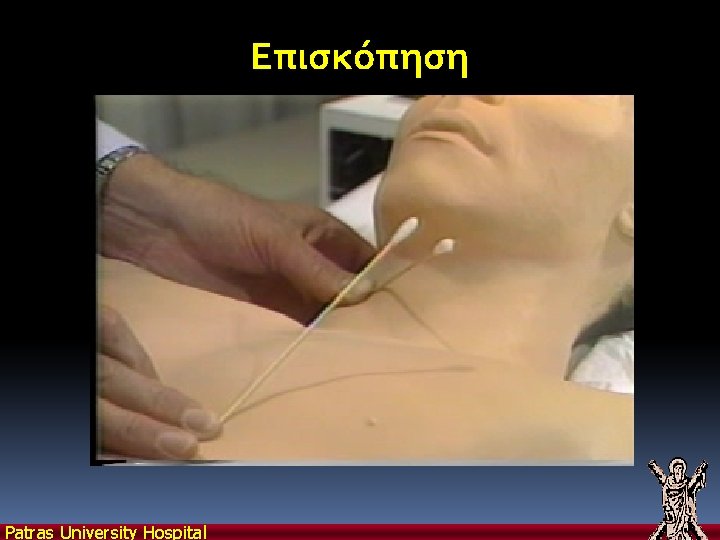
Επισκόπηση Patras University Hospital
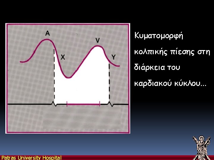
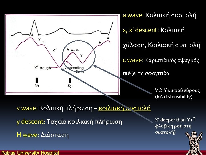
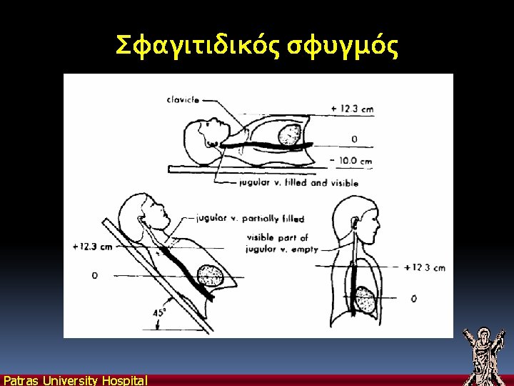
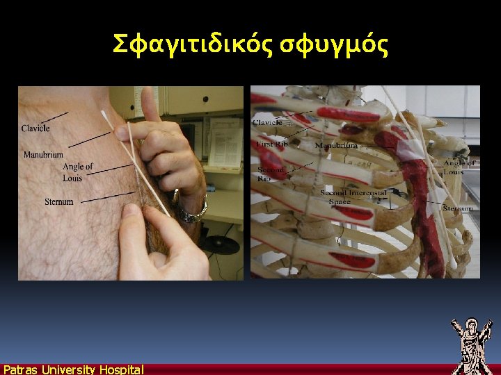
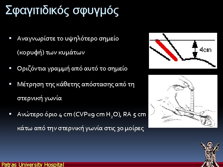
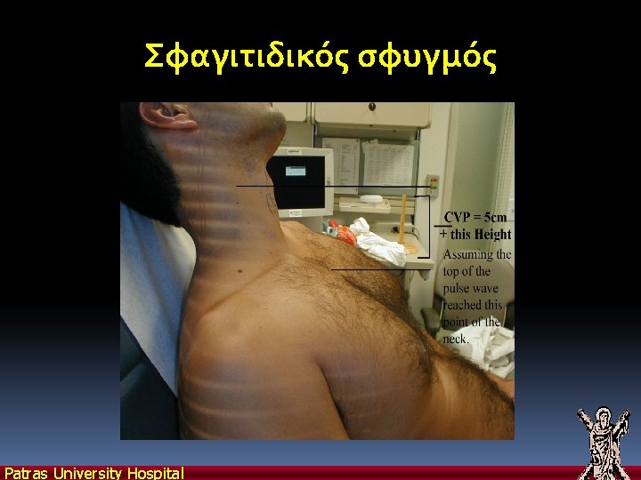
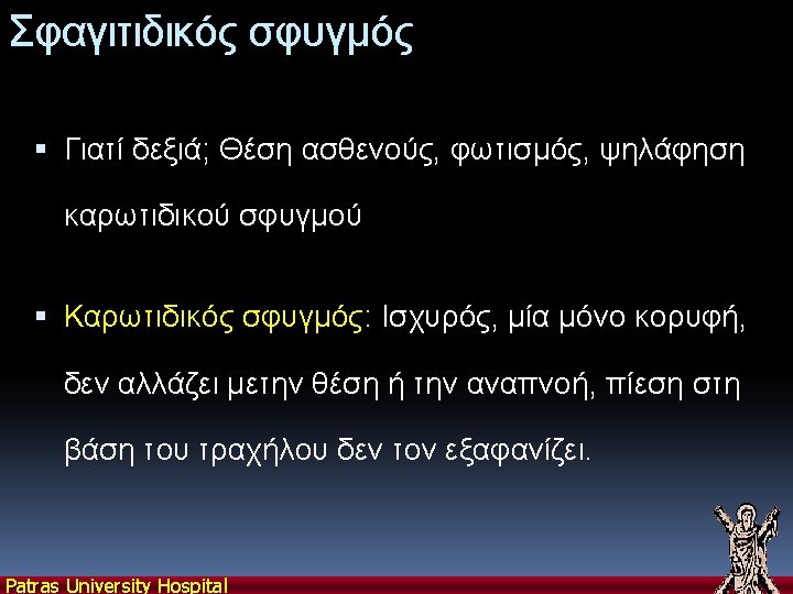
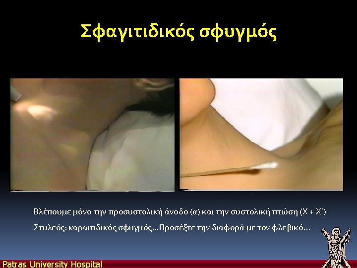
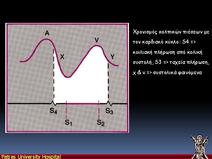
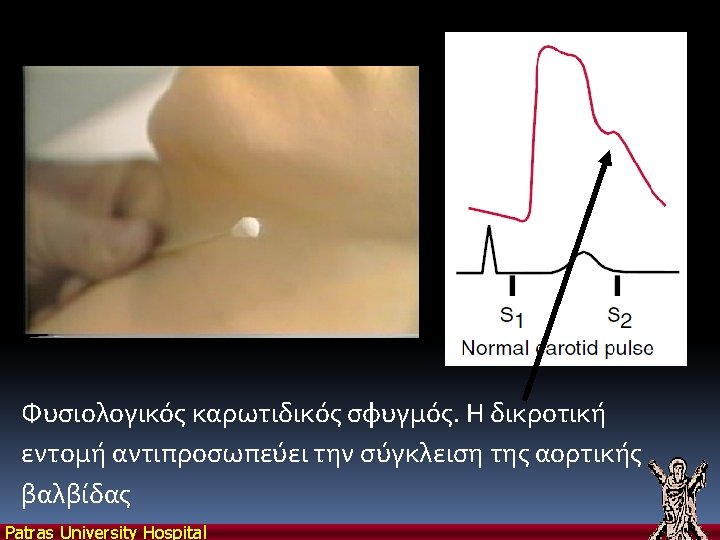
Σφαγιτιδικός σφυγμός Patras University Hospital
Σφαγιτιδικός σφυγμός Patras University Hospital
Σφαγιτιδικός σφυγμός Patras University Hospital
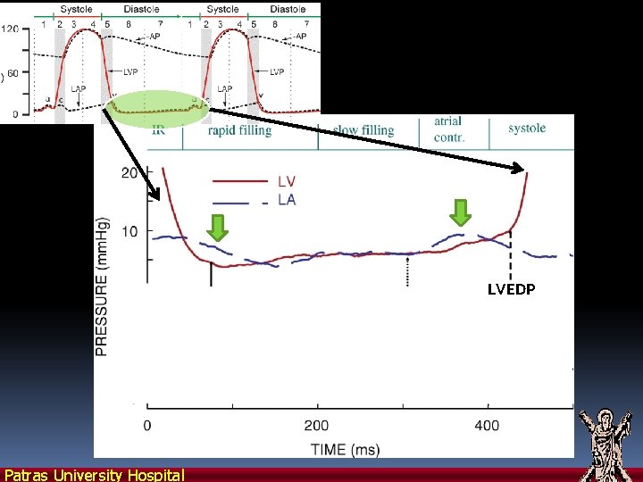
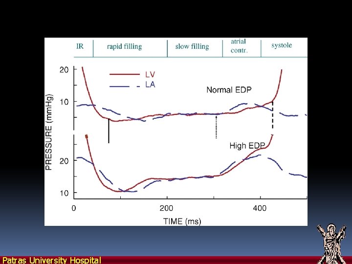
The 4 phases of diastole are marked in relation to high-fidelity pressure recordings from the left atrium (LA) and left ventricle (LV) in anesthetized dogs. LVEDP Patras University Hospital
The 4 phases of diastole are marked in relation to high-fidelity pressure recordings from the left atrium (LA) and left ventricle (LV) in anesthetized dogs. Patras University Hospital
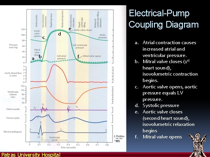
c d a b Patras University Hospital Electrical-Pump Coupling Diagram e f a. Atrial contraction causes increased atrial and ventricular pressure. b. Mitral valve closes (1 st heart sound), isovolumetric contraction begins. c. Aortic valve opens, aortic pressure equals LV pressure. d. Systolic pressure e. Aortic valve closes (second heart sound), isovolumetric relaxation begins f. Mitral valve opens
Patras University Hospital
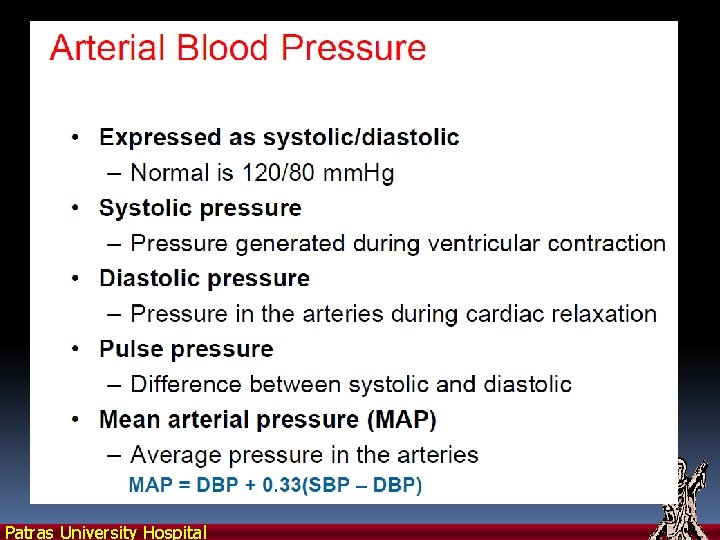
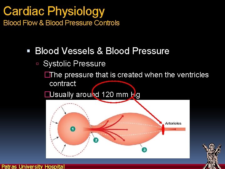
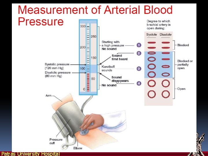
Cardiac Physiology Blood Flow & Blood Pressure Controls Blood Vessels & Blood Pressure Systolic Pressure �The pressure that is created when the ventricles contract �Usually around 120 mm Hg Patras University Hospital
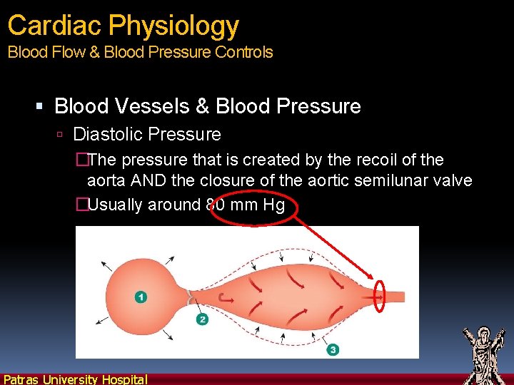
Cardiac Physiology Blood Flow & Blood Pressure Controls Blood Vessels & Blood Pressure Diastolic Pressure �The pressure that is created by the recoil of the aorta AND the closure of the aortic semilunar valve �Usually around 80 mm Hg Patras University Hospital
Patras University Hospital
Patras University Hospital
Patras University Hospital
Patras University Hospital
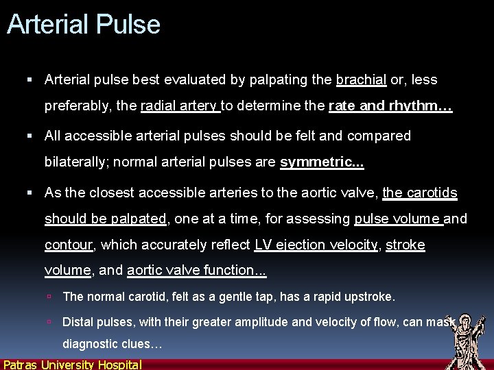
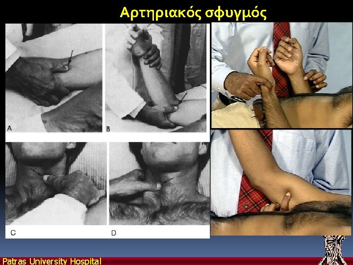
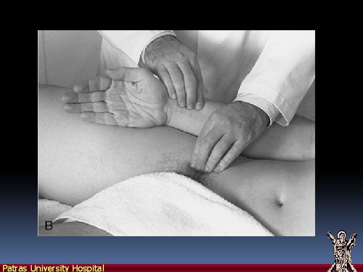
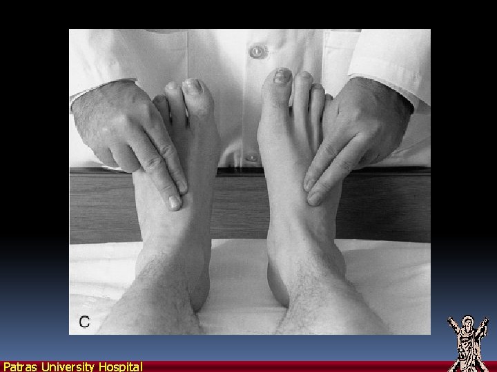
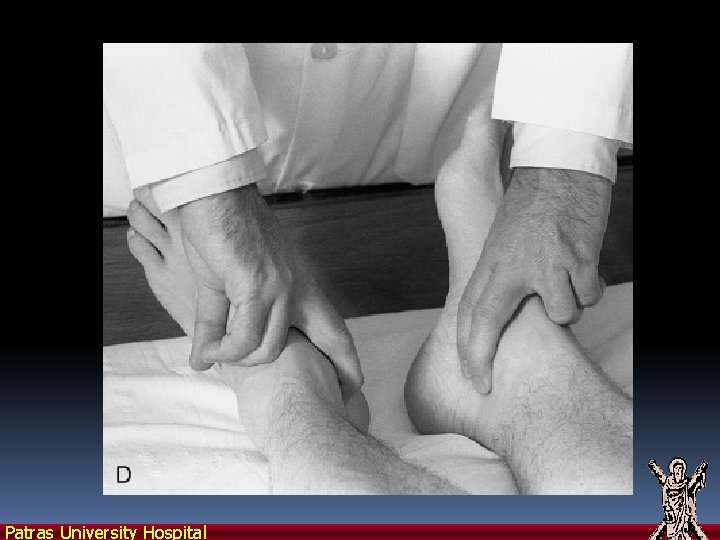
Arterial Pulse Arterial pulse best evaluated by palpating the brachial or, less preferably, the radial artery to determine the rate and rhythm… All accessible arterial pulses should be felt and compared bilaterally; normal arterial pulses are symmetric. . . As the closest accessible arteries to the aortic valve, the carotids should be palpated, one at a time, for assessing pulse volume and contour, which accurately reflect LV ejection velocity, stroke volume, and aortic valve function. . . The normal carotid, felt as a gentle tap, has a rapid upstroke. Distal pulses, with their greater amplitude and velocity of flow, can mask diagnostic clues… Patras University Hospital
Αρτηριακός σφυγμός Patras University Hospital
Patras University Hospital
Patras University Hospital
Patras University Hospital
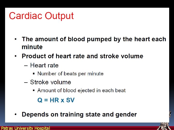
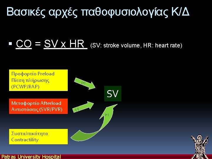
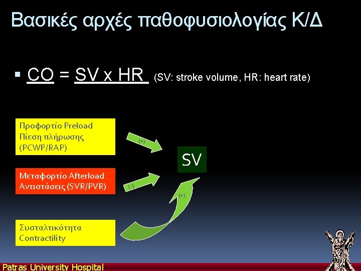
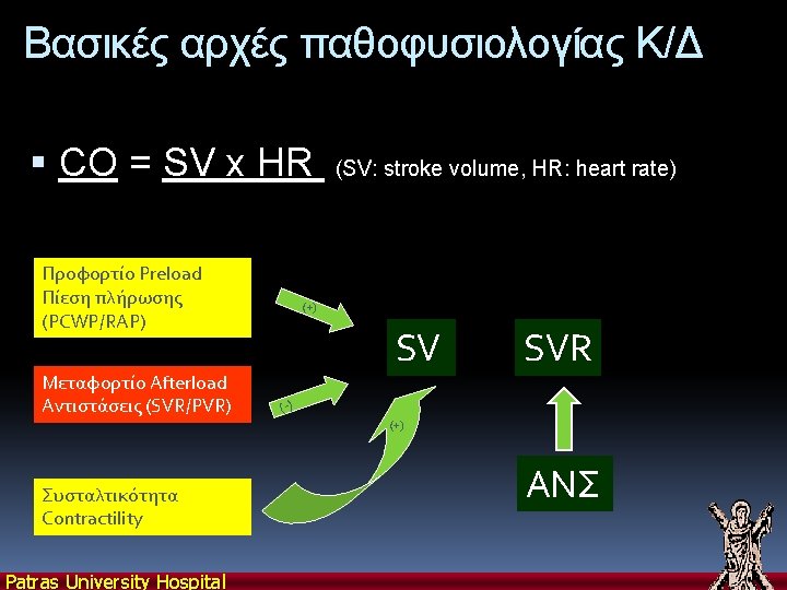
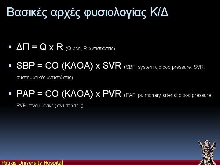
ΚΑΡΔΙΑΚΗ ΛΕΙΤΟΥΡΓΙΑ Primary function of heart: generate and sustain arterial blood pressure necessary to provide adequate perfusion of organs Stroke Volume (SV) Cardiac Output (CO) = SV x HR Cardiac index = CO/m 2 (2. 6 to 4. 2 L/min/m 2) Patras University Hospital
Starling’s Law of the Heart The heart adjusts its pumping rate to the rate of blood return. How? More blood returning stretches atria and ventricles more Stretching SA node muscle causes faster rhythmicity Stretching heart muscle causes faster conduction Stretching heart muscle causes stronger, more complete contraction… Patras University Hospital
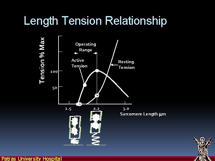
Tension % Max Length Tension Relationship Operating Range 100 Active Tension Resting Tension 50 1. 5 Patras University Hospital 2. 2 3. 0 Sarcomere Length mm
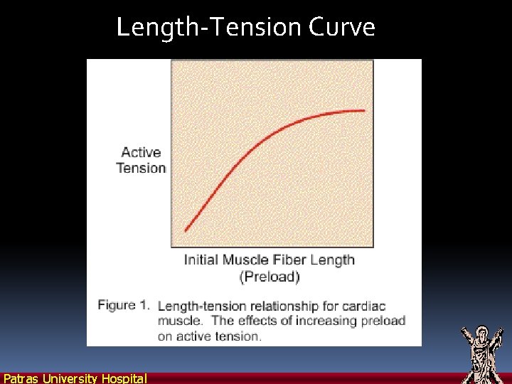
Length-Tension Curve Patras University Hospital
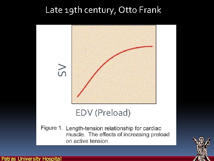
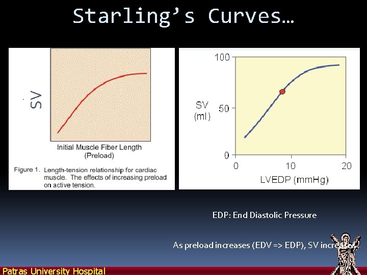
SV Late 19 th century, Otto Frank EDV (Preload) Patras University Hospital
Patras University Hospital
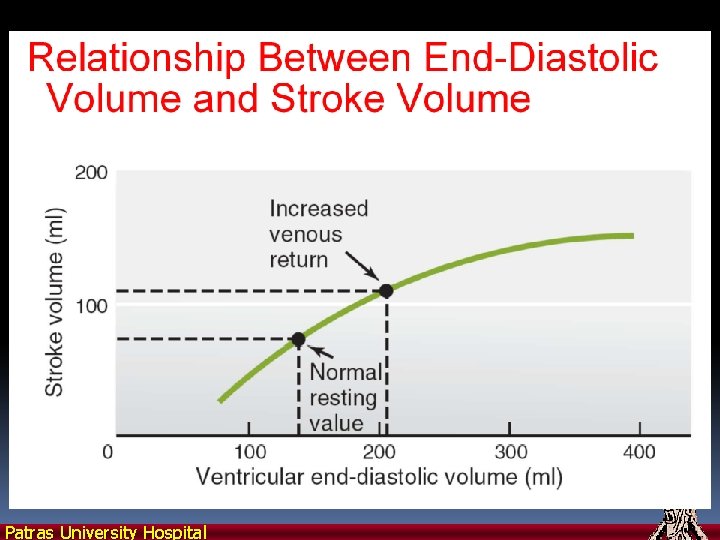
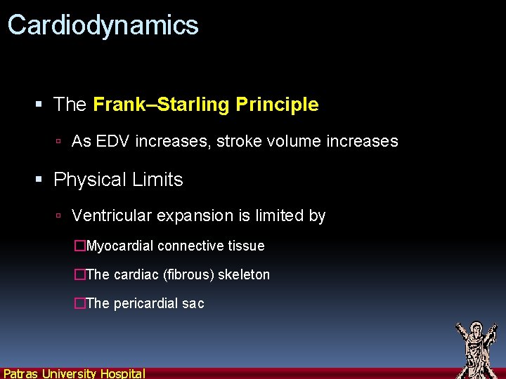
Cardiodynamics The Frank–Starling Principle As EDV increases, stroke volume increases Physical Limits Ventricular expansion is limited by �Myocardial connective tissue �The cardiac (fibrous) skeleton �The pericardial sac Patras University Hospital
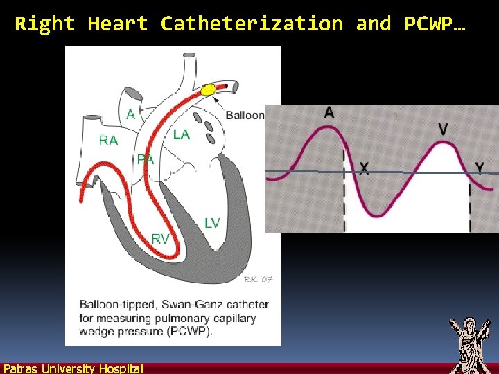
Right Heart Catheterization and PCWP… Patras University Hospital
Patras University Hospital
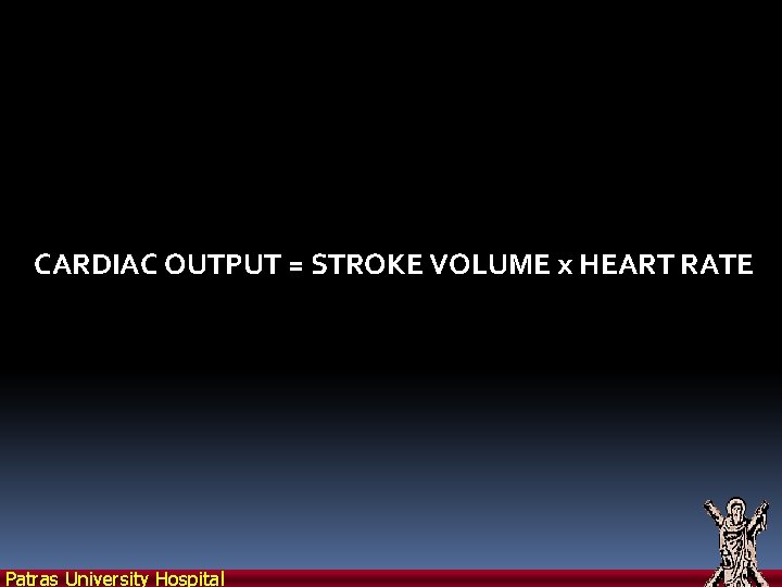
CARDIAC OUTPUT = STROKE VOLUME x HEART RATE Patras University Hospital
Patras University Hospital
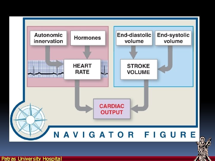
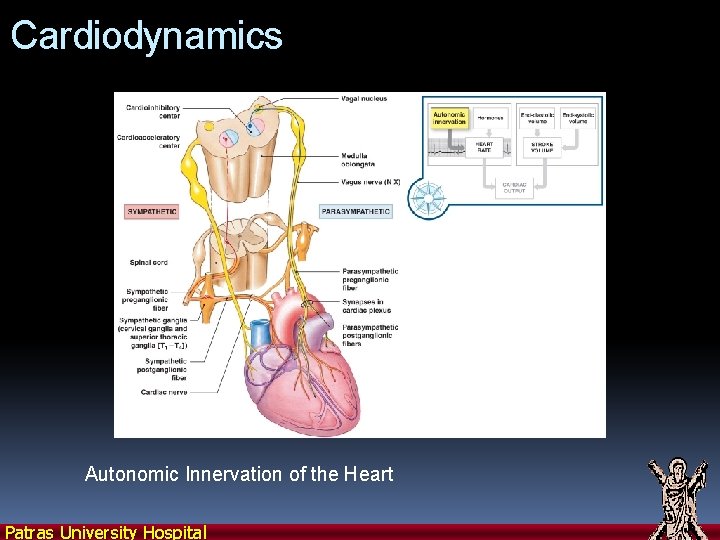
Cardiodynamics Autonomic Innervation of the Heart Patras University Hospital
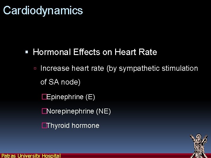
Cardiodynamics Hormonal Effects on Heart Rate Increase heart rate (by sympathetic stimulation of SA node) �Epinephrine (E) �Norepinephrine (NE) �Thyroid hormone Patras University Hospital
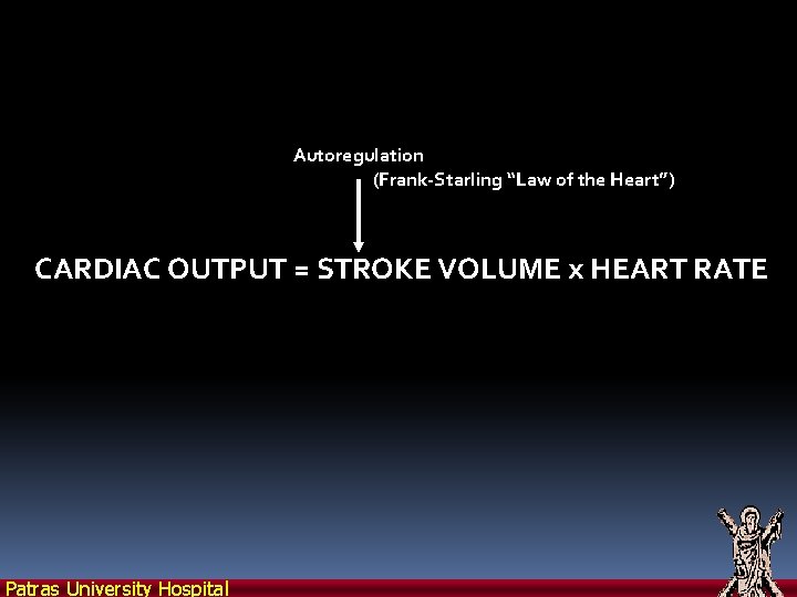
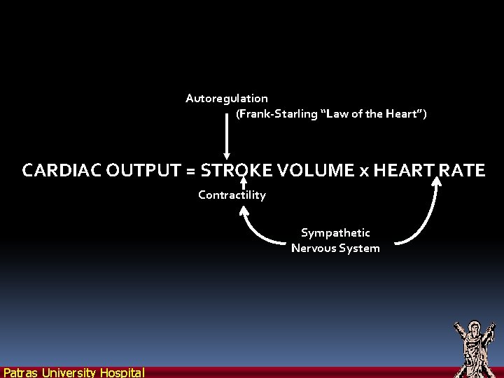
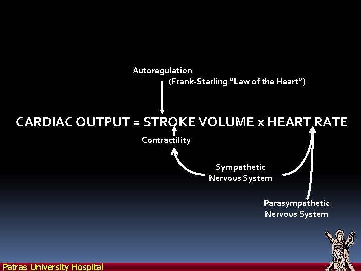
Autoregulation (Frank-Starling “Law of the Heart”) CARDIAC OUTPUT = STROKE VOLUME x HEART RATE Patras University Hospital
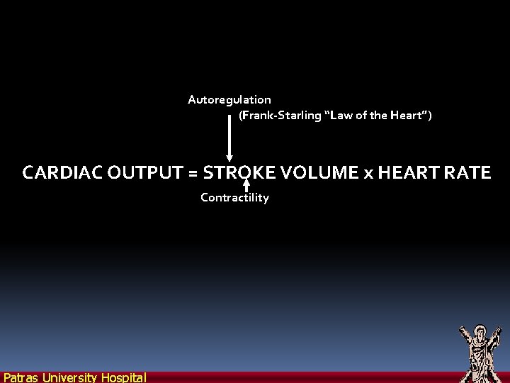
Autoregulation (Frank-Starling “Law of the Heart”) CARDIAC OUTPUT = STROKE VOLUME x HEART RATE Contractility Patras University Hospital
Autoregulation (Frank-Starling “Law of the Heart”) CARDIAC OUTPUT = STROKE VOLUME x HEART RATE Contractility Sympathetic Nervous System Patras University Hospital
Autoregulation (Frank-Starling “Law of the Heart”) CARDIAC OUTPUT = STROKE VOLUME x HEART RATE Contractility Sympathetic Nervous System Parasympathetic Nervous System Patras University Hospital
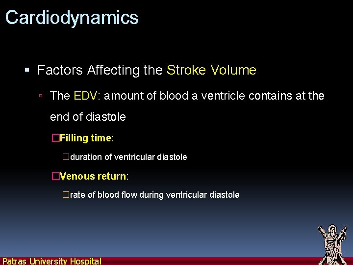
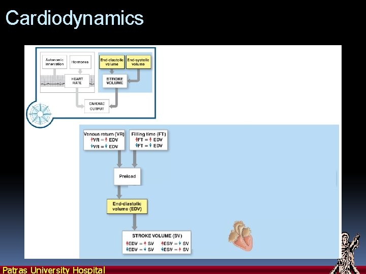
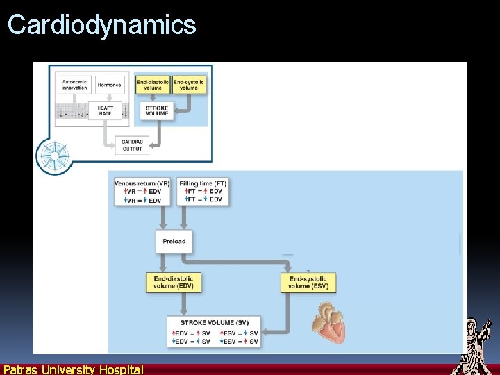
Cardiodynamics Factors Affecting the Stroke Volume The EDV: amount of blood a ventricle contains at the end of diastole �Filling time: �duration of ventricular diastole �Venous return: �rate of blood flow during ventricular diastole Patras University Hospital
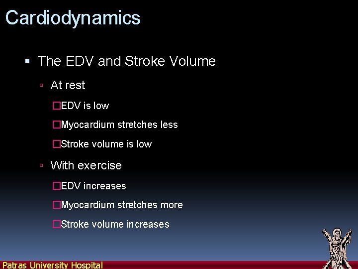
Cardiodynamics The EDV and Stroke Volume At rest �EDV is low �Myocardium stretches less �Stroke volume is low With exercise �EDV increases �Myocardium stretches more �Stroke volume increases Patras University Hospital
Cardiodynamics Patras University Hospital
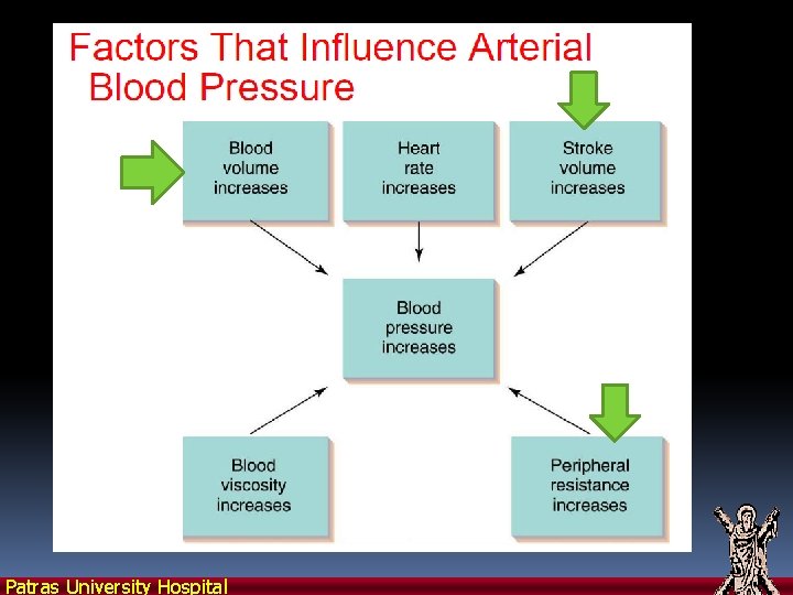
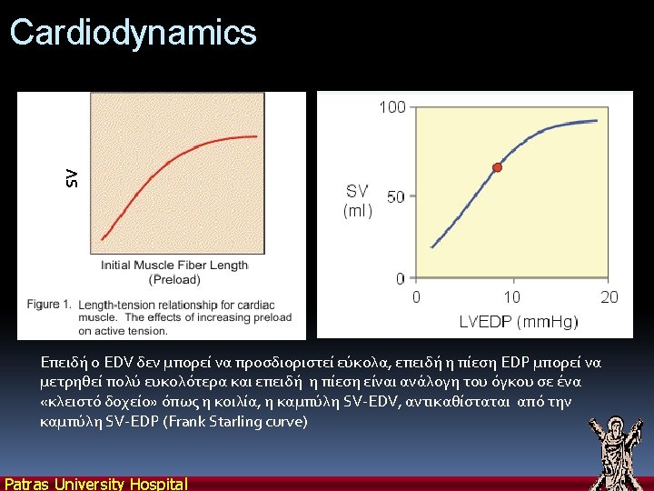
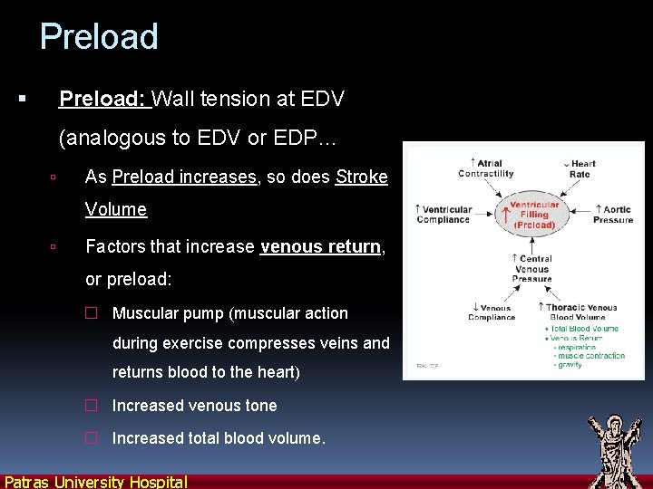
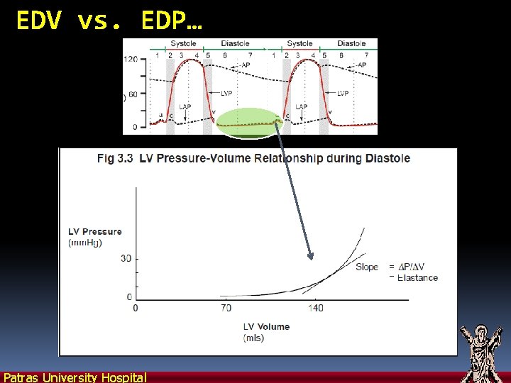
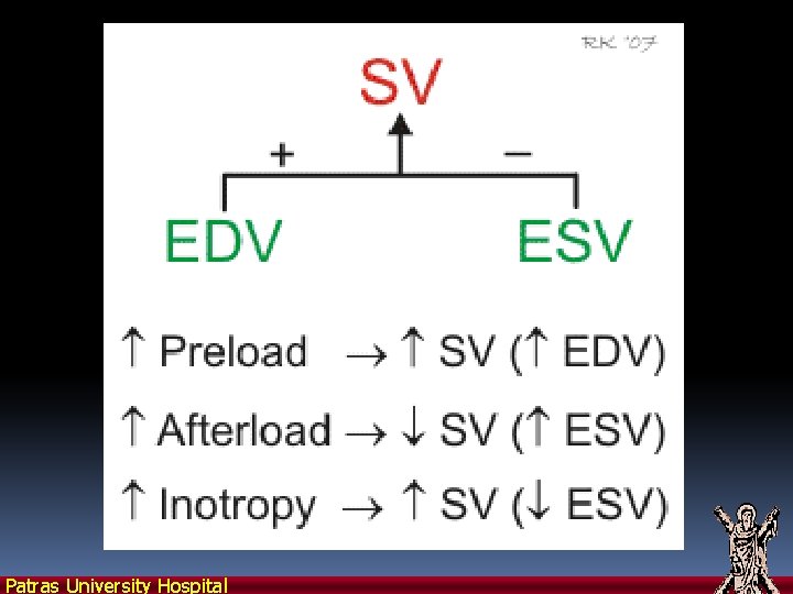
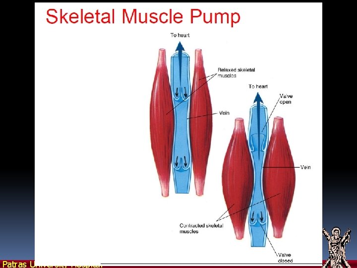
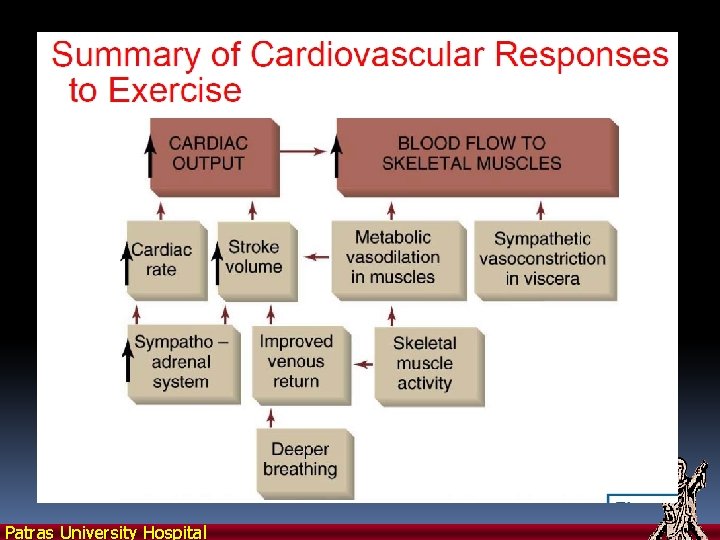
Preload: Wall tension at EDV (analogous to EDV or EDP… As Preload increases, so does Stroke Volume Factors that increase venous return, or preload: � Muscular pump (muscular action during exercise compresses veins and returns blood to the heart) � Increased venous tone � Increased total blood volume. Patras University Hospital
EDV vs. EDP… Patras University Hospital
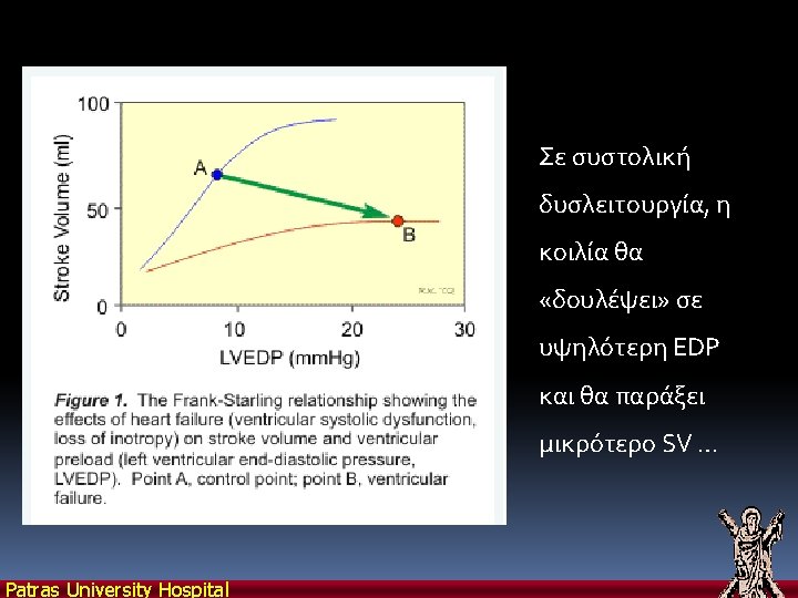
SV Starling’s Curves… EDP: End Diastolic Pressure As preload increases (EDV => EDP), SV increases Patras University Hospital
Cardiodynamics Patras University Hospital
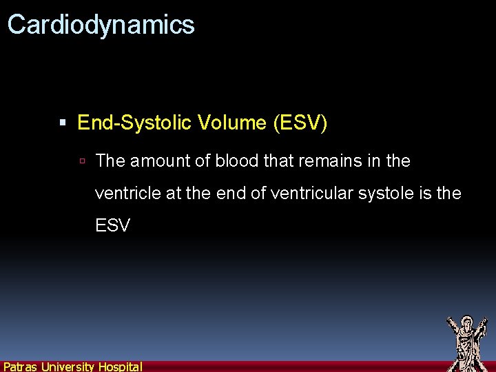
Cardiodynamics End-Systolic Volume (ESV) The amount of blood that remains in the ventricle at the end of ventricular systole is the ESV Patras University Hospital
Patras University Hospital
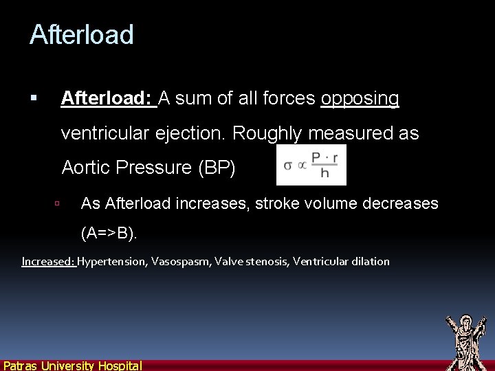
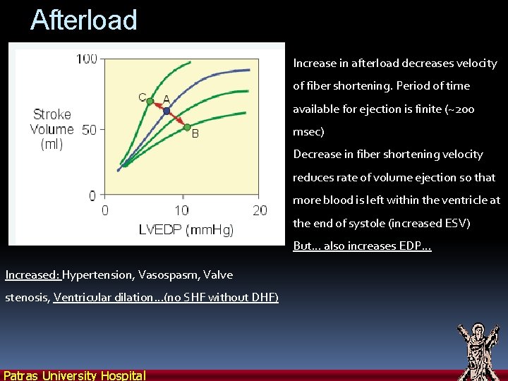
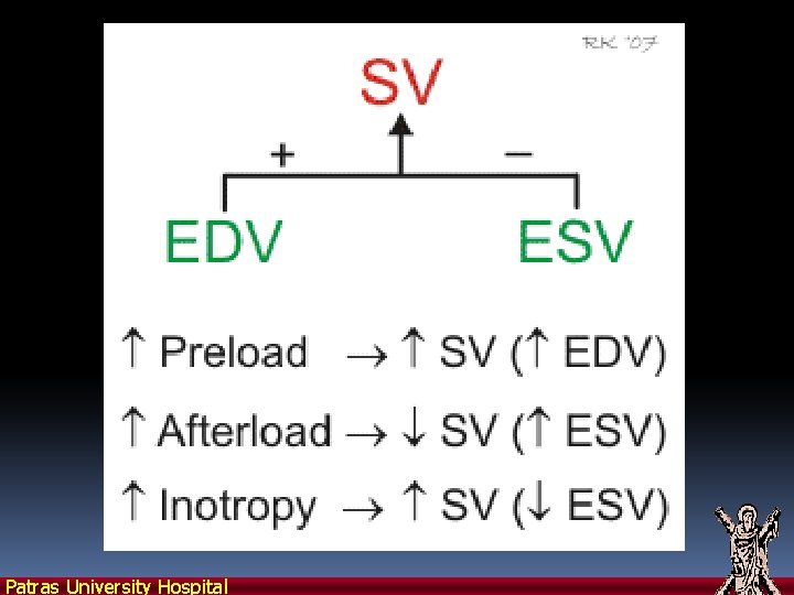
Afterload: A sum of all forces opposing ventricular ejection. Roughly measured as Aortic Pressure (BP) As Afterload increases, stroke volume decreases (A=>B). Increased: Hypertension, Vasospasm, Valve stenosis, Ventricular dilation Patras University Hospital
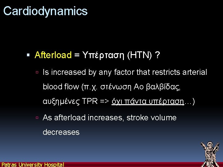
Cardiodynamics Afterload = Υπέρταση (HTN) ? Is increased by any factor that restricts arterial blood flow (π. χ. στένωση Αο βαλβίδας, αυξημένες TPR => όχι πάντα υπέρταση…) As afterload increases, stroke volume decreases Patras University Hospital
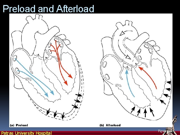
Preload and Afterload Patras University Hospital Figure 18. 21
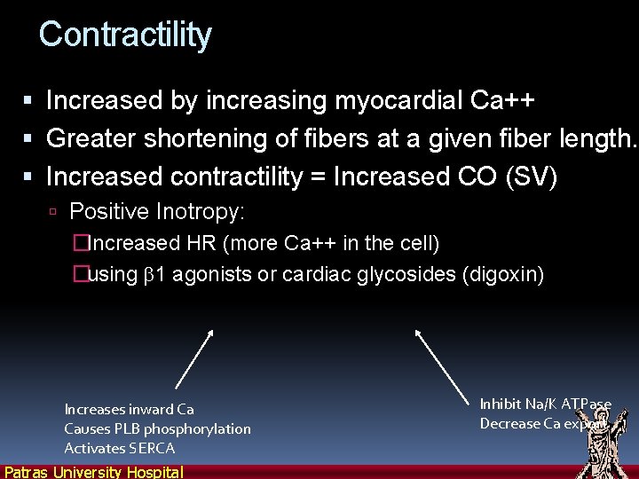
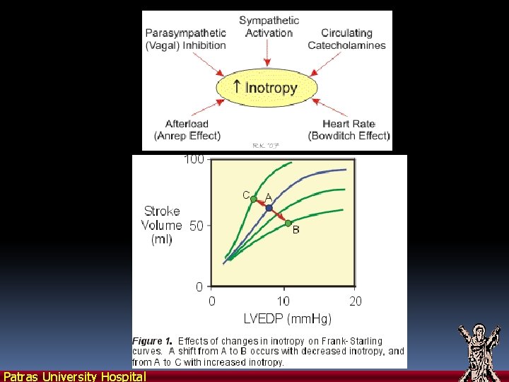
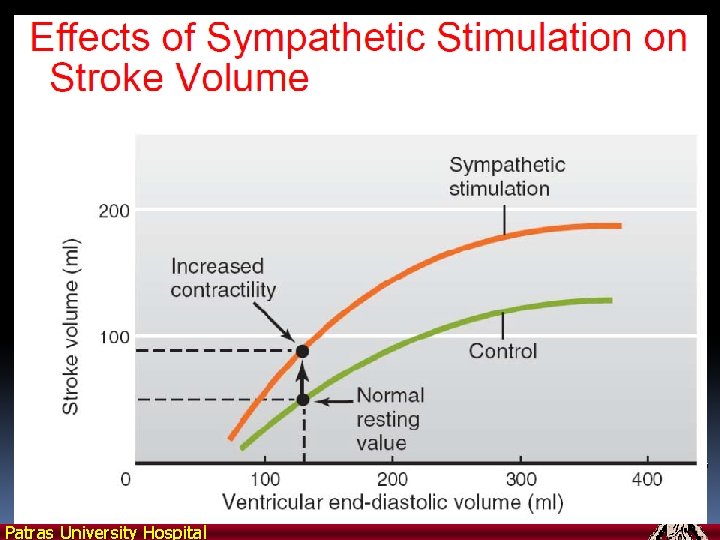
Contractility Increased by increasing myocardial Ca++ Greater shortening of fibers at a given fiber length. Increased contractility = Increased CO (SV) Positive Inotropy: �Increased HR (more Ca++ in the cell) �using b 1 agonists or cardiac glycosides (digoxin) Increases inward Ca Causes PLB phosphorylation Activates SERCA Patras University Hospital Inhibit Na/K ATPase Decrease Ca export
Measuring Contractility Patras University Hospital
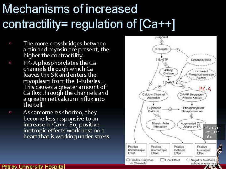
Mechanisms of increased contractility= regulation of [Ca++] The more crossbridges between actin and myosin are present, the higher the contractility. PK-A phosphorylates the Ca channels through which Ca leaves the SR and enters the myoplasm from the T-tubules. . This causes a greater amount of Ca flux through the channels and a greater net calcium influx into the cell. As sarcomeres shorten, they become less responsive to an increase in Ca++. So, positive inotropic effects work best on a heart that is working under stress. Patras University Hospital PK-A More Ca++ avail. for later.
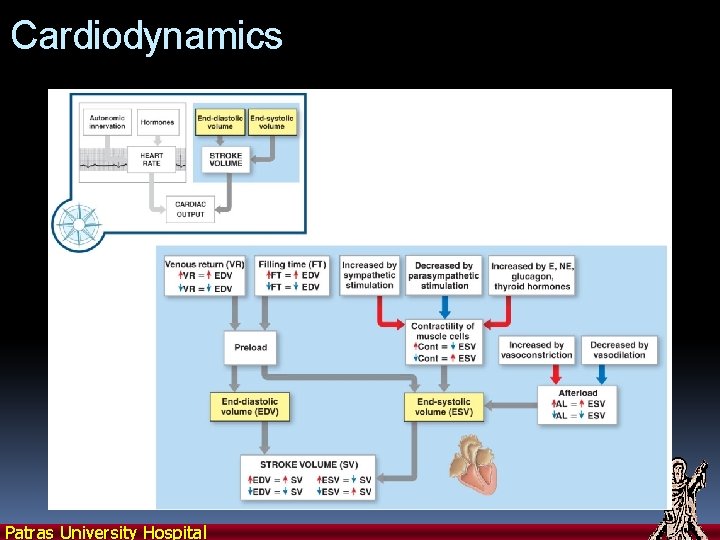
Cardiodynamics Patras University Hospital
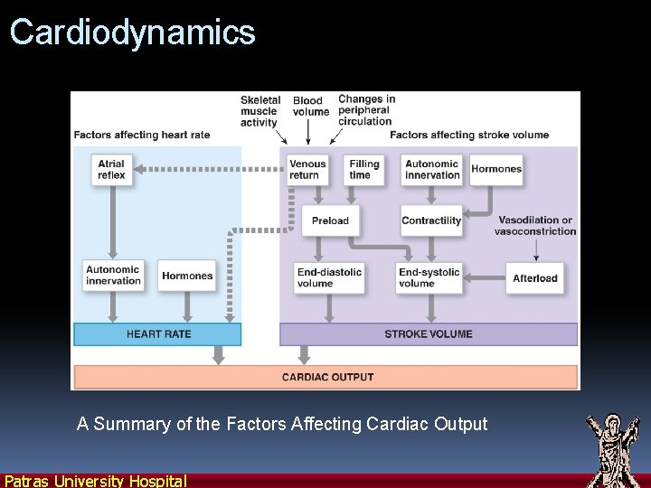
Cardiodynamics A Summary of the Factors Affecting Cardiac Output Patras University Hospital
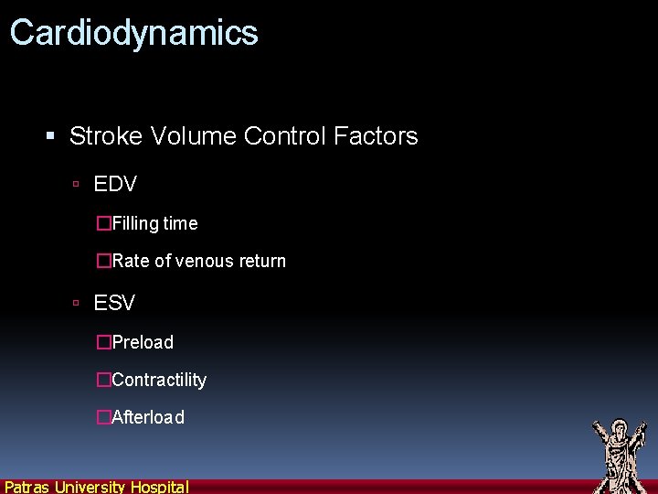
Cardiodynamics Stroke Volume Control Factors EDV �Filling time �Rate of venous return ESV �Preload �Contractility �Afterload Patras University Hospital
Cardiodynamics Cardiac Reserve The difference between resting and maximal cardiac output Patras University Hospital
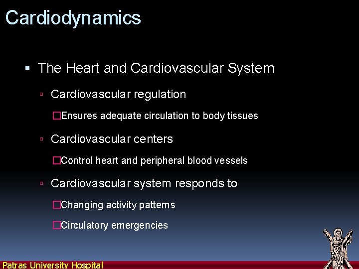
Cardiodynamics The Heart and Cardiovascular System Cardiovascular regulation �Ensures adequate circulation to body tissues Cardiovascular centers �Control heart and peripheral blood vessels Cardiovascular system responds to �Changing activity patterns �Circulatory emergencies Patras University Hospital
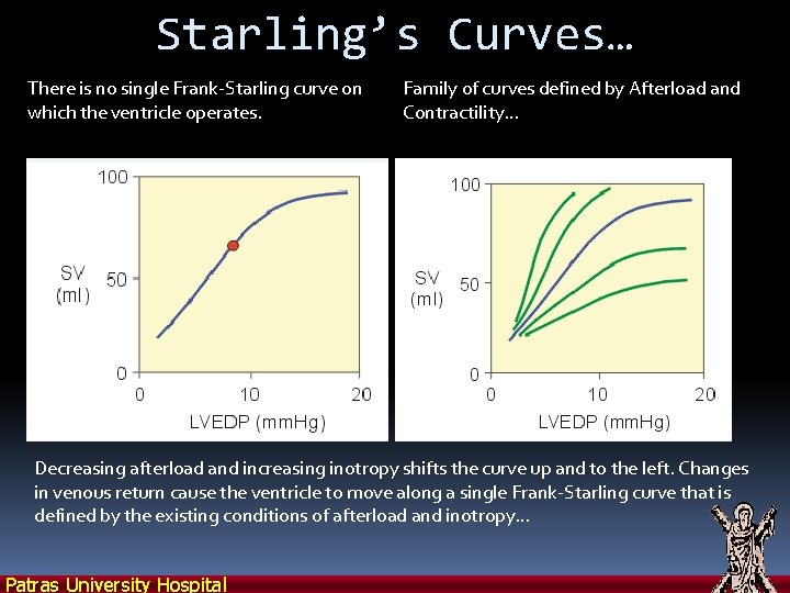
Starling’s Curves… There is no single Frank-Starling curve on which the ventricle operates. Family of curves defined by Afterload and Contractility… Decreasing afterload and increasing inotropy shifts the curve up and to the left. Changes in venous return cause the ventricle to move along a single Frank-Starling curve that is defined by the existing conditions of afterload and inotropy… Patras University Hospital
Afterload Increase in afterload decreases velocity of fiber shortening. Period of time available for ejection is finite (~200 msec) Decrease in fiber shortening velocity reduces rate of volume ejection so that more blood is left within the ventricle at the end of systole (increased ESV) But… also increases EDP… Increased: Hypertension, Vasospasm, Valve stenosis, Ventricular dilation…(no SHF without DHF) Patras University Hospital
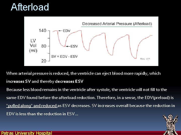
Afterload When arterial pressure is reduced, the ventricle can eject blood more rapidly, which increases SV and thereby decreases ESV Because less blood remains in the ventricle after systole, the ventricle will not fill to the same EDV found before the afterload reduction. Therefore, in a sense, the EDV(preload) is "pulled along" and reduced as ESV decreases. SV increases overall because the reduction in EDV is less than the reduction in ESV. . . Patras University Hospital
Patras University Hospital
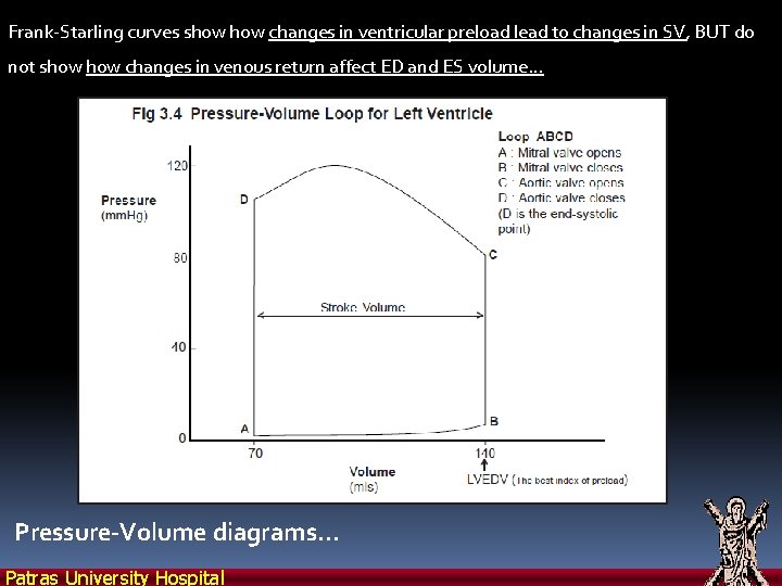
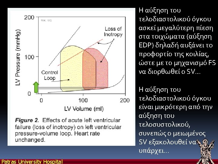
Frank-Starling curves show changes in ventricular preload lead to changes in SV, BUT do not show changes in venous return affect ED and ES volume… Pressure-Volume diagrams… Patras University Hospital
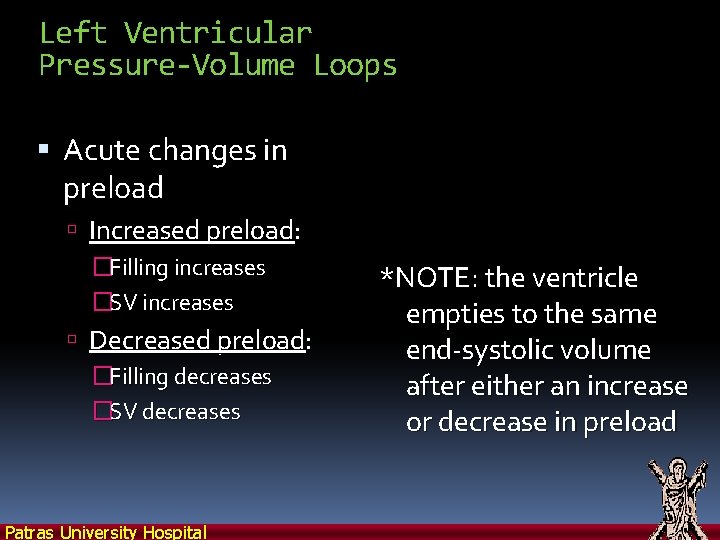
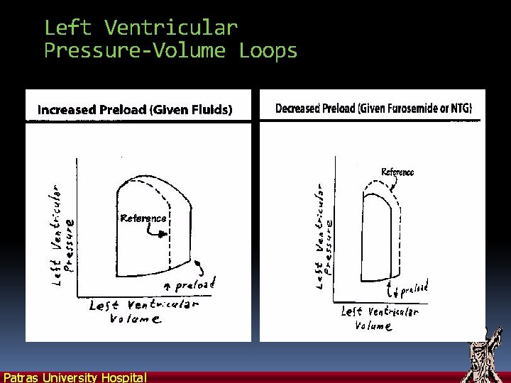
Left Ventricular Pressure-Volume Loops Acute changes in preload Increased preload: �Filling increases �SV increases Decreased preload: �Filling decreases �SV decreases Patras University Hospital *NOTE: the ventricle empties to the same end-systolic volume after either an increase or decrease in preload
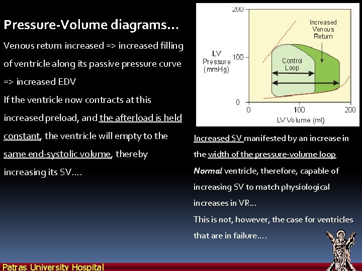
Pressure-Volume diagrams… Venous return increased => increased filling of ventricle along its passive pressure curve => increased EDV If the ventricle now contracts at this increased preload, and the afterload is held constant, the ventricle will empty to the Increased SV manifested by an increase in same end-systolic volume, thereby the width of the pressure-volume loop increasing its SV…. Normal ventricle, therefore, capable of increasing SV to match physiological increases in VR… This is not, however, the case for ventricles that are in failure…. Patras University Hospital
Left Ventricular Pressure-Volume Loops Patras University Hospital
Left Ventricular Pressure-Volume Loops Acute changes in Afterload Increased afterload: �Ventricle empties less completely �SV decreases �Increase in BP (shifts up and right) Patras University Hospital Decreased afterload: �Ventricle empties more completely �SV increases �Decrease in BP (shifts down and left)
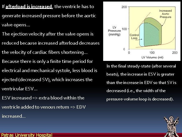
If afterload is increased, the ventricle has to generate increased pressure before the aortic valve opens… The ejection velocity after the valve opens is reduced because increased afterload decreases the velocity of cardiac fibers shortening… Because there is only a finite time period for electrical and mechanical systole, less blood is ejected (decreased SV), which increases the In the final steady-state (after several beats), the increase in ESV is greater than the increase in EDV so that SV is ventricular ESV… decreased (i. e. , the width of the ESV increased => extra blood within the pressure-volume loop is decreased). ventricle added to venous return => EDV increased… Patras University Hospital
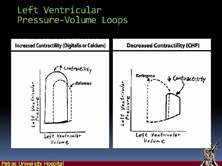
Left Ventricular Pressure-Volume Loops Altered contractility Increased contractility: �Ventricle empties more completely �SV increases �BP increases (shifts up and left) Patras University Hospital Decreased contractility: �Ventricle empties less completely �SV decreases �BP decreases (shifts down and right)
Left Ventricular Pressure-Volume Loops Patras University Hospital
Patras University Hospital
Patras University Hospital
Patras University Hospital
Patras University Hospital
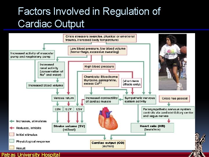
Factors Involved in Regulation of Cardiac Output Patras University Hospital
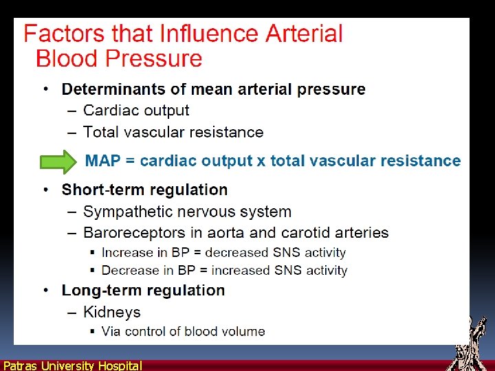
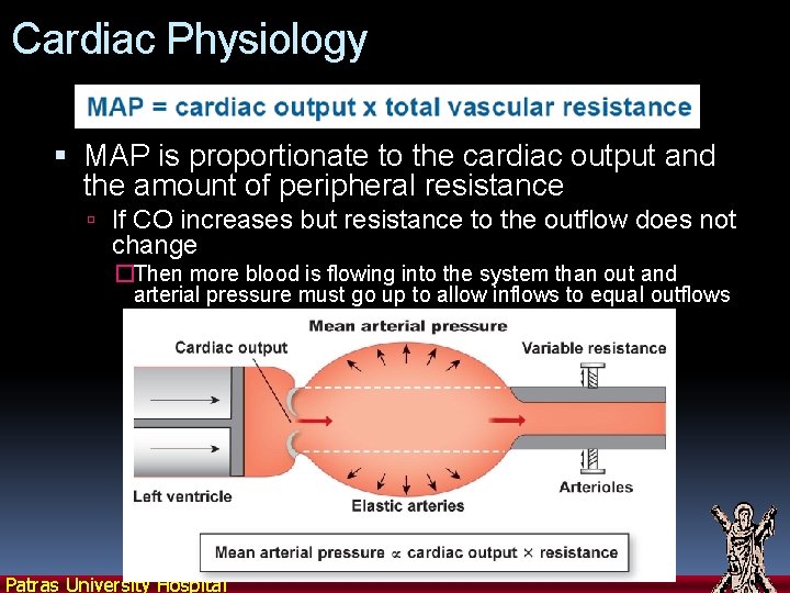
Cardiac Physiology MAP is proportionate to the cardiac output and the amount of peripheral resistance If CO increases but resistance to the outflow does not change �Then more blood is flowing into the system than out and arterial pressure must go up to allow inflows to equal outflows Patras University Hospital
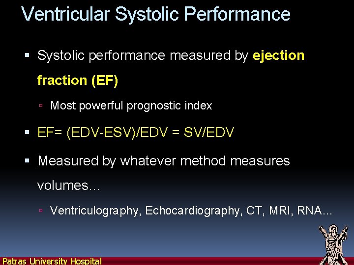
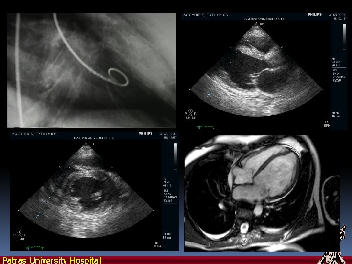
Ventricular Systolic Performance Systolic performance measured by ejection fraction (EF) Most powerful prognostic index EF= (EDV-ESV)/EDV = SV/EDV Measured by whatever method measures volumes… Ventriculography, Echocardiography, CT, MRI, RNA… Patras University Hospital
Patras University Hospital