Mass Spectrometry Frequently Asked Questions Dr Markus Wunderlin
- Slides: 61
Mass Spectrometry Frequently Asked Questions Dr. Markus Wunderlin, Seminar 07. 2004
Overview ØMass Spectrometry in a Nutshell - Facts and Basics ØMass Resolution and Mass Accuracy ØFragmentation – Dissozation – Adduct Formation ØImpurities - Contamination - Artefacts ØFTICR-MS: The „Ferrari Age“ Of MS
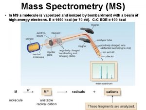
Facts and Basics Mass Spectrometry A technique for measuring and analyzing molecules, that involves introducing enough energy into a (neutral) target molecule to cause its ionization and disintegration. The resulting primary ions and their fragments are then analyzed, based on their mass/ charge ratios, to produce a "molecular fingerprint. "
Facts and Basics Difference Between Spectrometric Methods: Ionization implies a chemical process induced by physical methods. The sample is consumed during the measurement. Their is no defined stimulation of molecular energy levels through interaction with electromagentic radiation, where you can get the sample back without modification.
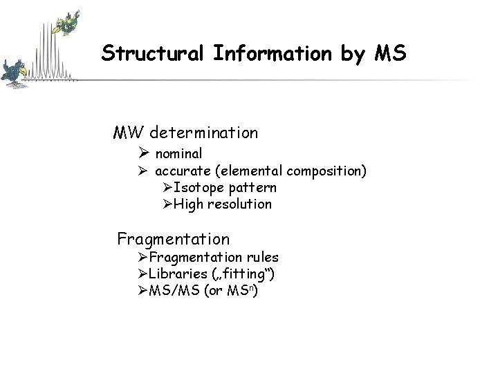
Structural Information by MS MW determination Ø nominal Ø accurate (elemental composition) ØIsotope pattern ØHigh resolution Fragmentation ØFragmentation rules ØLibraries („fitting“) ØMS/MS (or MSn)
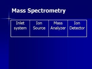
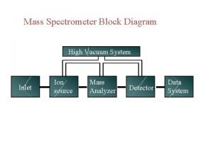
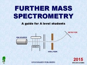
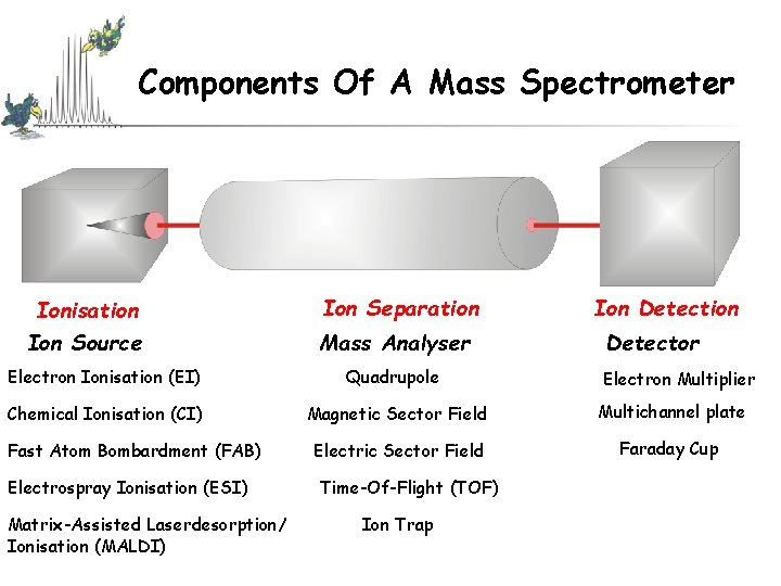
Components Of A Mass Spectrometer Ionisation Ion Source Ion Separation Mass Analyser Ion Detection Detector Electron Ionisation (EI) Quadrupole Chemical Ionisation (CI) Magnetic Sector Field Multichannel plate Electric Sector Field Faraday Cup Fast Atom Bombardment (FAB) Electrospray Ionisation (ESI) Matrix-Assisted Laserdesorption/ Ionisation (MALDI) Time-Of-Flight (TOF) Ion Trap Electron Multiplier
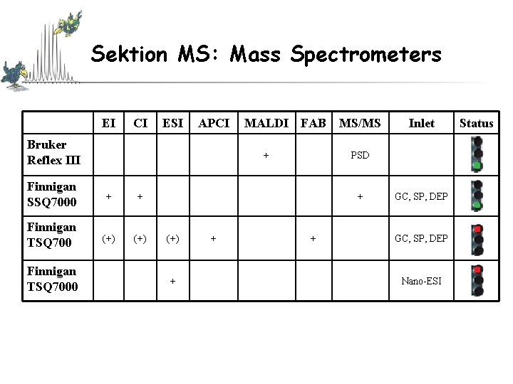
Sektion MS: Mass Spectrometers EI CI ESI APCI Bruker Reflex III + Finnigan SSQ 7000 + + Finnigan TSQ 700 (+) Finnigan TSQ 7000 MALDI FAB MS/MS PSD + (+) + + Inlet + GC, SP, DEP Nano-ESI Status
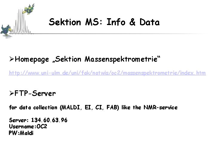
Sektion MS: Info & Data ØHomepage „Sektion Massenspektrometrie“ http: //www. uni-ulm. de/uni/fak/natwis/oc 2/massenspektrometrie/index. htm ØFTP-Server for data collection (MALDI, EI, CI, FAB) like the NMR-service Server: 134. 60. 63. 96 Username: OC 2 PW: Maldi
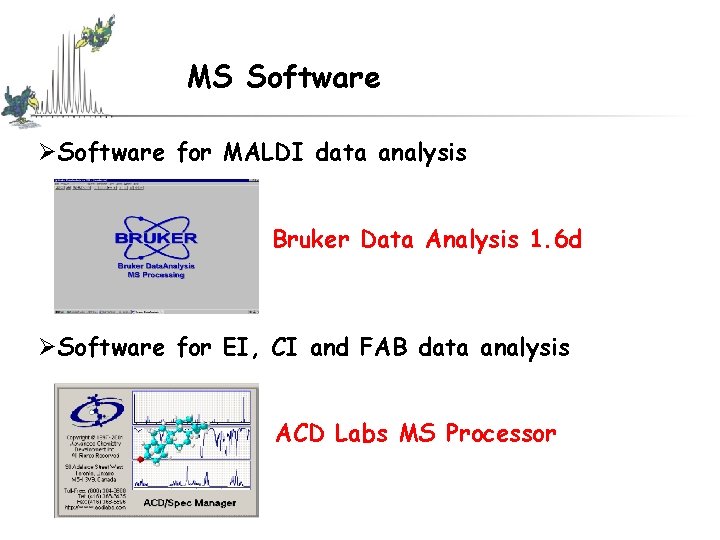
MS Software ØSoftware for MALDI data analysis Bruker Data Analysis 1. 6 d ØSoftware for EI, CI and FAB data analysis ACD Labs MS Processor
What type of analysis is needed ? Ionization methods: MALDI, EI, CI, (FAB), (ESI) – I will select the ionization method unless • you have previous success with a method • duplicating literature methods - Analyses are low resolution • confirms presence of analyte • for high mass compounds (m/w >10000) I try to obtain the best resolution possible • for high mass accuracy internal calibration (standard: external calibration)
What type of analysis is needed ? Which MS method is best for the compound I want to analyze ? ØMolecular weigth? ØSolvent & solubility? ØPurity? ØReactivity? ØWould it distill or sublime under Hi. Vac ? ØOne compound or mixture? ØAcidic? Basic? ØIonic?
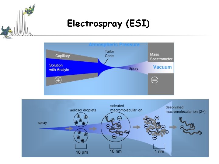
Ionization Methods Neutral species Charged species • Removal/addition of electron(s) – M + e- (M+. )* + 2 e • electron ionization • Removal/addition of proton(s) – M + (Matrix)-H MH+ + (Matrix) • chemical ionization (CI) • atmospheric pressure CI (APCI) • fast atom bombardment (FAB) • electrospray ionization (ESI) • matrix assisted laser desorption/ionization (MALDI)
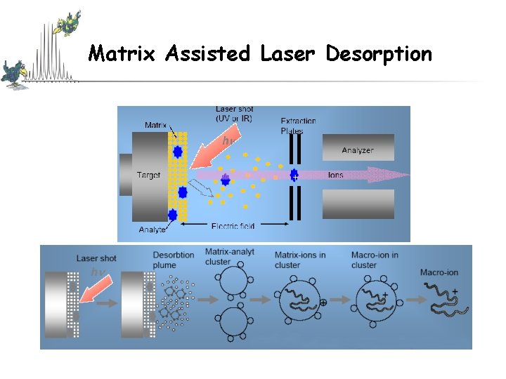
Matrix Assisted Laser Desorption
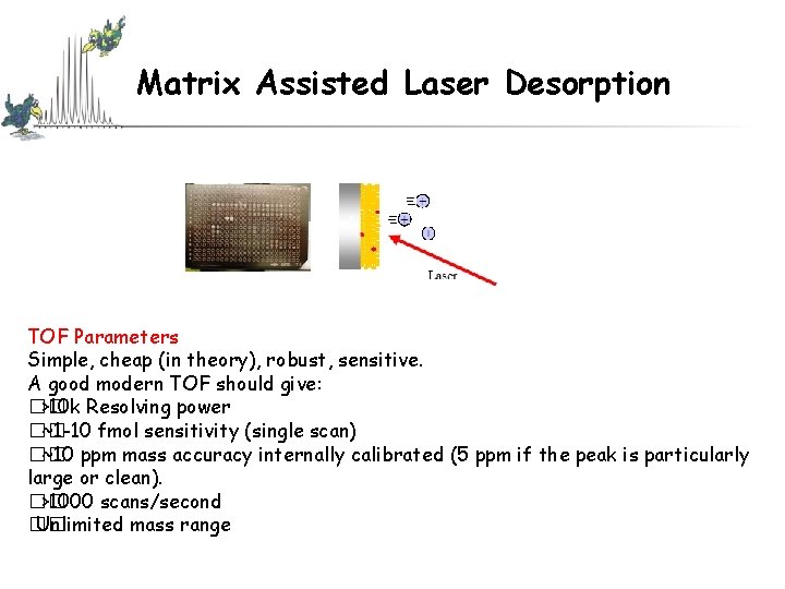
Matrix Assisted Laser Desorption TOF Parameters Simple, cheap (in theory), robust, sensitive. A good modern TOF should give: �� >10 k Resolving power �� ~1 -10 fmol sensitivity (single scan) �� ~10 ppm mass accuracy internally calibrated (5 ppm if the peak is particularly large or clean). �� >1000 scans/second �� Unlimited mass range
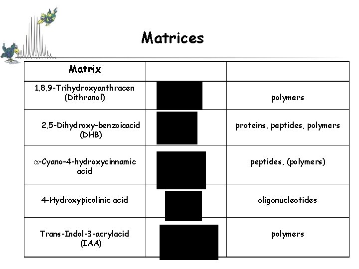
Matrices Matrix 1, 8, 9 -Trihydroxyanthracen (Dithranol) 2, 5 -Dihydroxy benzoicacid (DHB) polymers proteins, peptides, polymers -Cyano-4 -hydroxycinnamic acid peptides, (polymers) 4 -Hydroxypicolinic acid oligonucleotides Trans-Indol-3 -acrylacid (IAA) polymers
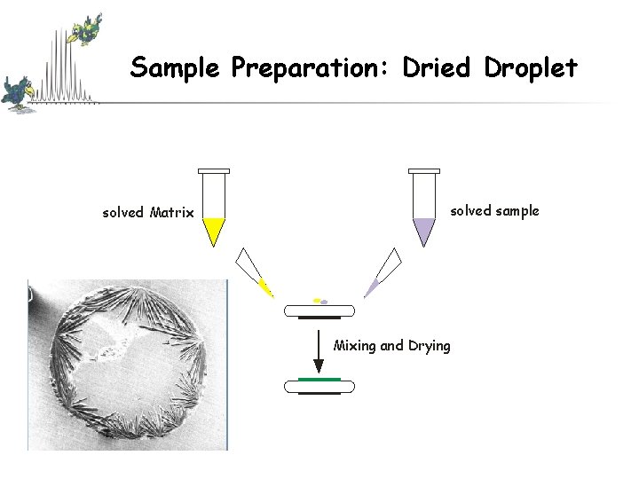
Sample Preparation: Dried Droplet solved Matrix solved sample Mixing and Drying
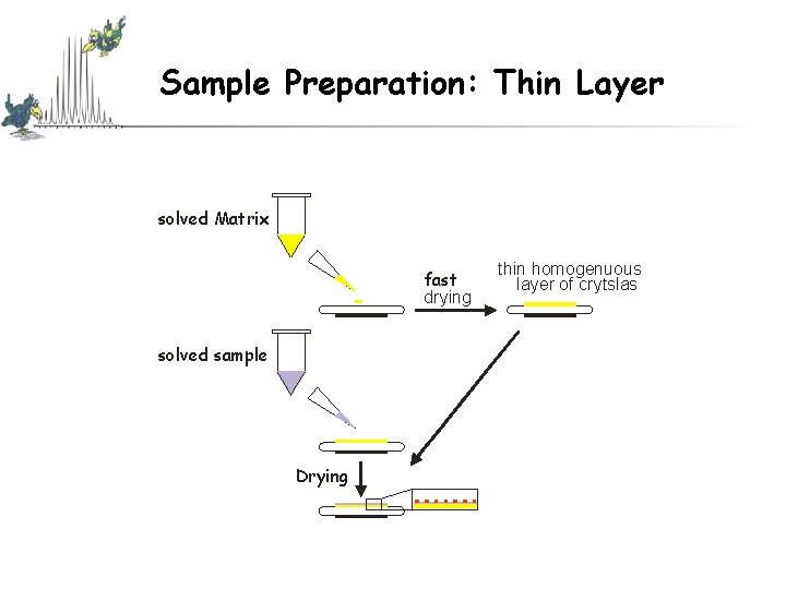
Sample Preparation: Thin Layer solved Matrix fast drying solved sample Drying thin homogenuous layer of crytslas
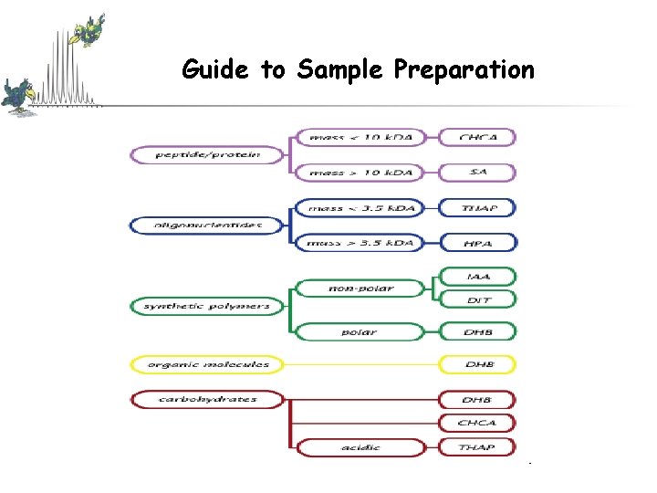
Guide to Sample Preparation
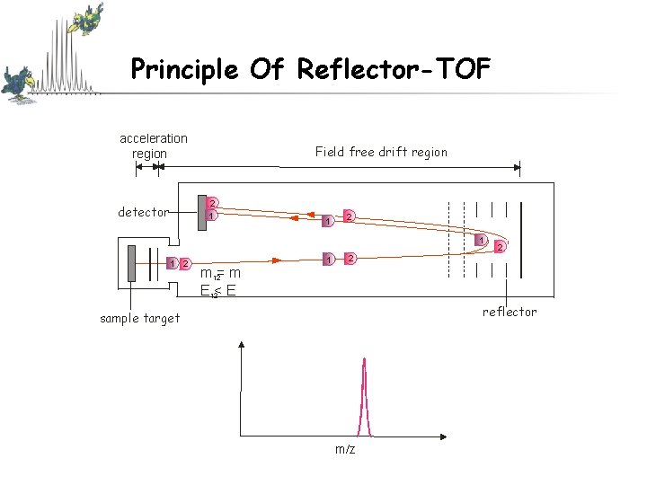
Reflector Ø Through ionisation there is an activation energy distribution (energy-, position- and time uncertainty, electronic repulsion energy, shielding effects) Ø Electric field after the field free drift region that reverses the direction of travel of the ion (reflects) Ø Ions with same m/z ratio but higher kinetic energy penetrate deeper into the reflector, delaying their time of arrival at the reflector relative to the slower low-energy ions Improved resolution, increase in mass accuracy
Principle Of Reflector-TOF acceleration region Field free drift region 2 1 detector 1 2 1 1 2 m 12= m E 12< E 1 2 2 reflector sample target m/z
Electrospray (ESI)
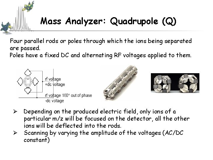
Mass Analyzer: Quadrupole (Q) Four parallel rods or poles through which the ions being separated are passed. Poles have a fixed DC and alternating RF voltages applied to them. Ø Depending on the produced electric field, only ions of a particular m/z will be focused on the detector, all the other ions will be deflected into the rods. Ø Scanning by varying the amplitude of the voltages (AC/DC constant)
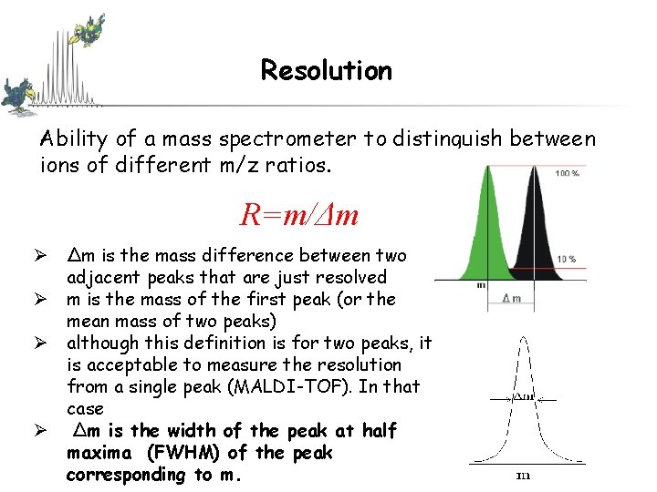
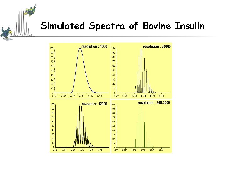
Resolution Ability of a mass spectrometer to distinguish between ions of different m/z ratios. R=m/Δm Ø Δm is the mass difference between two adjacent peaks that are just resolved Ø m is the mass of the first peak (or the mean mass of two peaks) Ø although this definition is for two peaks, it is acceptable to measure the resolution from a single peak (MALDI-TOF). In that case Ø Δm is the width of the peak at half maxima (FWHM) of the peak corresponding to m.
Resolution If we have 5000 resolution on a mass spectrometer, we can separate m/z 50. 000 from m/z 50. 010, or separate m/z 100. 000 from m/z 100. 020, or separate m/z 1000. 000 from m/z 1000. 200 (all down to a 10% valley between the two peaks).
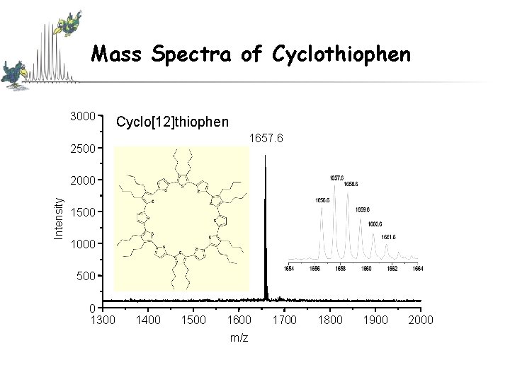
Mass Spectra of Cyclothiophen 3000 Cyclo[12]thiophen 1657. 6 2500 Intensity 2000 1500 1000 500 0 1300 1400 1500 1600 m/z 1700 1800 1900 2000
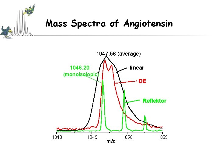
Mass Spectra of Angiotensin 1047. 56 (average) 1046. 20 (monoisotopic) linear DE Reflektor 1040 1045 m/z 1050 1055
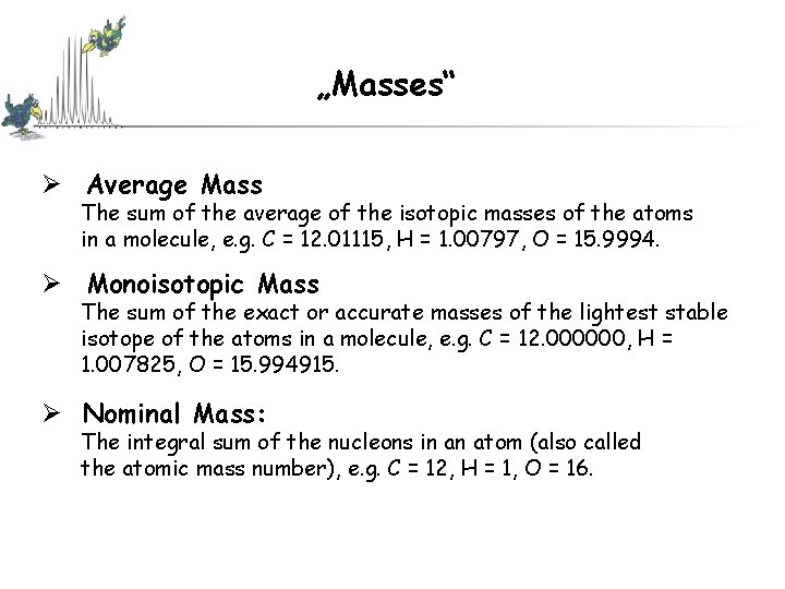
„Masses“ Ø Average Mass The sum of the average of the isotopic masses of the atoms in a molecule, e. g. C = 12. 01115, H = 1. 00797, O = 15. 9994. Ø Monoisotopic Mass The sum of the exact or accurate masses of the lightest stable isotope of the atoms in a molecule, e. g. C = 12. 000000, H = 1. 007825, O = 15. 994915. Ø Nominal Mass: The integral sum of the nucleons in an atom (also called the atomic mass number), e. g. C = 12, H = 1, O = 16.
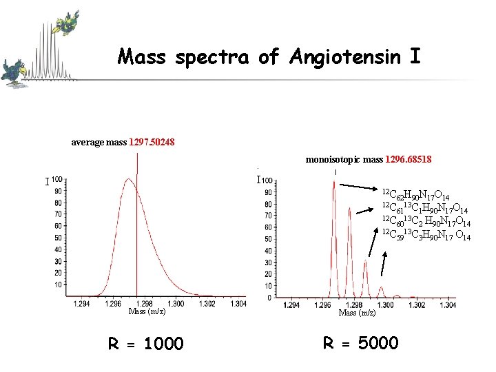
Mass spectra of Angiotensin I average mass 1297. 50248 . I I monoisotopic mass 1296. 68518 12 C 62 H 90 N 17 O 14 12 C 13 C H N O 61 1 90 17 14 12 C 13 C H N O 60 2 90 17 14 12 C 13 C H N O 59 3 90 17 14 Mass (m/z) R = 1000 Mass (m/z) R = 5000
Simulated Spectra of Bovine Insulin
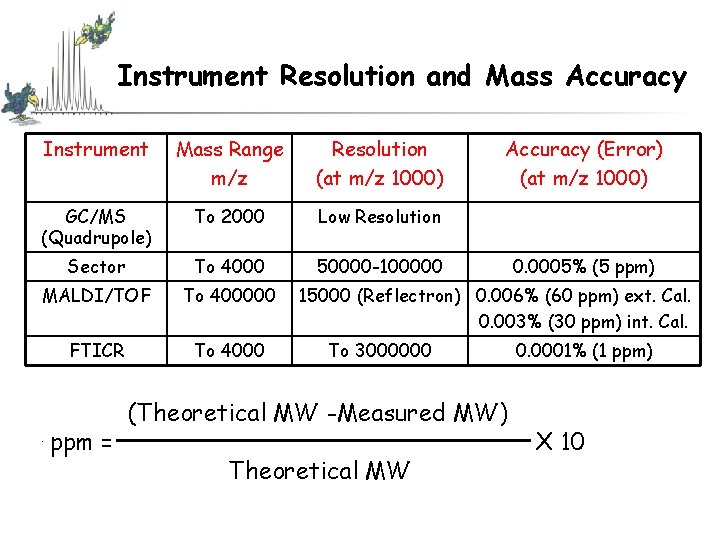
Instrument Resolution and Mass Accuracy Instrument Mass Range m/z Resolution (at m/z 1000) GC/MS (Quadrupole) To 2000 Low Resolution Sector To 4000 50000 -100000 MALDI/TOF To 400000 FTICR To 4000 ppm = Accuracy (Error) (at m/z 1000) 0. 0005% (5 ppm) 15000 (Reflectron) 0. 006% (60 ppm) ext. Cal. 0. 003% (30 ppm) int. Cal. To 3000000 (Theoretical MW -Measured MW) Theoretical MW 0. 0001% (1 ppm) X 10
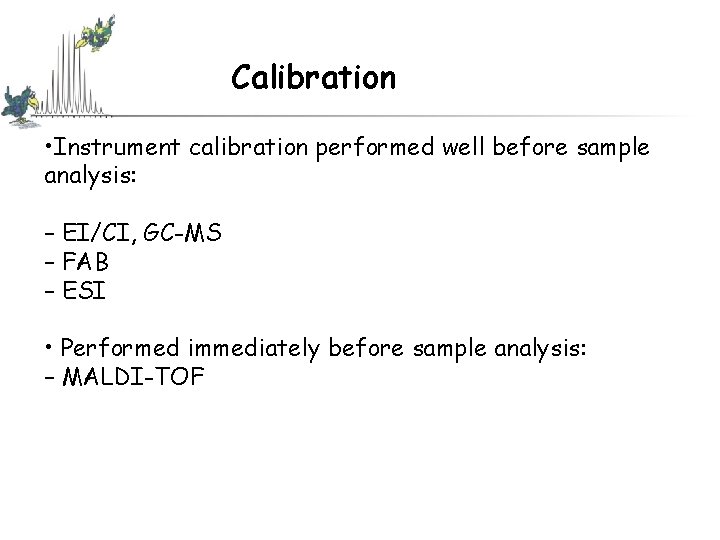
Calibration • Instrument calibration performed well before sample analysis: – EI/CI, GC-MS – FAB – ESI • Performed immediately before sample analysis: – MALDI-TOF
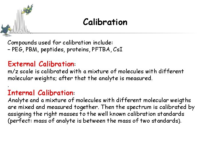
Calibration Compounds used for calibration include: – PEG, PBM, peptides, proteins, PFTBA, Cs. I External Calibration: m/z scale is calibrated with a mixture of molecules with different molecular weights; after that the analyte is measured. . Internal Calibration: Analyte and a mixture of molecules with different molecular weigths are mixed and measured together. Then the spectrum is calibrated by assigning the right masses to the well known calibration standards (perfect: mass of analyte is between the mass of two standards ).
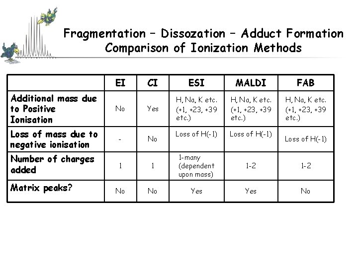
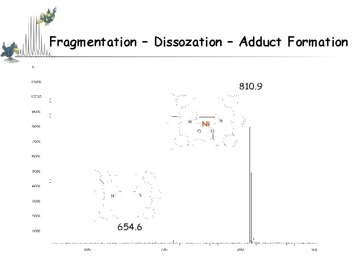
Fragmentation – Dissozation – Adduct Formation Comparison of Ionization Methods EI CI ESI MALDI FAB H, Na, K etc. (+1, +23, +39 etc. ) Loss of H(-1) 1 -2 Yes No Additional mass due to Positive Ionisation No Yes Loss of mass due to negative ionisation - No Number of charges added Matrix peaks? 1 1 1 -many (dependent upon mass) No No Yes Loss of H(-1)
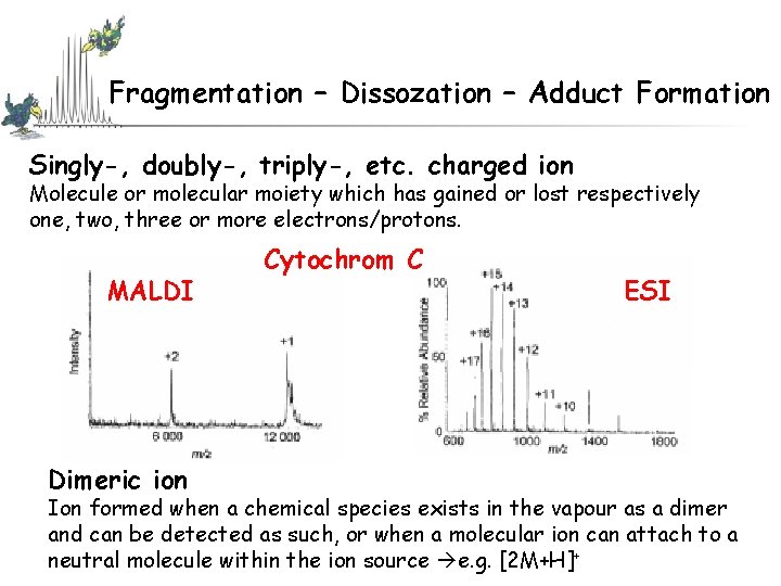
Fragmentation – Dissozation – Adduct Formation Singly-, doubly-, triply-, etc. charged ion Molecule or molecular moiety which has gained or lost respectively one, two, three or more electrons/protons. MALDI Dimeric ion Cytochrom C ESI Ion formed when a chemical species exists in the vapour as a dimer and can be detected as such, or when a molecular ion can attach to a neutral molecule within the ion source e. g. [2 M+H]+
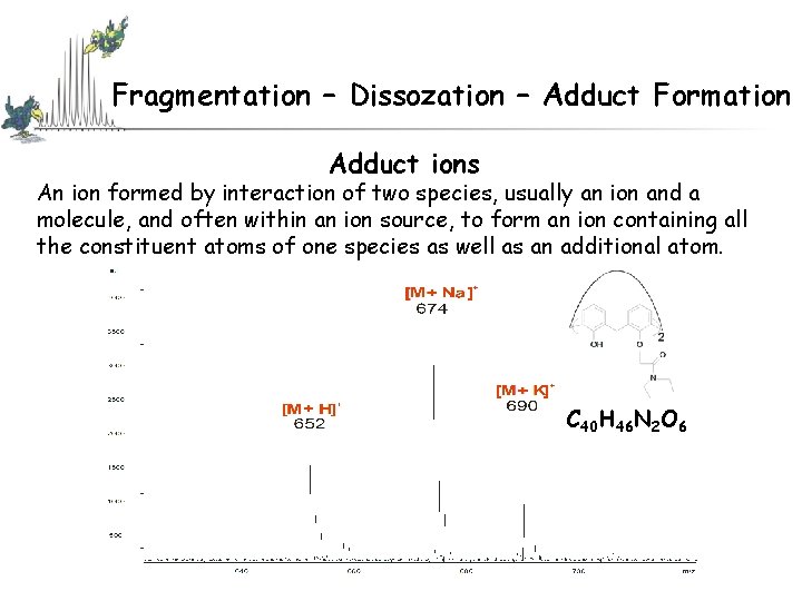
Fragmentation – Dissozation – Adduct Formation Adduct ions An ion formed by interaction of two species, usually an ion and a molecule, and often within an ion source, to form an ion containing all the constituent atoms of one species as well as an additional atom. C 40 H 46 N 2 O 6
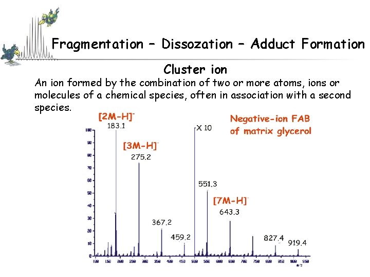
Fragmentation – Dissozation – Adduct Formation Cluster ion An ion formed by the combination of two or more atoms, ions or molecules of a chemical species, often in association with a second species.
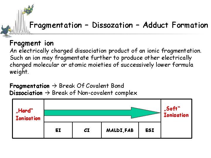
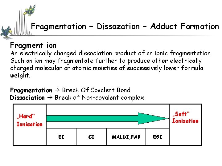
Fragmentation – Dissozation – Adduct Formation Fragment ion An electrically charged dissociation product of an ionic fragmentation. Such an ion may fragmentate further to produce other electrically charged molecular or atomic moieties of successively lower formula weight. Fragmentation Break Of Covalent Bond Dissociation Break of Non-covalent complex „Soft“ Ionisation „Hard“ Ionisation EI CI MALDI, FAB ESI
Fragmentation – Dissozation – Adduct Formation Fragment ion An electrically charged dissociation product of an ionic fragmentation. Such an ion may fragmentate further to produce other electrically charged molecular or atomic moieties of successively lower formula weight. Fragmentation Break Of Covalent Bond Dissociation Break of Non-covalent complex „Soft“ Ionisation „Hard“ Ionisation EI CI MALDI, FAB ESI
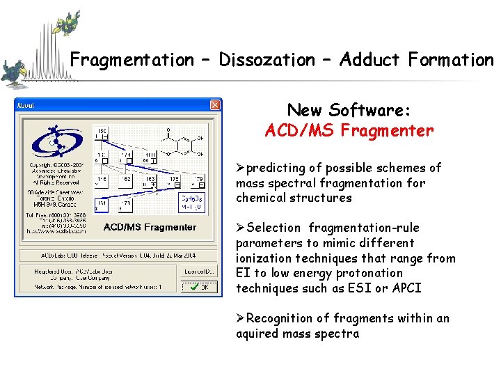
Fragmentation – Dissozation – Adduct Formation New Software: ACD/MS Fragmenter Øpredicting of possible schemes of mass spectral fragmentation for chemical structures ØSelection fragmentation-rule parameters to mimic different ionization techniques that range from EI to low energy protonation techniques such as ESI or APCI ØRecognition of fragments within an aquired mass spectra
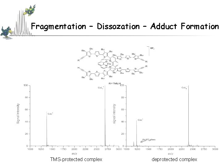
Fragmentation – Dissozation – Adduct Formation TMS-protected complex deprotected complex
Fragmentation – Dissozation – Adduct Formation
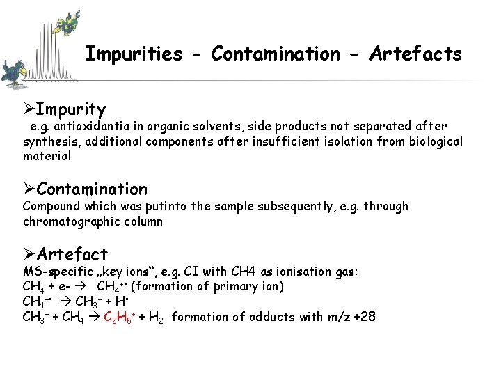
Impurities - Contamination - Artefacts ØImpurity e. g. antioxidantia in organic solvents, side products not separated after synthesis, additional components after insufficient isolation from biological material ØContamination Compound which was putinto the sample subsequently, e. g. through chromatographic column ØArtefact MS-specific „key ions“, e. g. CI with CH 4 as ionisation gas: CH 4 + e- CH 4+ • (formation of primary ion) CH 4+ • CH 3+ + H • CH 3+ + CH 4 C 2 H 5+ + H 2 formation of adducts with m/z +28
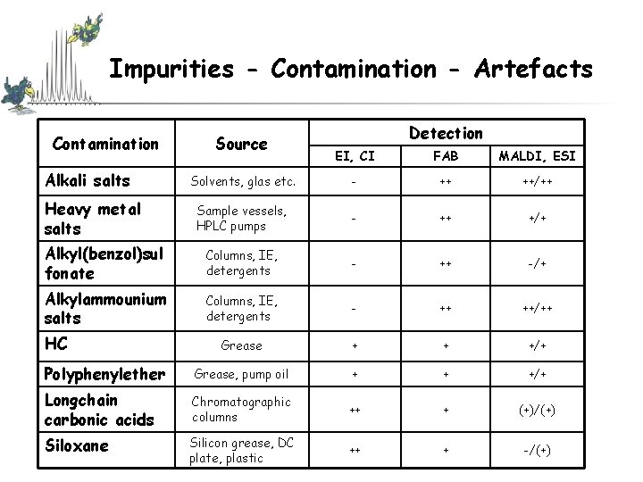
Impurities - Contamination - Artefacts Contamination Source Detection EI, CI FAB MALDI, ESI Solvents, glas etc. - ++ ++/++ Sample vessels, HPLC pumps - ++ +/+ Alkyl(benzol)sul fonate Columns, IE, detergents - ++ -/+ Alkylammounium salts Columns, IE, detergents - ++ ++/++ Grease + + +/+ Polyphenylether Grease, pump oil + + +/+ Longchain carbonic acids Chromatographic columns ++ + (+)/(+) Siloxane Silicon grease, DC plate, plastic ++ + -/(+) Alkali salts Heavy metal salts HC
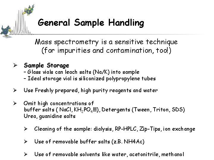
General Sample Handling Mass spectrometry is a sensitive technique (for impurities and contamination, too!) Ø Sample Storage – Glass vials can leach salts (Na/K) into sample – Ideal storage vial is siliconized polypropylene tubes Ø Use Freshly prepared, high purity reagents and water Ø Omit high concentrations of buffer salts ( Na. Cl, KH 2 PO 4!!!), Detergents (Tween, Triton, SDS) Urea, guanidine salts Ø Cleaning of the sample: dialysis, RP-HPLC, Zip-Tips, ion exchange Ø Use of removable buffer salts (z. B. NH 4 Ac) Ø Use of removable solvents like water, acetonitrile, methanol
General Sample Handling Ø Use Freshly prepared, high purity reagents and water Ø Omit high concentrations of buffer salts ( Na. Cl, KH 2 PO 4!!!), Detergents (Tween, Triton, SDS) Urea, guanidine salts Ø Cleaning of the sample: dialysis, RP-HPLC, Zip-Tips, ion exchange Ø Use of removable buffer salts (z. B. NH 4 Ac) Ø Use of removable solvents like water, acetonitrile, methanol
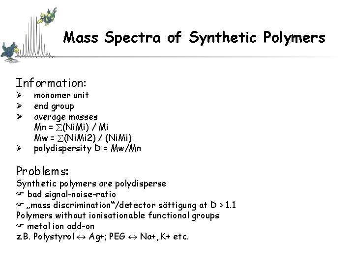
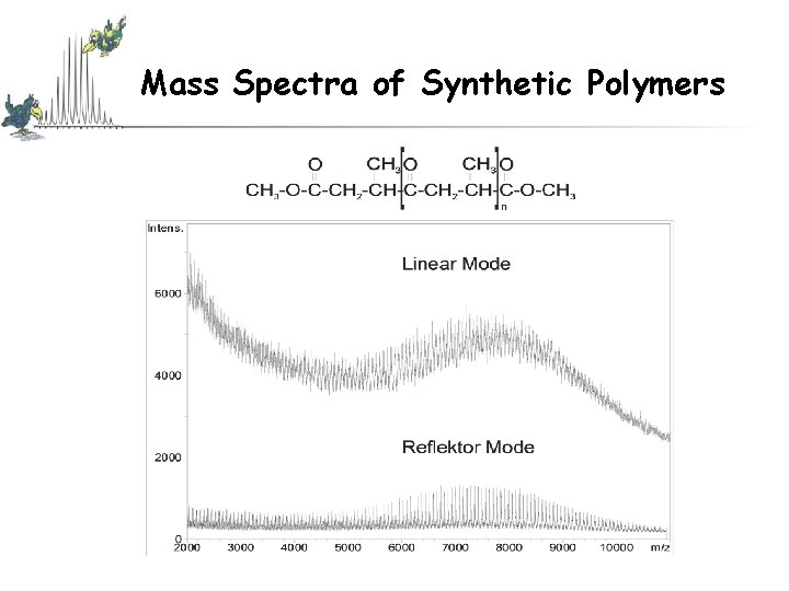
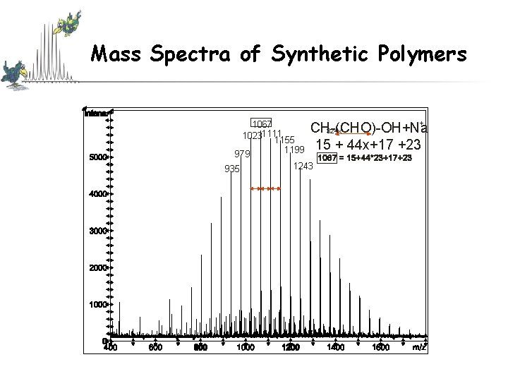
Mass Spectra of Synthetic Polymers Information: Ø Ø monomer unit end group average masses Mn = (Ni. Mi) / Mi Mw = (Ni. Mi 2) / (Ni. Mi) polydispersity D = Mw/Mn Problems: Synthetic polymers are polydisperse bad signal-noise-ratio „mass discrimination“/detector sättigung at D > 1. 1 Polymers without ionisationable functional groups metal ion add-on z. B. Polystyrol Ag+; PEG Na+, K+ etc.
Mass Spectra of Synthetic Polymers
Mass Spectra of Synthetic Polymers 1067 10231111 1155 979 935 1199 + CH-(CHO)-OH+Na 324 x 15 + 44 x+17 +23 1243
New aspects in mass spectrometry: Hybrid Mass Spectrometers Perhaps hundreds of hybrids have been explored. Some of the more successful: Triple quadrupole IT-TOF Quadrupole-FTMS TOF/TOF
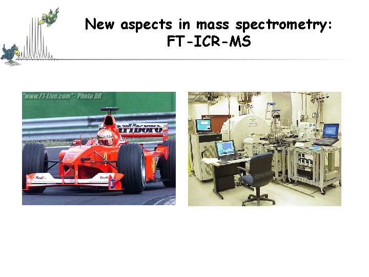
New aspects in mass spectrometry: FT-ICR-MS
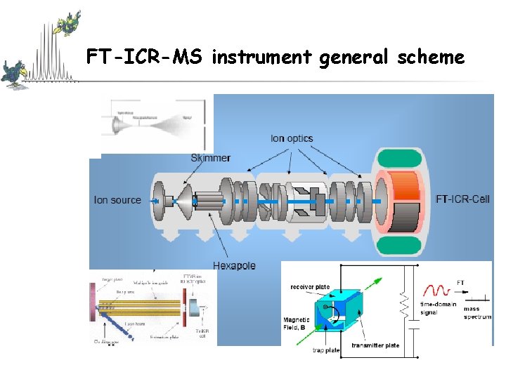
FT-ICR-MS instrument general scheme
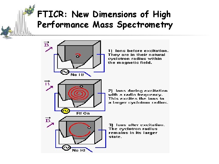
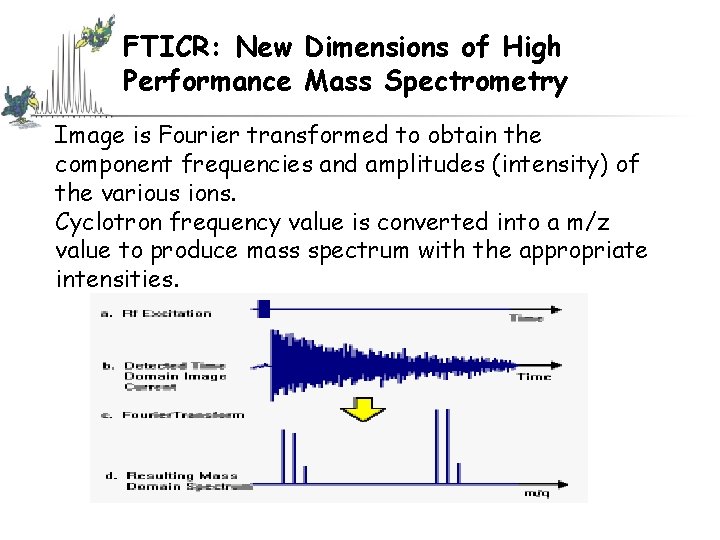
Fouriertransform-ICR: New Dimensions of High Performance Mass Spectrometry Ø A high-frequency mass spectrometer in which the cyclotron motion of ions, having different m/z ratios, in a constant magnetic field, is excited essentially simultaneously and coherently by a pulse of a radiofrequency electric field applied perpendicularly to the magnetic field. Ø The excited cyclotron motion of the ions is subsequently detected on receiver plates as a time domain signal that contains all the cyclotron frequencies excited. Ø Fourier transformation of the time domain signal results in the frequency domain FT-ICR signal which, on the basis of the inverse proportionality between frequency and m/z ratio, can be converted to a mass spectrum. Ø The ions are to be detected, with a selected m/z ratio, absorb maximum energy through the effect of a high-frequency field and a constant magnetic field perpendicular to it. Maximum energy is gained by ions that satisfy the cyclotron resonance condition and as a result these are separated from ions of different mass/charge.
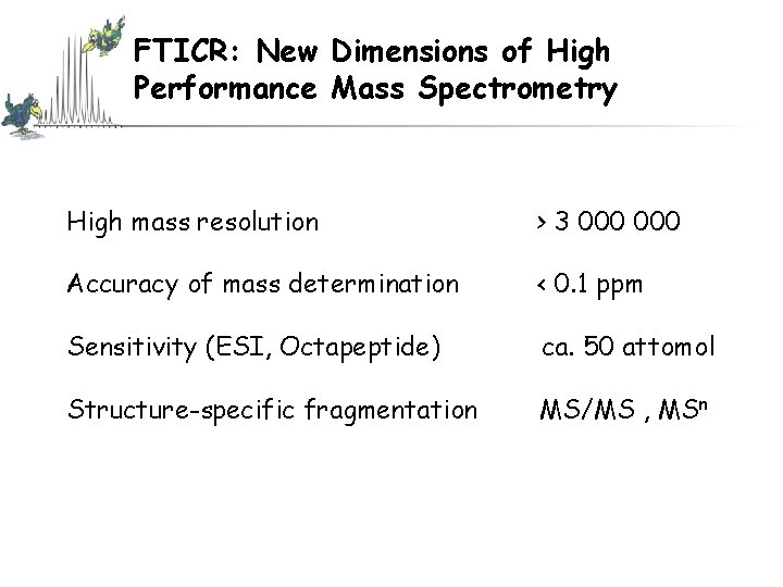
FTICR: New Dimensions of High Performance Mass Spectrometry High mass resolution > 3 000 Accuracy of mass determination < 0. 1 ppm Sensitivity (ESI, Octapeptide) ca. 50 attomol Structure-specific fragmentation MS/MS , MSn
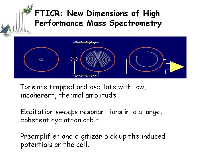
FTICR: New Dimensions of High Performance Mass Spectrometry Ions are trapped and oscillate with low, incoherent, thermal amplitude Excitation sweeps resonant ions into a large, coherent cyclotron orbit Preamplifier and digitizer pick up the induced potentials on the cell.
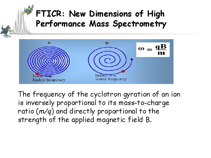
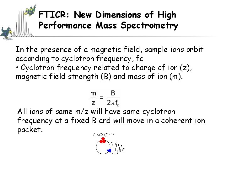
FTICR: New Dimensions of High Performance Mass Spectrometry The frequency of the cyclotron gyration of an ion is inversely proportional to its mass-to-charge ratio (m/q) and directly proportional to the strength of the applied magnetic field B.
FTICR: New Dimensions of High Performance Mass Spectrometry
FTICR: New Dimensions of High Performance Mass Spectrometry In the presence of a magnetic field, sample ions orbit according to cyclotron frequency, fc • Cyclotron frequency related to charge of ion (z), magnetic field strength (B) and mass of ion (m). All ions of same m/z will have same cyclotron frequency at a fixed B and will move in a coherent ion packet.
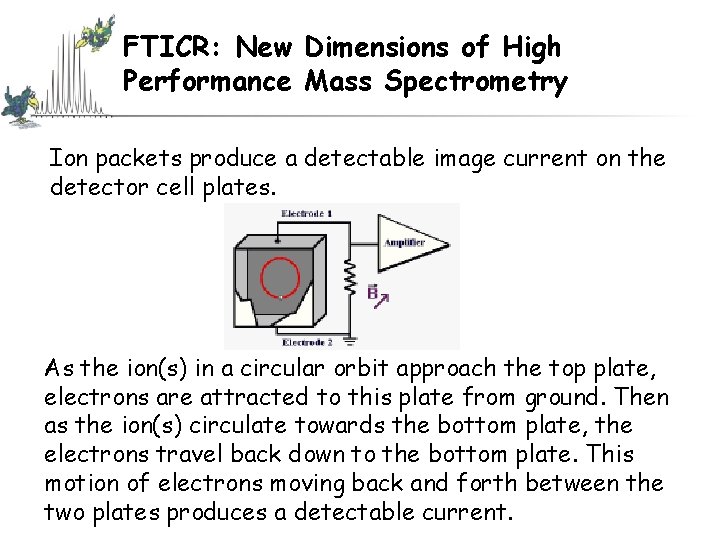
FTICR: New Dimensions of High Performance Mass Spectrometry Ion packets produce a detectable image current on the detector cell plates. As the ion(s) in a circular orbit approach the top plate, electrons are attracted to this plate from ground. Then as the ion(s) circulate towards the bottom plate, the electrons travel back down to the bottom plate. This motion of electrons moving back and forth between the two plates produces a detectable current.
FTICR: New Dimensions of High Performance Mass Spectrometry Image is Fourier transformed to obtain the component frequencies and amplitudes (intensity) of the various ions. Cyclotron frequency value is converted into a m/z value to produce mass spectrum with the appropriate intensities.
FTICR: New Dimensions of High Performance Mass Spectrometry
The End