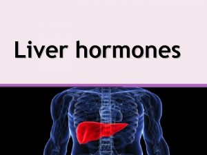
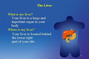
Liver Position of the liver it occupies the


- Slides: 29


Liver

• Position of the liver • it occupies • the whole of the right hypochondrium • parts of the Subcostal plane epigastrium and left hypochondrium ** Weight - Male about 1. 5 kg - Female about 1. 3 kg. Intertubercular plane

Wedge-shaped ** Surfaces: - It has 5 surfaces - Anterior - Posterior - Superior - Inferior - Right. - No sharp borders between them superi or Anteri or Righ t Posterior Inferio r

Surface anatomy of the liver CC 8 th. CC 1 cm below the costal margin at the right midaxillary line 9 th. CC transpyloric plane at the median plane

Surface anatomy 1 - Superior border of the liver: represented by a line joining the following points directed to the right 1 - A point in the left 5 th intercostal space (in the left mid-clavicular line). 2 - A point at the xiphisternal junction (in the median plane). 3 - A point on the right 5 th costal cartilage (the right mid-clavicular line). 4 - A point on the right 7 th rib (in the right midaxillary line). 2 - Inferior border: represented by a line joining the following points directed to the right 1 - A point in the left 5 th intercostal space (left mid-clavicular line). 2 - The tip of the left 8 th costal cartilage. 3 - The transpyloric plane in the median plane (midway between the umbilicus and xiphisternal junction. 4 - The tip of the right 9 th costal cartilage (right midclavicular line). 5 - 1 cm below the costal margin at the right midaxillary line. 3 - Right border: represented by a line convex to the right between the right 7 th rib to 1 cm below the costal margin in the right midaxillary line

Anatomical Lobes and fissures of the liver

Lt. 1/5 Rt 4/5 right lobe lef tl fa lci f or Anterior view Anatomical Lobes of the liver m ob e lig am en t.

Fissure for the ligamentum venosum (Post) (obliterated ductus venosus) Posteroinferior view Lt. 1/5 Fissure for ligamentum teres or round ligament (Inf) (obliterated left umbilical vein) Rt 4/5

IVC Right lobe proper Caudate lobe Fissure for the ligamentum venosum Fissure for ligamentum teres Quadrate lobe Gall bladder

Caudate lobe 2 1 Papillary process 3 (left) 8 7 4 5 Porta hepatis Posterior view 6 Caudate process § Porta hepatis (hilum of the liver) ** Structures passing through the porta hepatis are (VAD): 1 - 2 branches of the Portal Vein: posterior in position. 2 - 2 branches of the Hepatic Artery: intermediate in position. 3 - 2 Hepatic Ducts: anterior in position. 4 Lymphatic nodes and sympathetic fibres around the artery

- The liver is divided into right and left lobes according to the pattern of the portal triad (VAD): portal vein, hepatic artery and common hepatic duct. - The line of demarcation between the right and left lobes occurs along a line extending from the gall bladder to the inferior vena cava. 1 - Caudate lobe of left lobe 2 - Superior segment of left lobe (lateral segment) 3 - Inferior segment of left lobe proper (lateral segment) 4 - Quadrate lobe of left lobe (medial segment) 5 - Inferior anterior segment of right lobe 6 - Inferior posterior segment of right lobe 7 - Superior posterior segment of right lobe 8 - Superior anterior segment of right lobe § Functional (surgical) Segmentations of the liver

Falciform ligament Upper layer of coronary ligament IVC left Triangular ligament Lesser omentum Lower layer of coronary ligament Right triangular ligament Ligaments of the liver

Falciform ligament Free margin containing Ligamentum teres and paraumbilical vein

Ligaments (Peritoneal folds) of the liver 1 - Falciform ligament: sickle-shaped. - Borders, a- Convex border attached to the diaphragm and anterior abdominal wall. b- A concave border attached to the liver. c- A straight free border which extends from the umbilicus to the liver. It contains ligamentum teres and paraumblical vein. 2 - Upper layer of coronary ligament. 3 - Lower layer of coronary ligament. 4 - Right triangular ligament, - The meeting of the two coronary ligaments at the apex of the bare area. 5 - Left triangular ligament, on the superior surface of the left lobe of the liver. 6 - Lesser omentum around the porta hepatis.

Fissure for the falciform ligament 7 - Bare areas of the liver Bare area Fissure for the ligamentum venosum Porta hepatis Fissure for ligamentum teres Fossa for IVC Fossa for gall bladder

Anterior relations of liver Anterior surface: a- Anterior abdominal wall. b- Diaphragm. c- Costal cartilages. • Anterior abdominal wall

Relations on the superior surface Heart, pericardium Small part of Left Pleura, lung Copula of diaphragm Right Pleura, lung

(Anterior) (Diaphragm) (2 nd) (1 st & T. colon) (R. colic flexure) Relations of the posterior & inferior

Blood supply 1 - Hepatic arteries (left and right branches). It carries oxygenated blood. 2 - Hepatic veins → into the inferior vena cava. 2 - Portal vein carries venous blood from the gastrointestinal tract. N. B; - Blood is mixed in the liver sinusoids. IVC

Lymphatic drainage The lymphatic drainage of the liver consists of two major pathways: 1 - Superficial lymphatic end into: a- The lymph nodes around the I. V. C. b- Hepatic lymph nodes in the porta hepatis. c- Para-cardiac nodes around the lower part of esophagus d- Coeliac lymph nodes around the coeliac trunk. 2 Deep lymphatic: divided into ascending and descending trunks a- Ascending trunk end in the lymph nodes around the I. V. C. b- Descending trunk drain into hepatic lymph nodes in porta hepatis. - These lymph nodes end in the intestinal trunk to cisterna chyli.

Biliary system

Biliary System • Right and left hepatic ducts from the right and left lobes of the liver unite to form common hepatic duct. It joins the cystic duct usually at an acute angle to form the common bile duct It fuses with main pancreatic duct forming hepatopancreatic duct that opens into major duodenal ampulla (ampulla of Vater) in the 2 nd part of the duodenum.

** Parts of the gall bladder Neck - Its capacity 50 cc - Cystic duct: Its mucosa is twisted and forms a spiral valve (Heister's valve). These spiral folds do not disappear when the duct is distended and Cystic duct helps to keep the cystic duct open - Hartmann’s pouch is a Body dilatation at the neck is a common site for impaction of Gallstones Fundus

** Surface anatomy of gall bladder - A point close to the tip of the right 9 th costal cartilage. - At the crossing of transpyloric plane with the right mid-clavicular plane. ** Nerve supply of the gall bladder: - Sympathetic from the coeliac trunk. - Parasympathetic from vagus nerve. - Sensory from the right phrenic nerve. • Clinical notes - Inflammation of gall bladder leading to referred pain to the right shoulder because a- Right phrenic nerve (C. 3, 4, 5). b- The skin of the right shoulder is supplied by the supraclavicular nerves (C. 3, 4). 9 th CC Transpyloric plane Right midclavicular line

Qu est ion s I/Azzam - 2004 Th ank you

** Arterial supply, cystic artery from the right branch of hepatic artery. ** Venous drainage, cystic vein end in the right branch of the portal vein. ** Lymphatic: drain into the cystic and hepatic lymph nodes Second part of duodenum Hepatic ducts Common hepatic duct Cystic duct Common Bile duct main pancreatic duct Ampulla of Vater

Gall Stones Blockage Contraction Inflammation The infundibulum of the gallbladder is the common site for stones impaction If the stone blocks A gallstone is a the infundibulum or cystic duct, concretion in the cholecystitis gallbladder, (inflammation of infundibulum the gallbladder) cystic duct, or occurs because of bile duct bile accumulation, composed chiefly causing of cholesterol enlargement of the crystals gallbladder.

CHOLESTASIS An arrest in the flow of bile The distal end of the hepatopancreatic ampulla is the If bile cannot leave narrowest part ofit the gallbladder, the biliarythe passages enters blood and iscauses another and common site for jaundice impaction of gallstones Liver Cholestasis - Duct Obstruction - Liver or Pancreatic Disease Result (Hyperbilirubinemia – Jaundice) Pancreas Duodenum
 Second position ballet
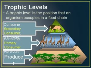
 What are trophic level
 Derived positions
 Fundamental position vs anatomical position
 Anything that occupies space
 The matter is anything that occupies
 A political community that occupies a definite territory
 K=pv
K=pv Pv k
Pv k It is anything that has mass and occupies space
 Within the meninges cerebrospinal fluid occupies the
 A sample of neon gas occupies a volume of 677 ml at 134 kpa
 A gas occupies 473 cm3 at 36°c. find its volume at 94°c
 It is anything that has mass and occupies space
 3 gas laws
 Are gasses highly compressible
 A gas occupies 473 cm3 at 36°c. find its volume at 94°c
 This is anything with mass that occupies space
 Trời xanh đây là của chúng ta thể thơ
 Phản ứng thế ankan
 Chó sói
 Thiếu nhi thế giới liên hoan
 Tia chieu sa te
 điện thế nghỉ
 Một số thể thơ truyền thống
 Thế nào là hệ số cao nhất
 Hệ hô hấp
 Ng-html
 Bảng số nguyên tố lớn hơn 1000
 đặc điểm cơ thể của người tối cổ

