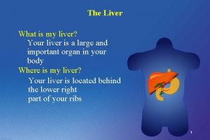
Position of the liver it occupies the whole


- Slides: 22


Position of the liver it occupies the whole of the right hypochondrium and parts of the epigastrium and left hypochondrium ** Weight in male about 1. 5 kg, in female about 1. 3 kg.

** Surfaces: - It has 5 surfaces anterior, posterior, superior, inferior and right lateral. - No sharp borders between them superior surface anterior surface right surfac e Wedge-shaped 5 surfaces,

Posterior surface inferior surface

IVC GB Fissur of ligamentum teres

Caudate lobe Papillary process Porta hepatis Caudate process

Fissures of the liver 1. Fissure for the ligamentum teres (obliterated left umbilical vein) - It presents in the inferior surface. It separates the left lobe from the quadrate lobe. 2. Fissure for the ligamentum venosum (obliterated ductus venosus). - It presents in the posterior surface. It separates the left lobe from the caudate lobe. 3 - Porta hepatis (hilum of the liver) in the posterior part of the inferior lobe. ** The structures passing through the porta hepatis are (VAD): 1 - 2 branches of the Portal Vein: posterior in position. 2 - 2 branches of the Hepatic Artery: intermediate in position. 3 - 2 Hepatic Ducts: anterior in position and lymph nodes. 4 - Fissure of the falciform ligament on the anterior surface

Ligaments of the liver 1 - Falciform ligament, sickle-shaped, a- Convex border attached to the diaphragm and anterior abdominal wall. b- A concave border attached to the liver. c- A straight free border which extends from the umbilicus to the liver containing ligamentum teres and paraumbilical vein between the umbilicus and left branch of the portal vein 2 - Upper and lower layers of coronary ligaments (boundaries of bare area). 3 - Right triangular ligament, meeting of the upper and lower layers of the coronary ligament at the apex of the bare area. 4 - Left triangular ligament, on the superior surface of the left lobe.

Fissure for the falciform ligament Bare areas of the liver Bare area Fissure for the ligamentum venosum Porta hepatis Fissure for ligamentum teres Fossa for IVC Fossa for gall bladder

Biliary System Hepatic ducts - Right and left hepatic ducts from the right and left lobes of the liver. - The two ducts unite to form common hepatic duct. Common hepatic duct joins the cystic duct of the gall bladder to form the common bile duct.

** Surface anatomy of the gall bladder (fundus) - A point close to the tip of the right 9 th costal cartilage. - At the crossing of transpyloric plane with the right mid-clavicular plane. ** Nerve supply of the gall bladder: - Sympathetic from the coeliac trunk. - Parasympathetic from vagus nerve. - Sensory from the right phrenic nerve. Clinical notes - Inflammation of gall bladder leading to referred pain to the right shoulder because Right phrenic nerve and supraclavicular nerves arise from (C. 3, 4, ). 9 th CC Transpyloric plane Right midclavicular line

** Parts of the gall bladder 1. Fundus: usually projects below the inferior border of the liver behind the anterior abdominal wall. - It is completely covered by peritoneum. 2 - Body; middle dilated part 3 - Neck: is the narrow part which is continuous with the cystic duct. - Its mucosa is twisted and forms a spiral valve. - The right side of the neck may present a small dilatation called Hartmann’s pouch the cystic duct of the gall bladder joins with Common hepatic duct to form the common bile duct.

Spleen is a lymphatic organ in the left hypochondriurm Upper border Notches Posterior end Tapering, medial Angle Lower border Anterior end Wide, lateral

• Spleen (Lien) - It is a lymphatic organ connected to the vascular system. ** Position: It lies in the left hypochondriurm. Features of the spleen A- 2 Ends 1 - Posterior end (tapering) directed upwards, backwards and medially. 2 - Anterior end (broad) directed downwards, forwards and laterally. B- 2 Borders 1 - Upper border, sharp. - It shows one or more Notches near its anterior end. - It meets the anterior end in the angle of the spleen. N. B; Notching of the upper border is an indication of foetal lobulation. 2 - Lower border, thick and round. C- 2 Surfaces diaphragmatic and visceral.

Upper border Axis Upper border Surface anatomy of Spleen Anterior end just behind Left midaxillary line

Surface anatomy - The upper border is parallel to the upper border of the 9 th rib. - The long axis of the spleen lies along the 10 th rib. - The lower border is parallel to the lower border of the 11 th rib. - The anterior end normally lies just behind the left midaxillary line. - The posterior end lies one and half inches lateral to the 10 th thoracic spine. N. B; the normal spleen is not palpable. - If the spleen is felt below the costal margin, it is enlarged at least 3 times of its normal size. ** How to place the spleen in the correct anatomical position 1 - Hold the spleen in your left hand with its convex surface applied to the palm, the round posterior end towards the wrist, the broad anterior end towards the tips of fingers and the notched upper border applied to the thumb. 2 - Put your hand behind the left midaxillary line with an angle 45 degrees with the horizontal.

Visceral surface Diaphragmatic surface Gastric Renal Hilum contains splenic vessels ic t ea r c n a P Colic

** Pancreas - Spleen is a combined exocrine and endocrine gland ** Position, transversely it lies across Pancreas the posterior abdominal wall. • It extends from the duodenum to the spleen, with a slight upward inclination from the right to the left sides. Duodenum

Parts of pancreas Neck

Transverse section of the body of pancreas Splenic A

1 - Main pancreatic duct (duct of Wirsung); runs along the long axis of the pancreas from the tail to the head. - It fuses with CBD forming hepato-pancreatic duct that opens into the major duodenal papilla (ampulla of Vater) in the 2 nd part of the duodenum. 2 - Accessory pancreatic duct, begins from the lower part of the head and uncinate process. - It opens into the 2 nd part of the duodenum on the minor duodenal papilla, one inch above the major duodenal papilla. Pancreatic ducts Minor duodenal papilla Major

Th ank Qu you est ion s I/Azzam - 2004
 Cdc whole school whole community whole child
 Second position echappe
 Throphic levels
 Massed practice
 Fundamental position vs anatomical position
 Fundamental starting position
 A gas occupies 473 cm3 at 36°c. find its volume at 94°c
 Within the meninges cerebrospinal fluid occupies the
 What is anything that has mass and occupies space
 Matter is anything that has
 A gas occupies 473 cm3 at 36°c. find its volume at 94°c
 3 gas laws
 A gas occupies 473 cm3 at 36°c. find its volume at 94°c
 Pv = k
 A political community that occupies a definite territory
 It is anything that has mass and occupies space
 A sample of neon gas occupies a volume of 677 ml at 134 kpa
 The matter is anything that occupies
 A gas occupies 473 cm3 at 36°c. find its volume at 94°c
 Matter has mass and occupies space
 Lp html
 Sơ đồ cơ thể người
 Số nguyên tố là gì


