Human Anatomy and Physiology II Unit IV Circulation


 � Right AV valve (tricuspid) �")


- Slides: 59
Human Anatomy and Physiology II Unit IV Circulation and Body Defense Part II The Heart
Unit Topics � � � Part 1 Blood Part 2 Heart Part 3 Blood Vessels Part 4 Lymphatic System Part 5 Immunity
The Cardiovascular System The cardiovascular system enables the rapid transport of nutrients, respiratory gases, waste products, and cells within the body.
Introduction � 100, 000 x beats per day �Right side vs. left side � 7, 000 -14, 000 L / day when inactive �What’s yours? �Cardiology study of the heart
Heart Topics 1. 2. 3. 4. 5. 6. 7. 8. Location of the Heart Chambers of the Heart Valves Coronary Circulation Cardiac Muscle Cardiac Conduction System Cardiac Cycle Cardiac Output
Location of the Heart � Size of fist � Located in the mediastinum. � Two-thirds lies to left of midline. � Upper third = great vessels
Location of the Heart Rests on the diaphragm � ◦ Phrenic nerve The heart is enclosed and held in place by the double layered pericardium. 1. Outer fibrous pericardium � 2. Inner serous pericardium A. Parietal layer B. Visceral layer
Location of the Heart � Surfaces of the heart
Pericardium
Pericardium
Pericardium Parietal pericardium Visceral Pericardium
Pericardial Diseases Pericarditis ◦ Inflammation of the pericardium � Pericardial effusion ◦ Build up of fluid in the pericardial cavity ◦ Often due to congestive heart failure � Cardiac tamponade ◦ Bleeding into the pericardial cavity �
Heart Topics 1. 2. 3. 4. 5. 6. 7. 8. Location of the Heart Chambers of the Heart Valves Coronary Circulation Cardiac Muscle Cardiac Conduction System Cardiac Cycle Cardiac Output
Location of the Heart � The wall of the four-chambered heart has three layers: 1. Epicardium 2. Myocardium 3. Endocardium
Layers of the Heart Wall
Inflammation of Heart Wall �Myocarditis ◦ Inflammation of myocardium ◦ Usually viral �Endocarditis ◦ Inflammation of endocardium ◦ Usually bacterial
Chambers of the Heart � Right atrium � Right ventricle � Left atrium � Left ventricle
Chambers of the Heart Right atrium and right ventricle pump blood to the lungs. � Left atrium and left ventricle pump blood to the rest of the body. � Auricles �
Coronary Sulci � Coronary sulcus � Anterior interventricular sulcus � Posterior interventricular sulcus
Right Atrium Forms the right border of the heart � Receives blood from: � ◦ Superior vena cava ◦ Inferior vena cava ◦ Coronary sinus Posterior atrial wall is smooth � Anterior wall is rough. � ◦ Pectinate muscle ◦ Right auricle
Posterior Wall
Pectinate Muscles
Right Atrium � In between the atria is the interatrial septum ◦ Fossa ovalis ◦ Foramen ovale
Atrial Septal Defects � Incomplete closure of the foramen ovale.
Right Atrium
Right Atrium Blood leaves the right atrium through the tricuspid valve � Forms most of the anterior surface of the heart �
Right Ventricle � The inside of the right ventricle contains muscular ridges called trabeculae carnae
Right Ventricle � Receives blood from the right atrium � Tricuspid valve ◦ Chordae tendinae ◦ Papillary muscles
Right Ventricle � Right and left ventricles are separated from each other by the interventricular septum
Ventricular Septal Defects � Incomplete formation of the interventricular septum.
Right Ventricle Blood leaves the right ventricle through the pulmonary semilunar valve � Pulmonary trunk � ◦ Right and left pulmonary arteries
Left Atrium Forms most of the base of the heart � Receives blood from the pulmonary veins � The left atrium is separated from the right atrium by the interatrial septum � The walls of the left atrium are smooth except for the auricle � ◦ � Fewer & smaller pectinate muscles Blood leaves the left atrium through the bicuspid valve
Left Ventricle � � � Forms the apex of the heart Thickest muscular chamber Receives blood from the left atrium Trabeculae carnae Bicuspid valve ◦ Chordae tendinae ◦ Papillary muscles
Left Ventricle � Blood leaves the left ventricle through the aortic semilunar valve into the branches of the aorta.
Left Ventricle � Ductus arteriosus � Ligamentum arteriosum
Myocardial Thickness and Function � Atrial walls are thinner than ventricular walls � Walls of the right ventricle thinner than walls of the left ventricle
Fibrous Skeleton � Four dense connective tissue rings wrapped around the four AV valves of the heart
Heart Topics 1. 2. 3. 4. 5. 6. 7. 8. Location of the Heart Chambers of the Heart Valves Coronary Circulation Cardiac Muscle Cardiac Conduction System Cardiac Cycle Cardiac Output
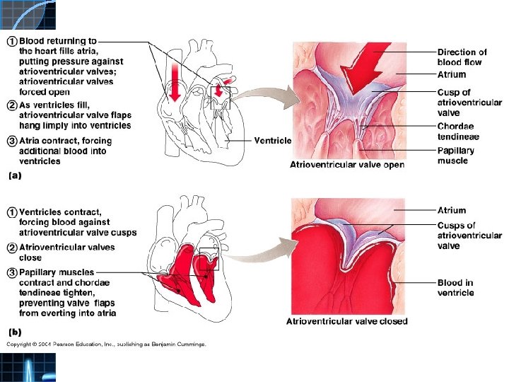
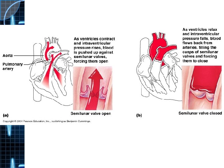
Heart Valves � Healthy heart valves ensure that blood flow is one way.
Heart Valves Left AV valve (bicuspid / mitral) � Right AV valve (tricuspid) � Pulmonary semilunar valve � Aortic semilunar valve �
AV Valves An AV valve opens towards the ventricle � One-way due to pressure gradient � Backflow prevented by papillary m. and chordae tendinae �
SL valves � An SL valve opens towards the artery � One-way due to pressure gradient � Backflow prevented by shape of valve
Heart Murmers � Valve incompetence � Valve stenosis
Valve Implants
Assigned Reading – Focused on in lab �Systemic, Pulmonary and Coronary Circulations (including coronary arteries and veins) Pages 698701 �Clinical connection on coronary artery disease and atherosclerosis = interest Lab Exercise 27 only �Heavily focuses on how you will be tested ◦ Label all diagrams ◦ Use heart models to locate all anatomical components (4 groups as only 4 hearts) ◦ Do this MANY times to become very comfortable with it. ◦ Follow the blood flow path through the heart – start at many different locations so you know it well. ◦ Complete RYK and UYK questions
Heart Topics 1. 2. 3. 4. 5. 6. 7. 8. Location of the Heart Chambers of the Heart Valves Coronary Circulation Cardiac Muscle Cardiac Conduction System Cardiac Cycle Cardiac Output
Blood Flow � Systemic circulation � Pulmonary circulation
Coronary Circulation � Lt. coronary a. ◦ Left anterior descending a. ◦ Circumflex a. � Rt. coronary a. ◦ Posterior descending coronary a.
Cardiac Veins Great � Middle � Small � Anterior �
Heart Topics 1. 2. 3. 4. 5. 6. 7. 8. Location of the Heart Chambers of the Heart Valves Coronary Circulation Cardiac Muscle Cardiac Conduction System Cardiac Cycle Cardiac Output
Cardiac vs. Skeletal Muscle
Cardiac Muscle
Desmosomes Gap Junctions �Plaque �P. M. and cadherins attach cells have small intercellular gaps = cell communication ◦ Plaque attaches to intermediate keratin �Connexins (membrane filaments of cytoskeleton proteins) form connexons �Prevents separation (tiny fluid-filled tunnels for under tension and during the diffusion of ions and contraction small molecules) connecting cells.
Cardiac Muscle
Figure 10. 7 Summary of the Events of Contraction and Relaxation in a Skeletal Muscle Fiber
Calcium Levels and the Heart � Hypercalcemia � Hypocalcemia � Regulated by parathyroid hormone ◦ Kidneys ◦ G. I tract ◦ Bones osteoclasts ◦ Cardiomyocytes =
SUMMARY DIFFERENCES SIMILARITIES Shorter in length Larger in diameter Y shaped – joined by intercalated discs, tightly interwoven Uni or Binucleate Involuntary Larger more numerous mitochondria Cannot regenerate Striated – sarcomeres Same sarcomere anatomy (wider + less T tubules) Same muscle contraction story (less SR so less Ca 2+ storage – parathyroid hormone) (SA node not somatic motor neuron)