Dental calculus Is a calcified deposit on the


 The inorganic")


 liberated from dental plaque, desquamated epithelia cells, or bacteria precipitates calcium")


- Slides: 23
Dental calculus Is a calcified deposit on the teeth and other solid structures in the oral cavity.
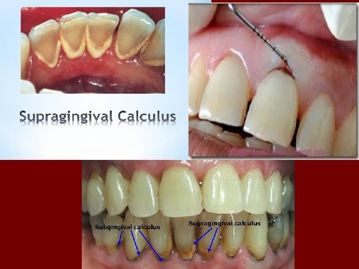
Clinical appearance and distribution of calculus Supragingival calculus is whitish- yellowish deposit, usually present along the gingival margins of the teeth. However, the color of which may be changed to brown due to secondary staining from tobacco and food pigments.
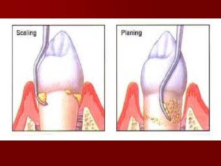
Subgingival calculus is brown to greenish black in color and it’s located below the crest of marginal gingiva, detection requires careful examination with an explorer. It is usually firmly attached to the tooth surface.
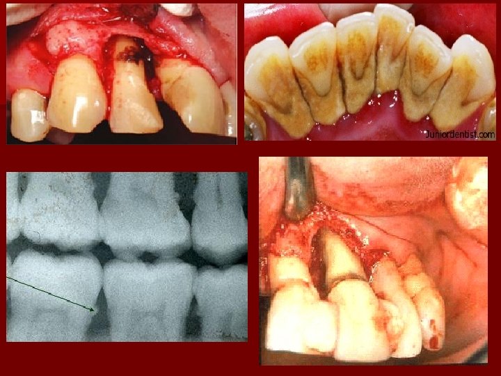
The primary effect of calculus is not due to mechanical irritation ( as was originally thought), but is related to its always being covered by bacteria and play a major role in maintaining P. D. disease by : n Keeping the plaque in close contact with the gingival tissue n Creating areas where plaque removal is impossible.
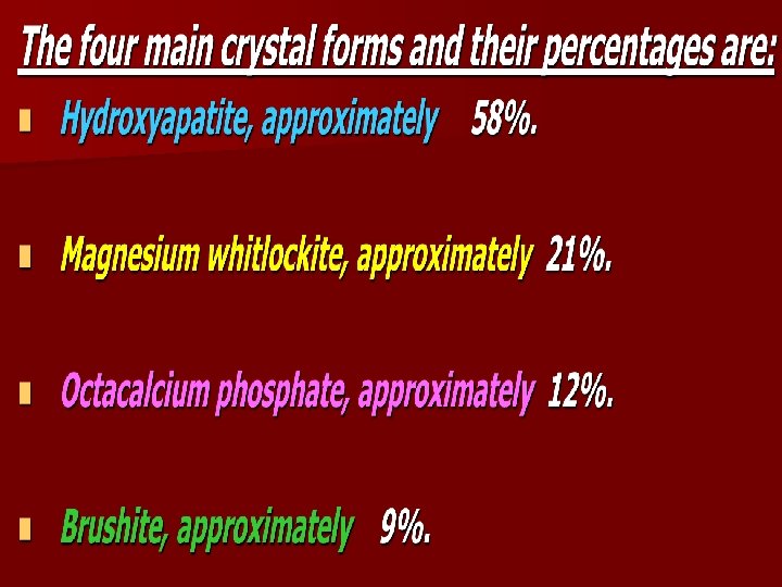
Composition of calculus Inorganic content: Sup. g. c. consists of inorganic (70%-90%) The inorganic portion consists of mainly of : n Calcium phosphate as (75. 9%). n Calcium carbonate (3. 1%). n Traces of magnesium phosphate and other metals.
At least two thirds of the inorganic component is crystalline in structure.
Organic contents: The organic component of calculus consists of a mixture of the followings: n Protein-polysaccharide complexes. n Desquamated epithelial cells. n Leukocytes. n Various types of microorganisms.
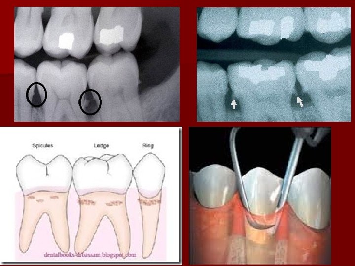
Attachment to the tooth surface n n Attachment by means of an organic pellicle which is also calcified. Penetration of calculus bacteria into cementum (this mode is not accepted by some investigators). Mechanical locking into surface irregularities, such as resorption lacunae and caries. Close adaptation of calculus undersurface depressions to the gently sloping mounds of the cementum surface.
Formation of calculus n The soft plaque is hardened between the first and the 14 th day of plaque formation; Calcifying plaques may become n 50% mineralized in 2 days and n 60% - 90% mineralized in 12 days. n
Crystals formation: n Initially in the intercellular matrix n On the bacterial surfaces n Finally within the bacteria. n Calcification begins along the inner surface of the sup. g. p. adjacent to the tooth surface.
The time required to reach the maximal level has been reported as n 10 n weeks, 18 weeks and 6 months. The decline from the maximal accumulation may be explained by the susceptibility of bulky calculus to mechanical wear from food and from cheeks, lips and tongue.
n Theoies of mineralizationof calculus: The theoretical mechanisms by which plaque becomes mineralized can be stratified into 2 categories. n 1. Mineral precipitation may be brought about in the following several ways: Incease in the p. H of the saliva causes precipitation of calcium phosphate salts The p. H may be elevated by the loss of carbon dioxide and the formation of ammonia by dental plaque bacteria or by protein degradation during stagnation. .
q. Colloidal proteins in saliva bind calcium and phosphate ions With stagnation of saliva, colloids settle out and the supersaturated state is no longer maintained, leading to precipitation of calcium phosphate salts.
1 - (Phosphatase) liberated from dental plaque, desquamated epithelia cells, or bacteria precipitates calcium phosphate 2 - (Esterase) is present in the cocci and filamentous organisms, leukocytes, macrophages, and desquamated epithelial cells of dental plaque. Esterase may initiate calcification by formation of fatty acids that calcium phosphate salts
n 2. Seeding agents induce small foci of calcification that enlarge and coalesce to form a calcified mass. it is suspected that the intercellular matrix of plaque plays an active role. The carbohydrate-protein complexes may initiate calcification by removing calcium from the saliva