BONE The most amazing story of bone histology

BONE The most amazing story of bone histology!
")
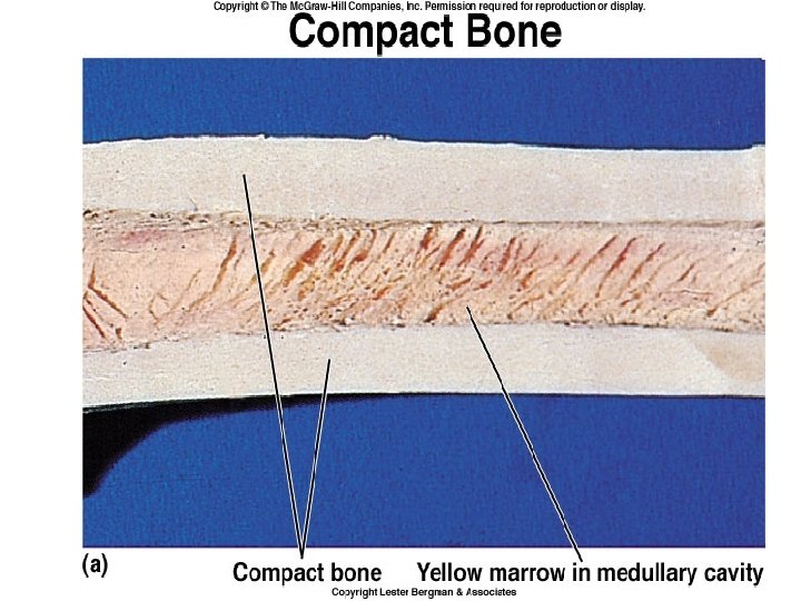
Functions of Bone • Supports soft tissue • Protects vital organs (cranium, thoracic cavity) • Contains bone marrow • Reservoir of Ca++, PO 4 to maintain constant concentrations in body fluids • Allows body to move

Specialized CT • Cells • _Osteogenic cells – Osteoblasts – Osteocytes – Osteoclasts (differ greatly from above 3) • Bone matrix – Calcified material, lacunae • And more…. – Canaliculi – Periosteum – Endosteum

Anatomy of a Long Bone
. These produce")
Osteogenic Cells • Stem cells that arise from mesenchyme (all ct does). These produce osteoblasts which in turn become osteocytes. • Only bone cells to undergo cell division • Located along inner portion of periosteum and in endosteum and in canals within bone that contain blood vessels.
 •")
Osteoblasts • Synthesize organic components of matrix (collagen type I, proteoglycans, glycoproteins. ) • Mature bone cells. • Synthesize and secrete collagen fibers. • Influence deposit of Ca++, PO 4. • Surround themselves with matrix and become trapped in their own secretions and become osteocytes.

Osteocytes

Osteocytes • Mature bone cells that sit in lacunae • Gap junctions between osteocytes provide nutrition (15 cells in a row) • Maintain bony matrix; long lived cells • Stimulated by calcitonin; inhibited by PTH

Osteocytes • Mature bone cells that sit in lacunae • Gap junctions between osteocytes provide nutrition (15 cells in a row) • Maintain bony matrix (metabolic functions) • Long lived cells • Stimulated by calcitonin; inhibited by PTH

Osteocyte with Cytoplasmic Extensions

Osteocytes with Canaliculi Photomicrograph of dried bone ground very thin. The lacunae and canaliculi filled with air deflect the light and appear dark, showing the communication between these structures through which nutrients derived from blood vessels flow. Medium magnification.
 •")
Osteoclasts • Derived from as many as 50 monocytes; engulf bony material (multinucleated) • Active osteoblasts stimulate osteoclast activity • Large, branched, motile cells with deeply folded plasma membrane called a ruffled border • Secrete powerful lysosomal enzymes that digest matrix. This is called resorption • Resorption normal necessary part of bone growth and maintainance


Osteoclasts

Bone Resorption

On this image, the deepest red color is bone while pink represents either fibrocartilage (i. e. , collagen within cartilage) or mineralized cartilage. The central clearing represents the invasion of bone into calcified cartilage. Osteoblasts are laying down new bone toward the left of the upper boundary of this cavity. osteoclasts are removing previously-formed bone. Remodeling

Remodeling

Bone Replacing Cartilage

Remodeling Bone

Bone Remodeling • http: //www. medes. fr/Eristo/Osteoporosis/B one. Remodeling. html • http: //www. siumed. edu/~dking 2/ssb/skeleto n. htm#bone

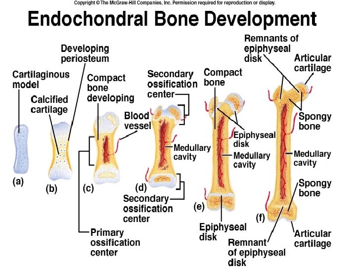
Endochondral Ossification

Previous slide • Photomicrograph of endochondral ossification. In the upper region is a row of osteoblasts with intense cytoplasmic basophilia, a feature to be expected in cells synthesizing a glycoprotein (collagen). Note an osteoblast being captured in the bone matrix (arrow). Between the layer of osteoblasts and the calcified bone matrix is a pale region made of noncalcified bone matrix called osteoid. PT stain. Medium magnification.

Mesenchyme Fibroblasts Osteoprogenitor cells Periosteum

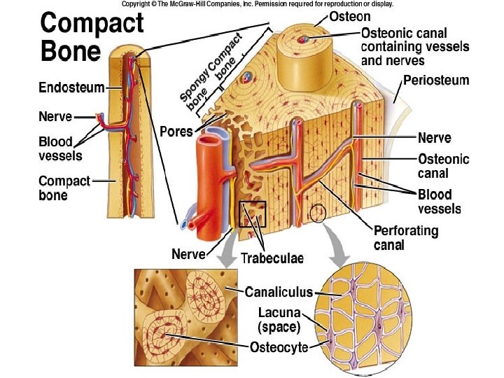

Osteon • Long cylinder parallel to long axis of diaphysis • Consists of: – Haversian canal with nerves, blood vessels; lamellae with osteocytes • Haversian canals communicate with marrow cavity, periosteum, other canals through Volkmann’s canals

Compact Bone
")
OSTEONS (wow!)
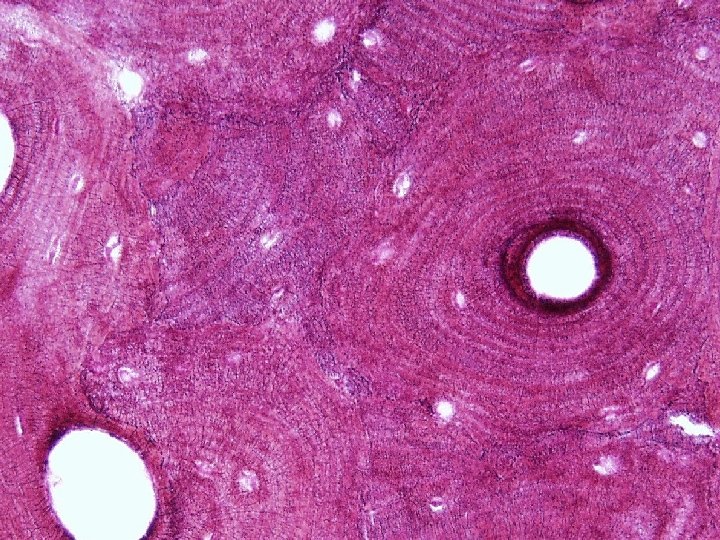

Canaliculi between Osteocytes
- Slides: 33