Chest Pain Evaluation Risk Assessment for Acute Coronary



- Slides: 47
Chest Pain Evaluation, Risk Assessment for Acute Coronary Syndrome & 2014 NSTEMI Guideline Update Arie Szatkowski, MD FACC Stern Cardiovascular Foundation Baptist Memorial Healthcare Corporation
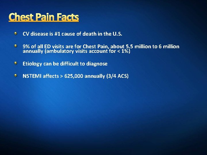
Chest Pain Facts CV disease is #1 cause of death in the U. S. 9% of all ED visits are for Chest Pain, about 5. 5 million to 6 million annually (ambulatory visits account for < 1%) Etiology can be difficult to diagnose NSTEMI affects > 625, 000 annually (3/4 ACS)
A Chest Pain Case A 65 year-old man with a past medical history significant for hypertension and dyslipidemia presents to clinic after 2 episodes of chest pain in past couple days. What do you want to know and do?
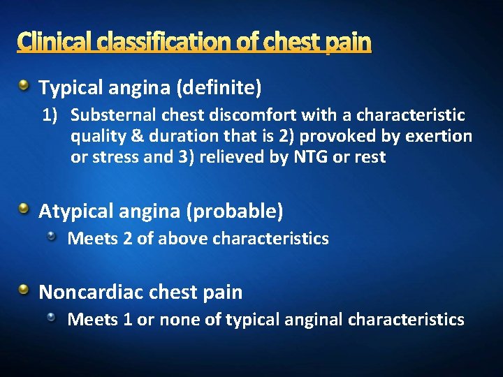
Clinical classification of chest pain Typical angina (definite) 1) Substernal chest discomfort with a characteristic quality & duration that is 2) provoked by exertion or stress and 3) relieved by NTG or rest Atypical angina (probable) Meets 2 of above characteristics Noncardiac chest pain Meets 1 or none of typical anginal characteristics
#1 Goal EXCLUDE Coronary artery disease and other life-threatening conditions
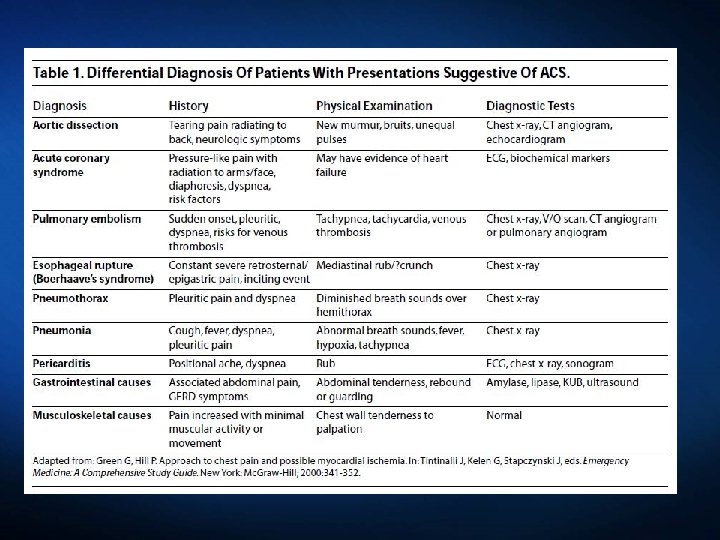
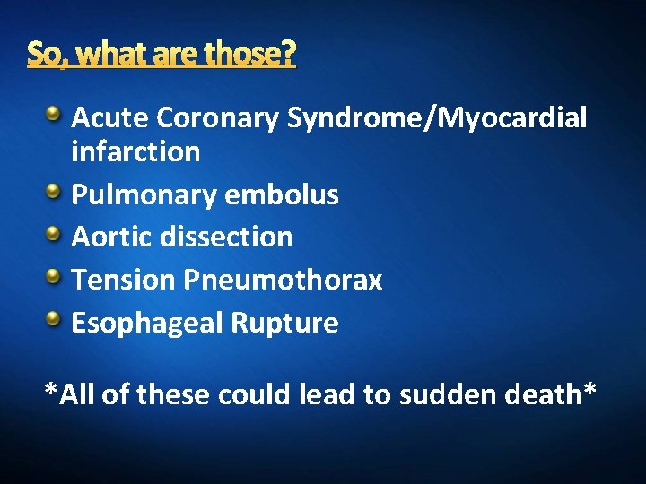
So, what are those? Acute Coronary Syndrome/Myocardial infarction Pulmonary embolus Aortic dissection Tension Pneumothorax Esophageal Rupture *All of these could lead to sudden death*
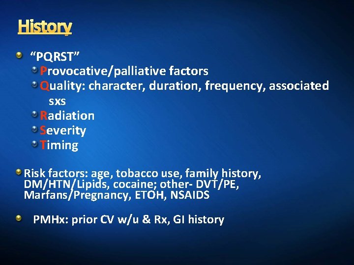
History “PQRST” Provocative/palliative factors Quality: character, duration, frequency, associated sxs Radiation Severity Timing Risk factors: age, tobacco use, family history, DM/HTN/Lipids, cocaine; other- DVT/PE, Marfans/Pregnancy, ETOH, NSAIDS PMHx: prior CV w/u & Rx, GI history
Provocation and Palliation Postprandial? GI or cardiac disease Exertion? Angina or esophageal pain Antacids or food? Gastroesophageal origin Sublingual nitro? Esophageal or cardiac Cold, emotional stress, sexual intercourse can promote ischemic pain “GI Cocktail” (viscous lidocaine and antacid)? GI or cardiac Worse with swallowing? Esophageal origin Cessation of activity/rest? Ischemic origin Body position, movement, deep breathing? Musculoskeletal origin Sitting up and leaning forward? Pericarditis
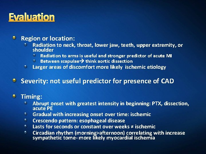
Evaluation Region or location: Radiation to neck, throat, lower jaw, teeth, upper extremity, or shoulder Radiation to arms is useful and stronger predictor of acute MI Between scapulae think aortic dissection Larger areas of discomfort more likely ischemic etiology Severity: not useful predictor for presence of CAD Timing: Abrupt onset with greatest intensity in beginning: PTX, dissection, acute PE Gradual with increasing onset over time: ischemic Crescendo pattern: esophageal disease Lasts for seconds or constant over weeks ≠ ischemic Circadian rhythm (morning>afternoon) correlating with increase sympathetic tome- more likely myocardial ischemia
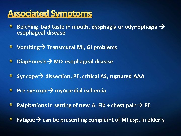
Associated Symptoms Belching, bad taste in mouth, dysphagia or odynophagia esophageal disease Vomiting Transmural MI, GI problems Diaphoresis MI> esophageal disease Syncope dissection, PE, critical AS, ruptured AAA Pre-syncope myocardial ischemia Palpitations in setting of new A. Fib + chest pain PE Fatigue can be presenting complaint of MI esp. in elderly
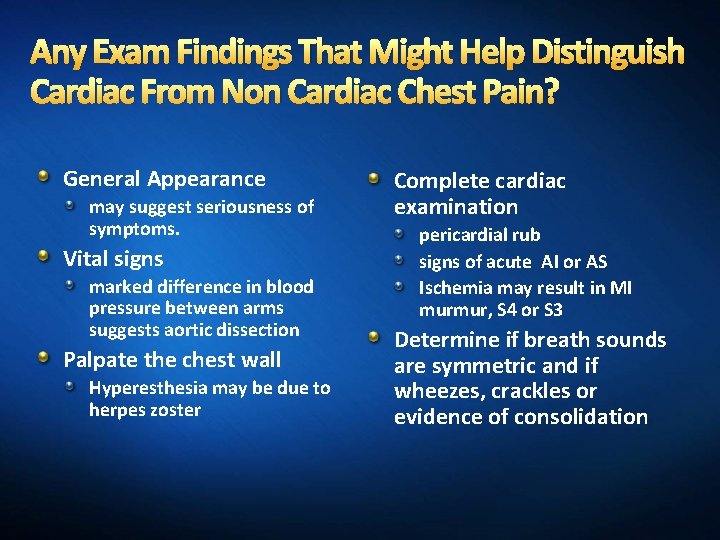
Any Exam Findings That Might Help Distinguish Cardiac From Non Cardiac Chest Pain? General Appearance may suggest seriousness of symptoms. Vital signs marked difference in blood pressure between arms suggests aortic dissection Palpate the chest wall Hyperesthesia may be due to herpes zoster Complete cardiac examination pericardial rub signs of acute AI or AS Ischemia may result in MI murmur, S 4 or S 3 Determine if breath sounds are symmetric and if wheezes, crackles or evidence of consolidation
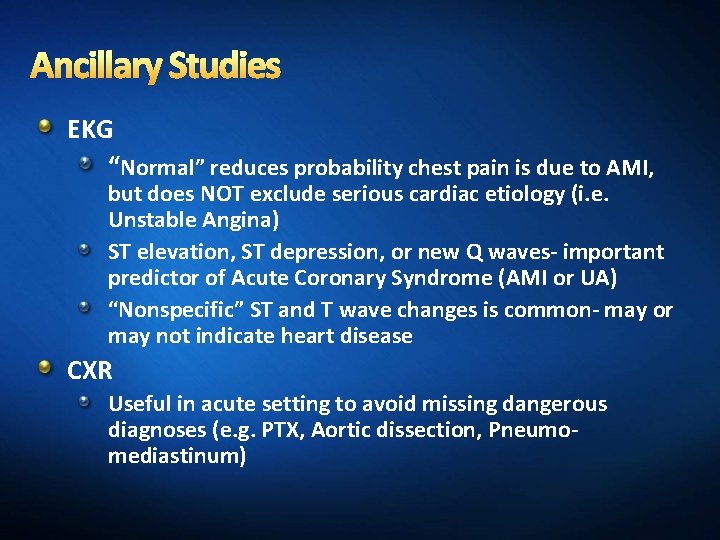
Ancillary Studies EKG “Normal” reduces probability chest pain is due to AMI, but does NOT exclude serious cardiac etiology (i. e. Unstable Angina) ST elevation, ST depression, or new Q waves- important predictor of Acute Coronary Syndrome (AMI or UA) “Nonspecific” ST and T wave changes is common- may or may not indicate heart disease CXR Useful in acute setting to avoid missing dangerous diagnoses (e. g. PTX, Aortic dissection, Pneumomediastinum)
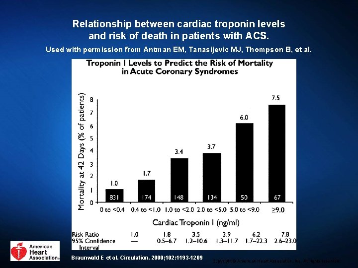
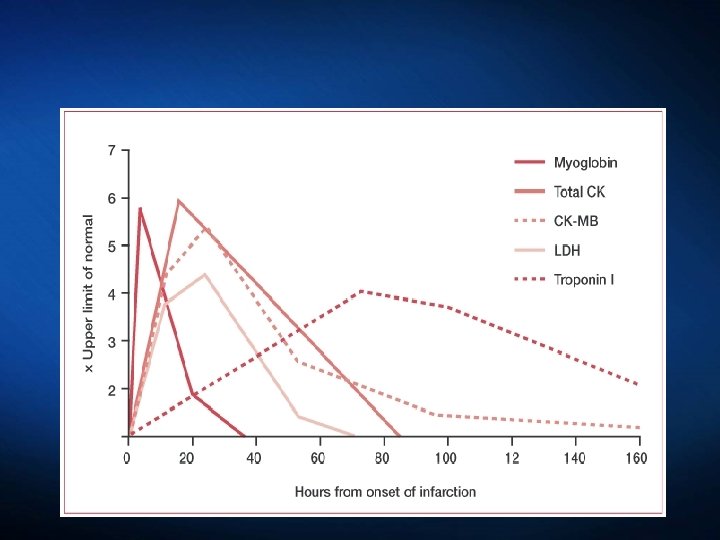
Relationship between cardiac troponin levels and risk of death in patients with ACS. Used with permission from Antman EM, Tanasijevic MJ, Thompson B, et al. Braunwald E et al. Circulation. 2000; 102: 1193 -1209 Copyright © American Heart Association, Inc. All rights reserved.
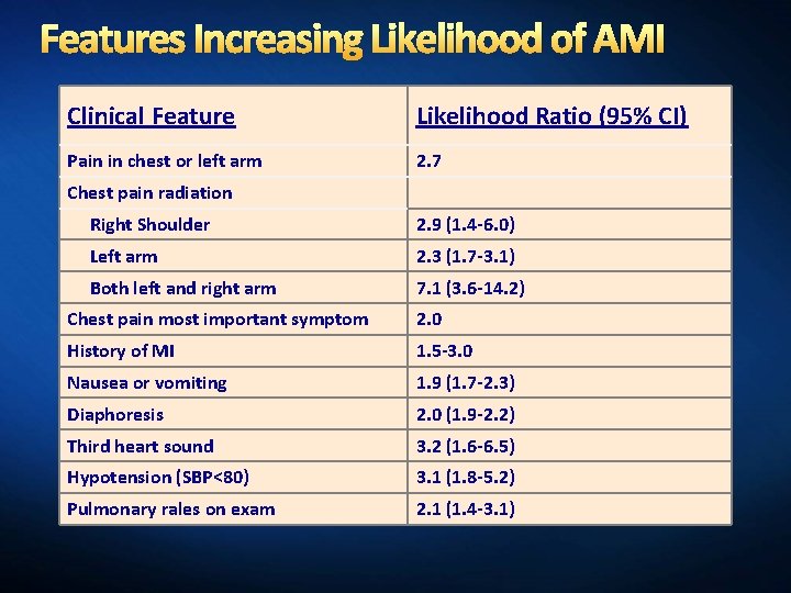
Features Increasing Likelihood of AMI Clinical Feature Likelihood Ratio (95% CI) Pain in chest or left arm 2. 7 Chest pain radiation Right Shoulder 2. 9 (1. 4 -6. 0) Left arm 2. 3 (1. 7 -3. 1) Both left and right arm 7. 1 (3. 6 -14. 2) Chest pain most important symptom 2. 0 History of MI 1. 5 -3. 0 Nausea or vomiting 1. 9 (1. 7 -2. 3) Diaphoresis 2. 0 (1. 9 -2. 2) Third heart sound 3. 2 (1. 6 -6. 5) Hypotension (SBP<80) 3. 1 (1. 8 -5. 2) Pulmonary rales on exam 2. 1 (1. 4 -3. 1)
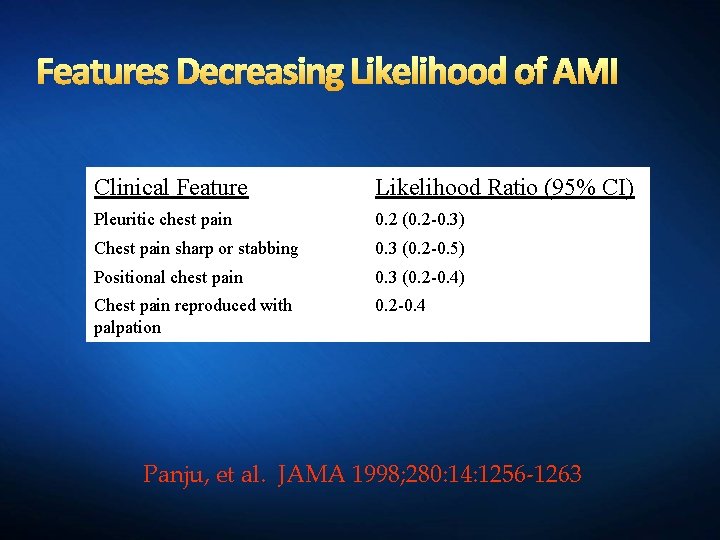
Features Decreasing Likelihood of AMI Clinical Feature Likelihood Ratio (95% CI) Pleuritic chest pain 0. 2 (0. 2 -0. 3) Chest pain sharp or stabbing 0. 3 (0. 2 -0. 5) Positional chest pain 0. 3 (0. 2 -0. 4) Chest pain reproduced with palpation 0. 2 -0. 4 Panju, et al. JAMA 1998; 280: 14: 1256 -1263
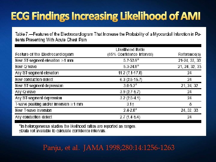
ECG Findings Increasing Likelihood of AMI Panju, et al. JAMA 1998; 280: 14: 1256 -1263
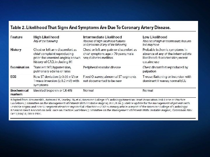
High Likelihood of ACS Worsening frequency, intensity, duration, timing (e. g. nocturnal pain, rest pain) of prior angina New onset SOB, nausea, sweating, extreme fatigue in patient with known h/o CVD Onset of typical anginal symptoms in pt without h/o CVD New murmur (or worsening of previously noted murmur), hypotension, diaphoresis, rales, pulmonary edema Transient ST deviation (≥ 1 mm) or TWI in multiple precordial leads
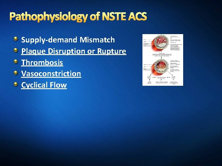
Pathophysiology of NSTE ACS Supply-demand Mismatch Plaque Disruption or Rupture Thrombosis Vasoconstriction Cyclical Flow
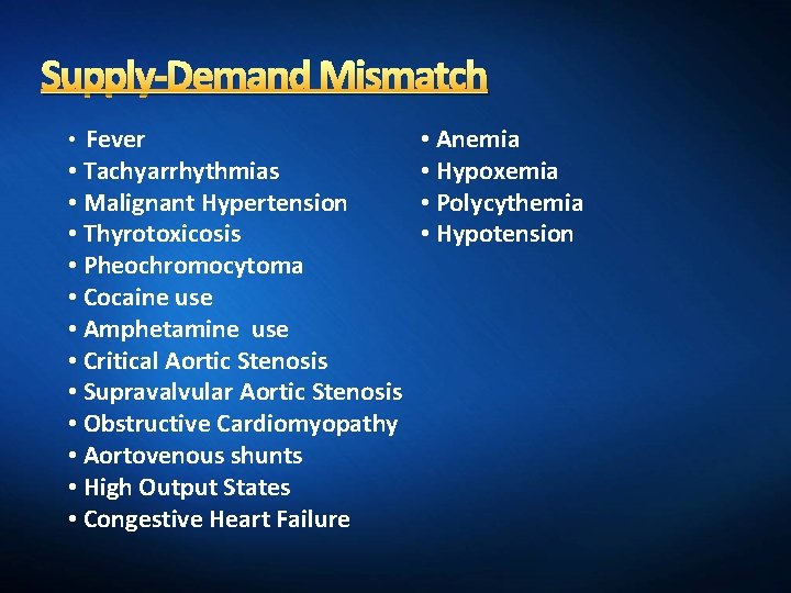
Supply-Demand Mismatch • Fever • Tachyarrhythmias • Malignant Hypertension • Thyrotoxicosis • Pheochromocytoma • Cocaine use • Amphetamine use • Critical Aortic Stenosis • Supravalvular Aortic Stenosis • Obstructive Cardiomyopathy • Aortovenous shunts • High Output States • Congestive Heart Failure • Anemia • Hypoxemia • Polycythemia • Hypotension
What’s New in the 2014 NSTEMI Guidelines?
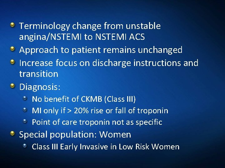
Terminology change from unstable angina/NSTEMI to NSTEMI ACS Approach to patient remains unchanged Increase focus on discharge instructions and transition Diagnosis: No benefit of CKMB (Class III) MI only if > 20% rise or fall of troponin Point of care troponin not as specific Special population: Women Class III Early Invasive in Low Risk Women
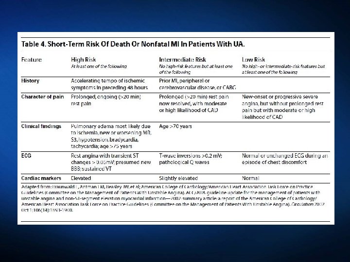
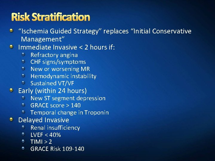
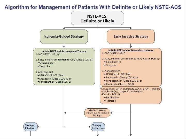
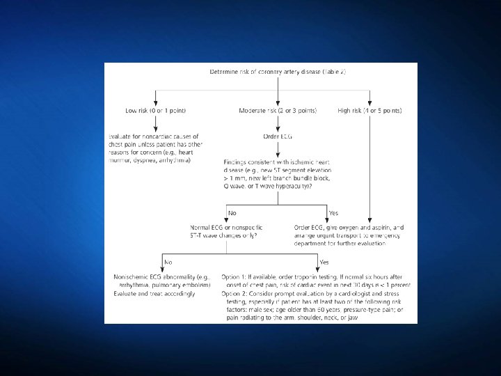
Risk Stratification “Ischemia Guided Strategy” replaces “Initial Conservative Management” Immediate Invasive < 2 hours if: Refractory angina CHF signs/symptoms New or worsening MR Hemodynamic instability Sustained VT/VF Early (within 24 hours) New ST segment depression GRACE score > 140 Temporal change in Troponin Delayed Invasive Renal insufficiency LVEF < 40% TIMI > 2 GRACE Risk 109 -140
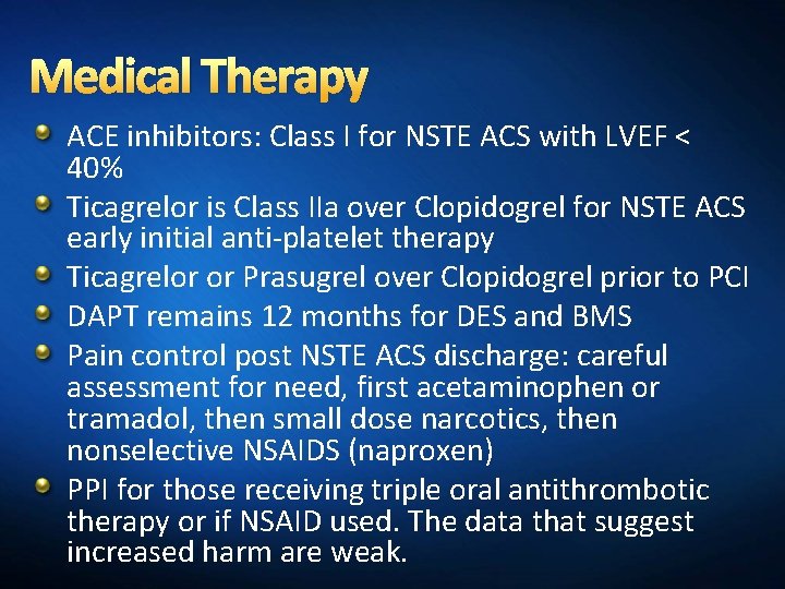
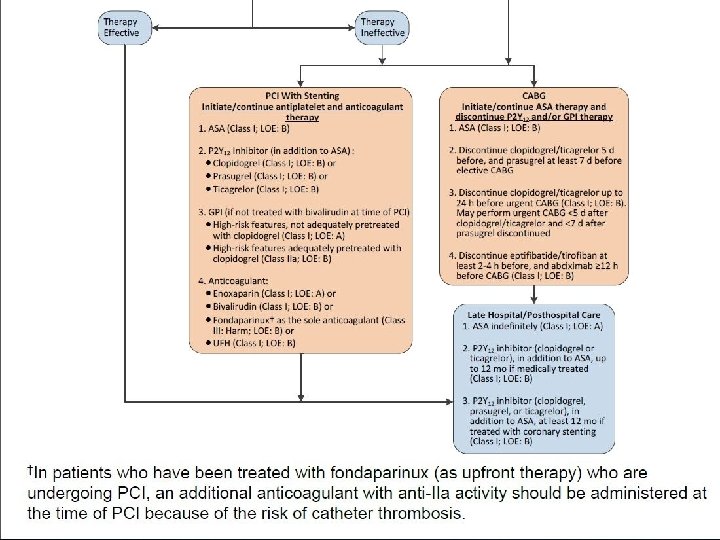
Medical Therapy ACE inhibitors: Class I for NSTE ACS with LVEF < 40% Ticagrelor is Class IIa over Clopidogrel for NSTE ACS early initial anti-platelet therapy Ticagrelor or Prasugrel over Clopidogrel prior to PCI DAPT remains 12 months for DES and BMS Pain control post NSTE ACS discharge: careful assessment for need, first acetaminophen or tramadol, then small dose narcotics, then nonselective NSAIDS (naproxen) PPI for those receiving triple oral antithrombotic therapy or if NSAID used. The data that suggest increased harm are weak.
What About Clinical Tools/Risk Scores to Guide Decisions?
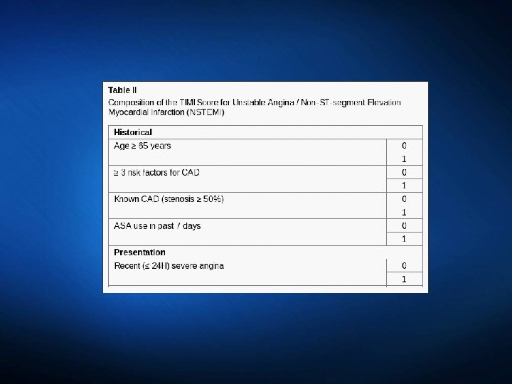
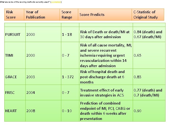
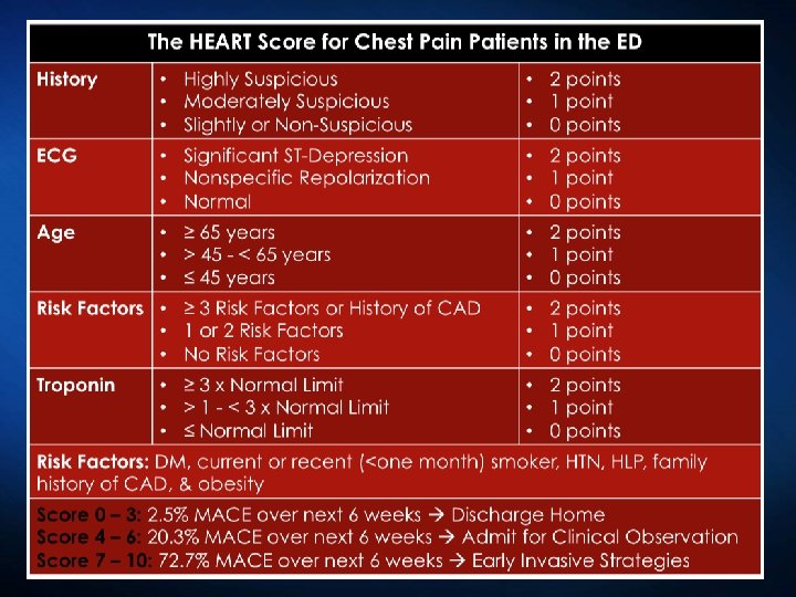
What are some of the scoring methods currently used? (22294968) Risk Score Year of Publication Score Range Score Predicts C-Statistic of Original Study PURSUIT 2000 1 - 18 Risk of Death or death/MI at 0. 84 (death) and 30 days after admission 0. 67 (death/MI) Risk of all cause mortality, MI, and severe recurrent ischemia requiring urgent 0. 65 revascularization within 14 days after admission Risk of hospital death and post-discharge death at 6 0. 83 months TIMI 2000 0 -7 GRACE 2003 1 - 372 FRISC 2004 0 -7 Treatment effect of early invasive strategies in ACS 0 - 10 Prediction of combined endpoint of MI, PCI, CABG or 0. 90 death within 6 weeks after presentation HEART 2008 0. 77 (death) and 0. 7 (death/MI)
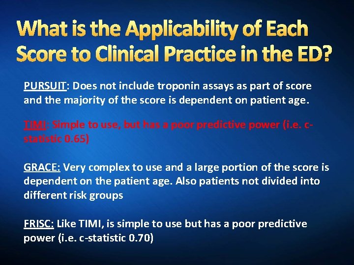
What is the Applicability of Each Score to Clinical Practice in the ED? PURSUIT: Does not include troponin assays as part of score and the majority of the score is dependent on patient age. TIMI: Simple to use, but has a poor predictive power (i. e. cstatistic 0. 65) GRACE: Very complex to use and a large portion of the score is dependent on the patient age. Also patients not divided into different risk groups FRISC: Like TIMI, is simple to use but has a poor predictive power (i. e. c-statistic 0. 70)
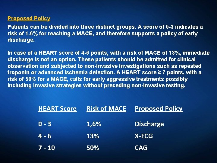
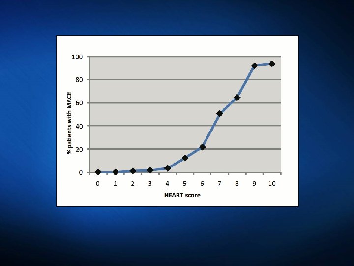
Proposed Policy Patients can be divided into three distinct groups. A score of 0 -3 indicates a risk of 1. 6% for reaching a MACE, and therefore supports a policy of early discharge. In case of a HEART score of 4 -6 points, with a risk of MACE of 13%, immediate discharge is not an option. These patients should be admitted for clinical observation and subjected to non-invasive investigations such as repeated troponin or advanced ischemia detection. A HEART score ≥ 7 points, with a risk of 50% for a MACE, calls for early aggressive treatments possibly including invasive strategies without preceding non-invasive testing. HEART Score Risk of MACE Proposed Policy 0 -3 1, 6% Discharge 4 -6 13% X-ECG 7 - 10 50% CAG
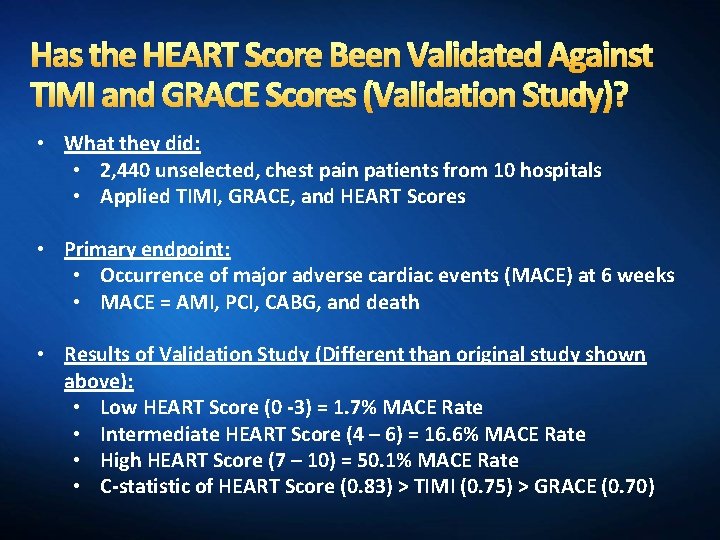
Has the HEART Score Been Validated Against TIMI and GRACE Scores (Validation Study)? • What they did: • 2, 440 unselected, chest pain patients from 10 hospitals • Applied TIMI, GRACE, and HEART Scores • Primary endpoint: • Occurrence of major adverse cardiac events (MACE) at 6 weeks • MACE = AMI, PCI, CABG, and death • Results of Validation Study (Different than original study shown above): • Low HEART Score (0 -3) = 1. 7% MACE Rate • Intermediate HEART Score (4 – 6) = 16. 6% MACE Rate • High HEART Score (7 – 10) = 50. 1% MACE Rate • C-statistic of HEART Score (0. 83) > TIMI (0. 75) > GRACE (0. 70)
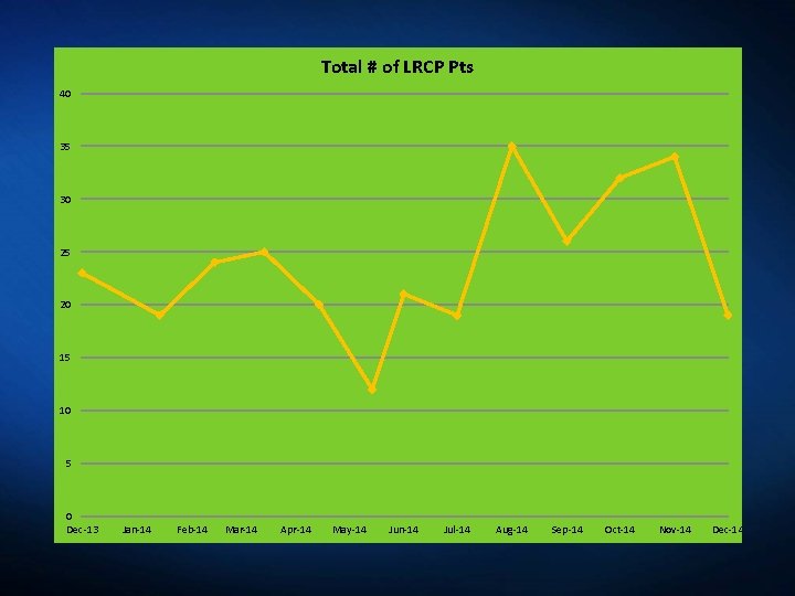
Total # of LRCP Pts 40 35 30 25 20 15 10 5 0 Dec-13 Jan-14 Feb-14 Mar-14 Apr-14 May-14 Jun-14 Jul-14 Aug-14 Sep-14 Oct-14 Nov-14 Dec-14
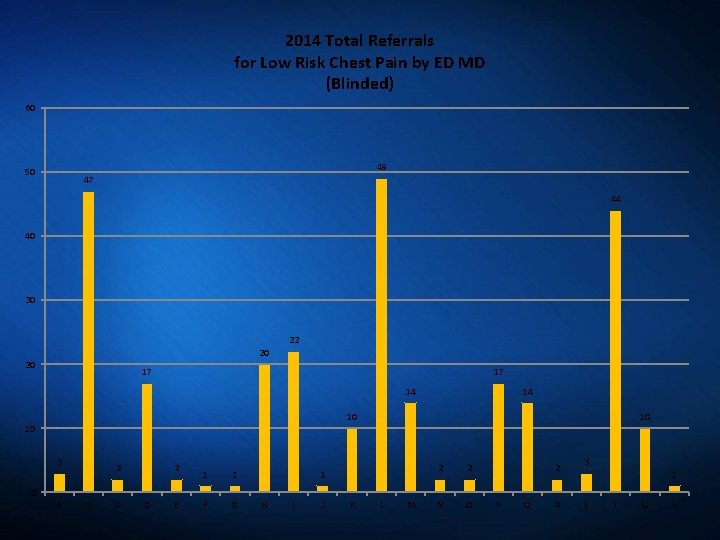
2014 Total Referrals for Low Risk Chest Pain by ED MD (Blinded) 60 49 50 47 44 40 30 22 20 20 17 17 14 14 10 10 3 2 2 1 1 F G 10 1 2 2 N O 2 3 1 0 A B C D E H I J K L M P Q R S T U V
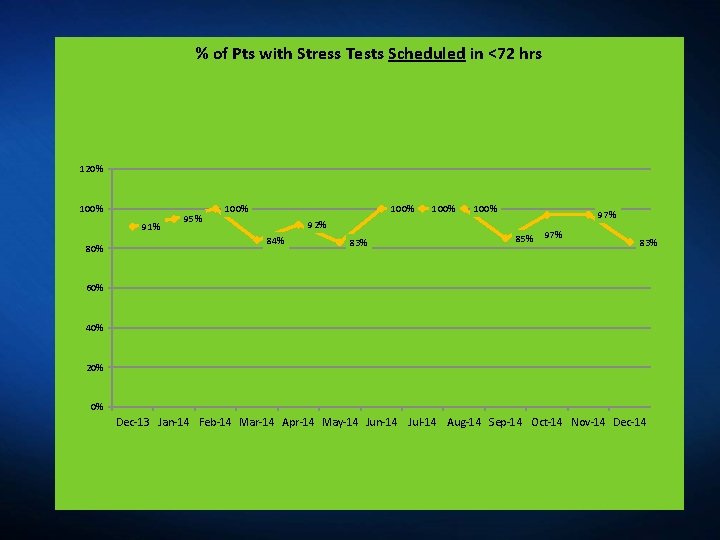
% of Pts with Stress Tests Scheduled in <72 hrs 120% 100% 91% 80% 95% 100% 97% 92% 84% 83% 85% 97% 83% 60% 40% 20% 0% Dec-13 Jan-14 Feb-14 Mar-14 Apr-14 May-14 Jun-14 Jul-14 Aug-14 Sep-14 Oct-14 Nov-14 Dec-14
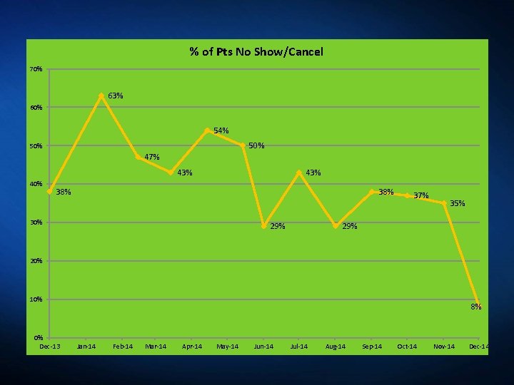
% of Pts No Show/Cancel 70% 63% 60% 54% 50% 47% 43% 40% 43% 38% 30% 29% 37% 35% 29% 20% 10% 0% Dec-13 8% Jan-14 Feb-14 Mar-14 Apr-14 May-14 Jun-14 Jul-14 Aug-14 Sep-14 Oct-14 Nov-14 Dec-14
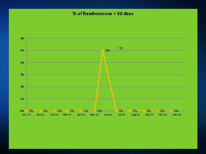
% of Readmissions < 30 days 6% 5% 5% 1 pt 4% 3% 2% 1% 0% 0% Dec-13 0% Jan-14 0% Feb-14 0% Mar-14 0% Apr-14 0% May-14 Jun-14 0% Jul-14 0% Aug-14 0% Sep-14 0% Oct-14 0% Nov-14
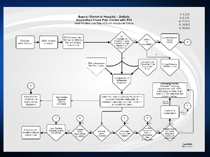
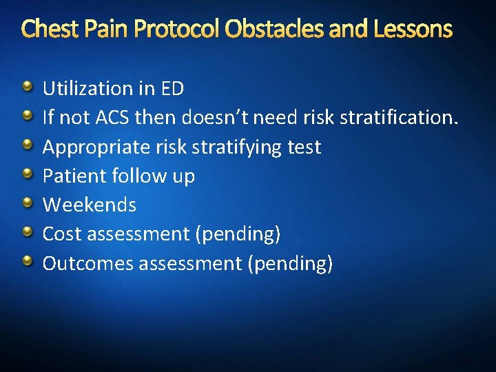
Chest Pain Protocol Obstacles and Lessons Utilization in ED If not ACS then doesn’t need risk stratification. Appropriate risk stratifying test Patient follow up Weekends Cost assessment (pending) Outcomes assessment (pending)
Take Home/Summary Focus on the life threatening causes first Know the indicators for immediate invasive therapy Use Risk Tools but Clinical judgment prevails Know the right test for the situation