Viral Hepatitis Essam M ALFahadawi Viral hepatitis is


 Acid at")
 • 1) is spread: – by the fecal-oral route by")


: - outer lipid")


 1) Transmission: – parenterally, – sexually, – vertically (from mother")


")


 - delta agent - SS, circular (-) RNA virus -")


 Virus binding and internalization, b) cytoplasmic release")


: it is")


 • Against Hepatitis B (recombinant) - Monovaccines")


- Slides: 57
Viral Hepatitis Essam M. AL-Fahadawi
Viral hepatitis is viral infection of the liver that causes hepatic inflammation which is followed by the classic icteric symptoms of jaundice and the release of liver enzymes
Periods and clinical symptoms of hepatitis 1. Incubation period. 2. Preicteric period. Hepatitis B, C or D also cause: - Skin rash - Arthralgia - Weight loss 3. Icteric period. 4. Recovery.
Icteric symptoms of liver damage
Jaundice/Icterus • Elevated levels of bilirubin in blood is associated with “dark colored” urine. • Obstructive jaundice causes impairment of excretion into the small intestine. • This results in the loss of the “normal yellowing” of the stool. • The stool is “white”.
Classification of Hepatitis Viruses Virus Family Nucleic acid Envelope HAV Picornaviridae ss +RNA No HBV Hepadnaviridae Incomplete circular ds –DNA Yes HCV Flaviviridae ss +RNA Yes HDV Genus Deltavirus ss –RNA Yes HEV Hepeviridae ss +RNA No
Viral hepatitis Transmission FOOD & WATER BORN BLOOD BORN Incubation period Hepatitis A: 2 -6 weeks Hepatitis B: 2 -6 months Hepatitis E: 2 -9 weeks Hepatitis C: 2 -26 weeks Hepatitis D: unlimited Type of hepatitis Acute, chronic
Family Picornaviridae. Hepatitis A virus • Small, non-enveloped RNA virus with cubical symmetry. • One stable serotype only viral protein genome-linked
Characteristics of HAV • Stable to: – – – Saltwater, groundwater (months) Acid at p. H 1 Solvents (ether, chloroform) Detergents Drying Temperature: • 40 C: weeks • 560 C for 30 minutes • 610 C for 20 minutes • Inactivated by: – Chlorine treatment of drinking water – Formalin
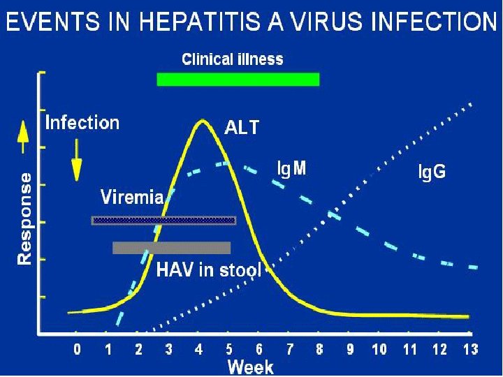
Hepatitis A (infectious hepatitis) • 1) is spread: – by the fecal-oral route by ingestion of contaminated food and water; • 2) contagious period extends 2 -3 weeks before and 8 -10 days after onset of jaundice. Virus may cause asymptomatic shedding. • 3) does not cause chronic liver disease. • 4) immunity is life-long.
Spread of HAV within the body HAV enters the body by ingestion and intestinal infection. Then it spreads by the bloodstream, to the liver, a target organ. Large numbers of virus particles are detectable in feces during the incubation period, beginning as early as 10 -14 days after exposure. Pathological changes appear exclusively in the liver.
Clinical finding • Swelling and tenderness of liver • Jaundice -yellow tinge in the skin and eyes • dark urine • transaminase, alkaline phosphatase levels increased
Laboratory diagnosis of hepatitis A • Biochemical tests to study liver function: - The Bilirubin. - Alanine aminotransferase (ALT). - Aspartate aminotransferase (AST). • Serology: ELISA – Acute infection: Ig. M. – Past Infection i. e. immunity: Ig. G. • Direct demonstration: – ELISA (detection of antigen in stool), – RT-PCR of faeces.
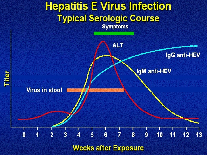
Family Hepeviridae. Hepatitis E virus • non-enveloped, cubic symmetry, 32 -34 nm, • ss + RNA virus • 4 genotypes
Epidemiology of HEV infection • Contagious period extends from before to after symptoms. • Transmission - via fecal-oral route by ingestion of contaminated food and water. • It causes only acute disease. • Life long immunity. • HEV infection is especially serious in pregnant women (mortality rate about 20 %). • Transmission through placenta results in death of fetus.
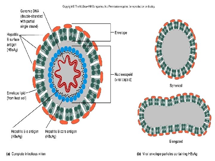
Family Hepadnaviridae. Hepatitis B virus The hepatitis B virion (Dane particle): - outer lipid envelope with the surface antigen (HBs. Ag). - an electron-dense core (nucleocapsid): ds circular DNA and polymerase surrounded by the core antigen (HBc. Ag). The HBs. Ag is produced in excess by the infected hepatocytes and is secreted in the form of spherical and filamentous particles. HBe. Ag is the soluble component of the core, with is released during active infection. HBx. Ag is a transactivator of viral transcription that appears to contribute to Hepatocellular carcinoma ( HCC ) by altering patterns of host gene expression.
1. Attachment to the cell surface receptor, fusion and releasing the core into the cytoplasm. 2. The core proteins dissociate from the DNA. 3. Completion of ds DNA by DNA polymerase and entering the nucleus. 4. Formation of a circular episome. 5. Transcription of m. RNAs. 6. Translation. 7. RNA is encapsidated by core proteins. 8. Inside the core, the RNA is transcribed to minus strand DNA by polymerase called reverse transcriptase. 9. Transcription the minus strand DNA into a plus strand DNA. 10. Budding the virus through the endoplasmic reticulum and/or Golgi Body membranes from which it acquires HBs. Ag. 11. Shedding of viruses embedded in a lipid bilayer and empty envelopes from HBs. Ag. • 1011 new virions are produced every day during acute infection Pathway for the replication of HBV
Characteristics of HBV • Stable to: – – Acid at p. H 2, 3 UV radiation Phenol Temperature: • 1000 C for 5 minutes retains infectious activity • 200 C for 3 months • 40 C (in a refrigerator) for 6 months – Survival: • in dried blood for 4 -5 months, • in frozen plasma for 15 -20 years, . • Inactivated by: – – – Autoclaving (1200 C for 20 min). Boiling for 60 minutes. Chloramine 1 -2% for 2 hours. Ethanol 70 -80% for 2 min. Formalin 1, 5 % for 2 min. Hydrogen peroxide
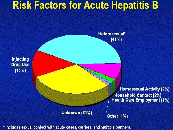
Hepatitis B (serum hepatitis) 1) Transmission: – parenterally, – sexually, – vertically (from mother to fetus), – close contact (hemocontact). 2) chronic hepatitis in 10% to 15% of adult patients, 30 -90% of young patients 3) is causally associated with primary hepatocellular carcinoma and cirrhosis.
Epidemiology of HBV infection • Virus is shed during both symptomatic and asymptomatic periods. • Transmission: - In blood, semen, vaginal secretions, saliva and mother’s milk. - Via transfusion, needle stick injury, injecting drug use, sex, child-birth, and breast-feeding.
Concentration of hepatitis B virus in body fluids High Moderate blood semen serum vaginal fluid wound exudates saliva breast milk Low/Not Detectable urine feces sweat tears Concentration HBV in the blood 109 virions per 1 ml
Spread of HBV in the body
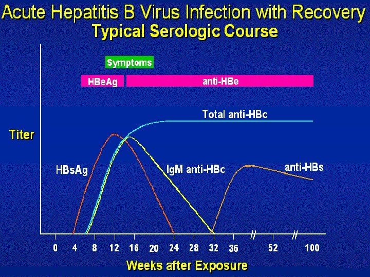
Symptoms of typical acute viral hepatitis B infection Right Upper Quadrant
HBV can multiply in monocytes, T- and B-lymphocytes, NK. Extrahepatic manifestation: glomerulonephritis, vasculitis and ARTHRITIS-DERMATITIS (a serum sickness-like syndrome). These patients experience rash, petechiae, purpura, arthralgia and arthritis. It involves mostly small joints of hands and knees and occurs in the prodromal period, before jaundice appears and resolves within a week.
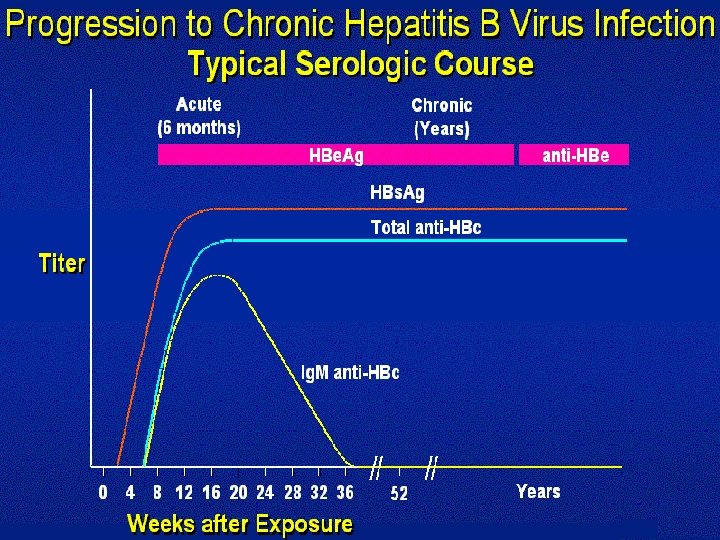
Clinical outcomes of acute hepatitis B infection
CLINICAL OUTCOMES OF HEPATITIS B Principal considerations: Asymptomatic carrier: the carrier patient never develops antibodies against HBs. Ag and harbors the virus without liver injury. Chronic persistent hepatitis: the patient has a low-grade hepatitis Chronic active hepatitis: the patient has an acute hepatitis state that continues without the normal recovery (last longer than 6 -12 months). Fulminant hepatitis: severe acute hepatitis with rapid destruction of the liver.
Hepatocellular carcinoma A section of a liver removed at liver transplantation showing a massive HCC (in white) and multiple satellite nodules
Pathology 1 - Symptoms are immune-mediated, resulting from inflammation and cellmediated (cytotoxic T cell) responses to HBs. Ag on the surface of hepatocytes. These resolve the disease. 2 -If the cell-mediated immune response is weak, symptoms are mild but the infection does not resolve and chronic hepatitis ensues. 3 - Chronic hepatitis leads to cirrhosis and hepatocellular carcinoma. Both of these are fatal in the absence of a liver transplant
HBs. Ag window • “HBs. Ag window” - the period from about 6 to 8 months when neither free HBs. Ag nor its antibody can be detected. • Anti-HBs. Ag are undetectable because they are complexed with the large amount of the antigen that is shed from infected cells. • The best tool for diagnosis of an acute HBV infection during the window is the presence of anti-HBc Ig. M
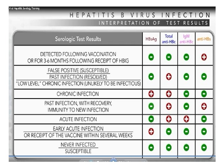
Laboratory markers for HBV infection and their interpretation • HBs. Ag – present in acute or chronic infection. It is detectable in the blood from 1 to 6 months after infection. • anti-HBs – marker of recovery and/or immunity to HBV infection (from about 8 months after infection). • anti-HBc Ig. M - marker of recent acute infection ≥ 6 months. • anti-HBc Ig. G - past or chronic infection. • HBe. Ag - indicates active replication of virus and therefore infectiveness (from 2 to 6 months after infection). • anti-HBe - virus no longer replicating. However, the patient can still be positive for HBs. Ag which is made by integrated HBV (from about 4 months after infection). • DNA - indicates active replication of virus.
Interpretation of Serological Markers of HBV Infection
Hepatitis D virus (HDV) - delta agent - SS, circular (-) RNA virus - HDV is coated with HBs. Ag which is needed for release from the host hepatocyte and for entry in the next round of infection. - 3 genotypes.
Epidemiology of HDV infection • HDV infects children and adults with underlying HBV infection. • Transmission: - parenterally and sexually; - vertically (rarely). - In blood, semen, and vaginal secretions; - Unsterilized equipment and infusion. Intravenous drug abusers, transfusion and organ recipients, and hemophiliacs are at highest risk for infection.
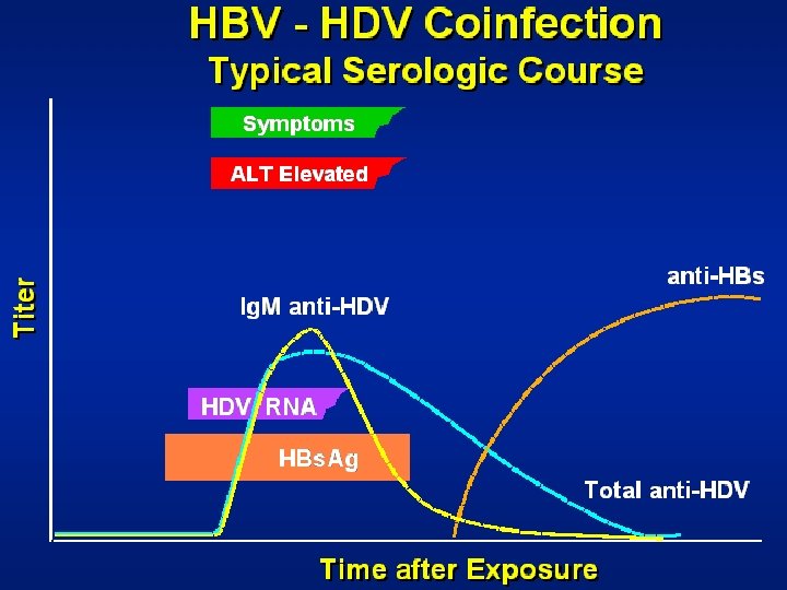
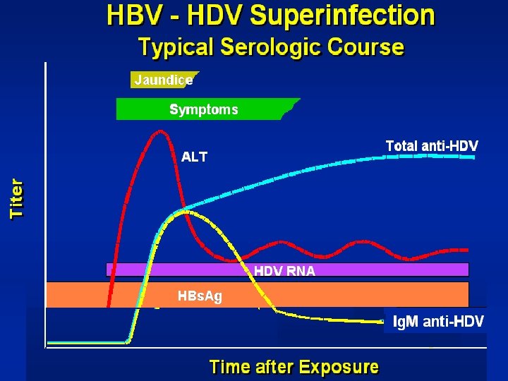
Consequences of delta virus infection • Co-infection by HDV and HBV exacerbates the acute disease (90%) and fulminant hepatitis (2 -4%) is more likely than with HBV alone. • Superinfection of a person already infected with HBV causes a more rapid, severe progression than co-infection because super-infection requires HBV replication before HDV replication can occur.
Laboratory markers for HDV infection • In an HBV-HDV co-infection: – HDV antigen is detected in about ¼ of patients. HDV antigen disappears when HBs. Ag appears, – anti-HDV Ig. G and Ig. M are usually seen, – anti-HDV decline as the symptoms resolve and, unlike anti-HBs, there are no antibodies to show that the patient was once HDV-infected, – anti-HBs Ig. G rises. • In a super-infection by HDV: – HBs. Ag and HDV RNA remain because super-infection usually leads to chronic infection. – anti-HDV Ig. G rises.
Family Flaviviridae. Hepatitis C and G viruses Enveloped ss+RNA viruses, 55 -65 nm diameter. 14 genotypes of HCV. 5 genotypes of HGV. Electron micrograph of the hepatitis C viruses
HCV life cycle • • • a) Virus binding and internalization, b) cytoplasmic release and uncoating, c) translation, d) RNA replication, e) packaging and assembly, f) virion maturation and release.
Epidemiology of HCV and HGV infection • 85 -90% patients will develop chronic disease. • About 50% of chronic carriers of HCV progress to cirrhosis or liver cancer in 1040 years. • Transmission: - parenterally (through sharing drug-injection equipment or from transfusion of unscreened blood or untreated clotting factors), - sexually, - perinatally. - In blood, semen, and vaginal secretions. - Contagious period extends 2 -3 weeks before and 10 weeks after onset of symptoms.
Routes of transmission of HCV
Risk factors of hepatitis C • • • Blood-to-blood contact (mainly transfusion): it is estimated that 90 %of persons with chronic HCV infection were infected through transfusion of unscreened blood or blood products. Unsterilized injection equipment and infusion: considered to be the primary sources of HCV infection. Sexual intercourse: although this is rare, and usually only occurs when HIV is also present and makes blood contact more likely. Dental equipment: People can be exposed to HCV via inadequately or improperly sterilized medical or dental equipment including needles or syringes, oral hygiene instruments, and jet air guns, etc. Hemodialysis: The basic problem is that proper sterilization methods are not followed. Occupational exposure to blood: Medical and dental personnel, first responders (e. g. surgeons, nurses and emergency medical technicians) can be exposed to HCV through needle sticks or blood spatter to the eyes or open wounds. Tattoos: Tattooing dyes, ink pots, piercing implements can transmit HCV-infected blood from one person to another if proper sterilization techniques are not followed. Shared personal care items: Such as razors, toothbrushes, cuticle scissors, and other manicuring or pedicure equipment can easily be contaminated with blood carrying the virus, and take care that HCV can live on dried blood for 15 days. Vertical transmission: refers to the transmission of a communicable disease from an infected mother to her child during delivery.
Pathogenesis of HCV infection • Hepatocytes are target cells for HCV. • Cytopathic effect (CPE ) – lysis of hepatocytes. • Sensitive cells: monocytes, macrophages, B-lymphocytes, neutrophiles, cells of bone marrow. • HCV can reproduce in cells of pancreas, adrenal glands, thyroid glands, spleen. • Immunomediated extrahepatic manifestations: – vasculitis (malaise, arthralgia, purpura); – glomerulonephritis; – altered nervous system (peripheral nerves, brain).
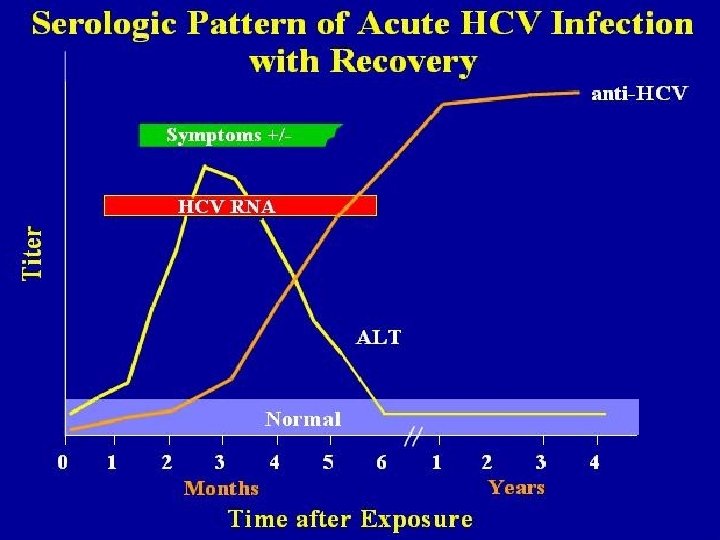
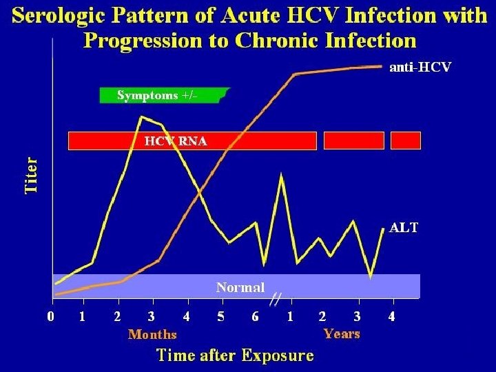
Clinical outcomes of hepatitis С infection Immunity is not strong, reinfection is possible
Laboratory diagnosis of viral hepatitis • Biochemical methods: Detection of the level of bilirubin and enzymes ALT, AST in serum. • Serology: ELISA - Detection of the Ag and antibodies • Detection of a viral genome: PCR • Rapid immunochromatographic test
Vaccines • Against Hepatitis A Havrix (inactivated) • Against Hepatitis B (recombinant) - Monovaccines (HBs. Ag – genetic engineering): Engerix B H-B-Vax Recombivax B - Divaccines: Twinrix (inactivated HAV + HBs. Ag)
Treatment • Hepatitis B: • Anti-HBV immunoglobuline • Alpha-interferon • Lamivudine • Adefovir • Hepatitis C: • Alpha-interferon + Ribavirine
The End