The foundation of the study of Genetics DNA


 is made up of long chains of nucleotides Nucleotide structure")
 nitrogen-containing base")


 of DNA are")


 of the cell cycle DNA replication is required")


 Transcription Messenger RNA (m. RNA) Ribosomal RNA (r.")

 codon for leucine (Leu)")
")


- Slides: 42
The foundation of the study of Genetics
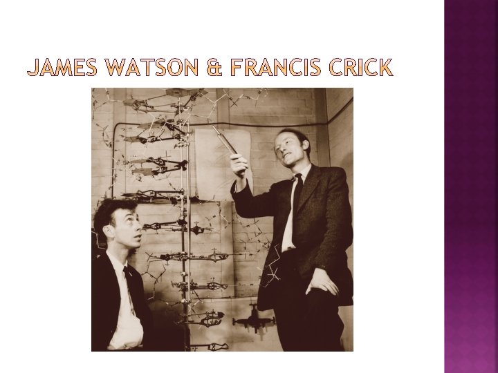
DNA structure was not discovered until the 1950’s After looking at an X-ray diffraction photo stolen from Rosalind Franklin by her coworker (Wilkins), James Watson and Francis Crick were able to create a model of DNA The model depicted a double helix of even width In 1953, James Watson, Francis Crick, and Maurice Wilkins received the Nobel Prize (no mention of Rosalind Franklin)
Franklin’s x-ray images suggested that DNA was a double helix of even width.
Erwin Chargaff, in his biochemical studies, discovered: The amount of Adenine in a DNA sample always equaled the amount of Thymine The amount of Cytosine, likewise, was equal to the amount of Guanine This became known as Chargaff’s rule This information further served to confirm the model created by James Watson and Francis Crick Adenine pairs with Thymine Cytosine pairs with Guanine
DNA (deoxyribonucleic acid) is made up of long chains of nucleotides Nucleotide structure Phosphate Group 5 carbon sugar (deoxyribose) Nitrogen Base (4 different bases) Adenine Thymine Guanine Cytosine
phosphate group deoxyribose (sugar) nitrogen-containing base
DNA is double stranded The two strands are equally spaced & twist This is why the structure of DNA is referred to as a double helix
The backbone of each side of DNA is held together by covalent bonds and is made up of the phosphate groups and deoxyribose sugars
The two complementary strands (the rungs of the DNA ladder) of DNA are held together by weak hydrogen bonds between the complementary nitrogen base pairs These attractions are easily broken and reformed Important for DNA replication and transcription
hydrogen bond covalent bond
Making copies of DNA
Occurs during Interphase (S phase) of the cell cycle DNA replication is required such that the two new daughter cells created will have a complete set of DNA Enzymes break the hydrogen bonds, separating the two strands Other enzymes bring free nucleotides in the nucleus to build the new strands of DNA
DNA replication is called a semiconservative process as the two double helices created consists of: One old strand of DNA One new strand of DNA The “old” sides of the DNA strands are used as the template (building instructions) for building the “new” strands of DNA Two double helices are created in the end
nucleotide The DNA molecule unzips in both directions.
original strand Two molecules of DNA new strand
The basis of gene expression
States that information stored in DNA flows in one direction from DNA, to RNA, to proteins
Transcription The 1 st phase of protein synthesis DNA can’t leave the nucleus, so a smaller molecule that can leave has to be made Occurs in the nucleus Translation The 2 nd and final phase of protein synthesis The molecule made in transcription is used to build a protein Occurs at the site of a ribosome
Nucleus- where DNA is located; where transcription occurs Ribosome- where proteins are built (site of protein synthesis); where translation occurs Rough ER- Newly made proteins enter this transport organelle Golgi Apparatus/Body-The protein is checked, modified, and re-packaged for transport to its final destination Vesicles-Transport vehicles for the protein around the cell
A segment of DNA called a gene with the information for building a protein is used as a template (building instructions) to build a messenger RNA (m. RNA) molecule Enzymes open the double helix Other enzymes build the m. RNA molecule from the gene of DNA Messenger RNA is a copy of the DNA instructions Like a secretary or a transcriptionist that records information
DNA
Double helix Nucleotide sugar: deoxyribose Nitrogen bases Single stranded Nucleotide sugar: ribose Nitrogen bases Adenine Thymine Uracil Cytosine Guanine Base Pair Rule A-T C-G Base Pair Rule A-U C-G DNA RNA
DNA Messenger RNA (m. RNA) Transcription Messenger RNA (m. RNA) Ribosomal RNA (r. RNA) Transfer RNA (t. RNA) Translation
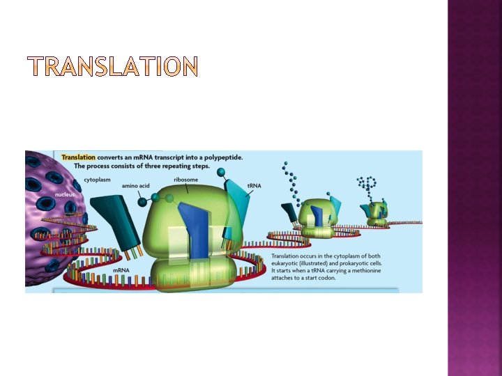
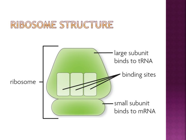
The m. RNA molecule leaves the nucleus and moves to a ribosome The m. RNA “sticks” to the small subunit of the ribosome, which is made up of a ribosomal RNA (r. RNA) molecule The m. RNA is fed between the small subunit of the ribosome and the large subunit The m. RNA message is “read” by the ribosome 3 nucleotides at a time Each set of 3 nucleotides on m. RNA is called a codon
codon for methionine (Met) codon for leucine (Leu)
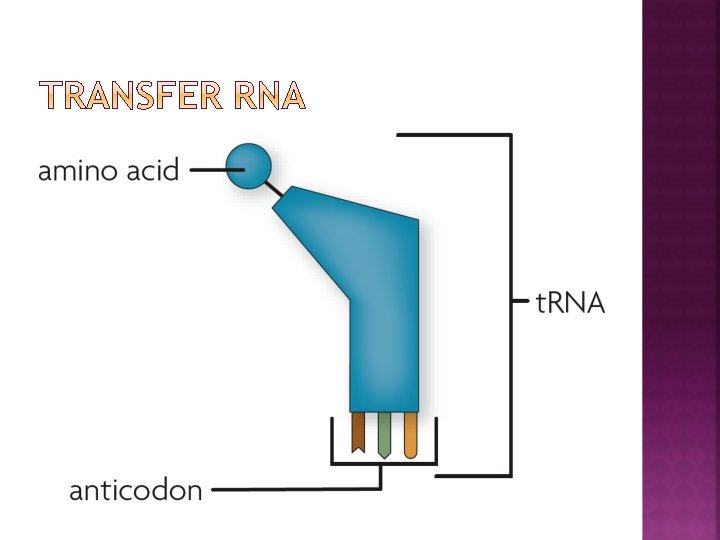
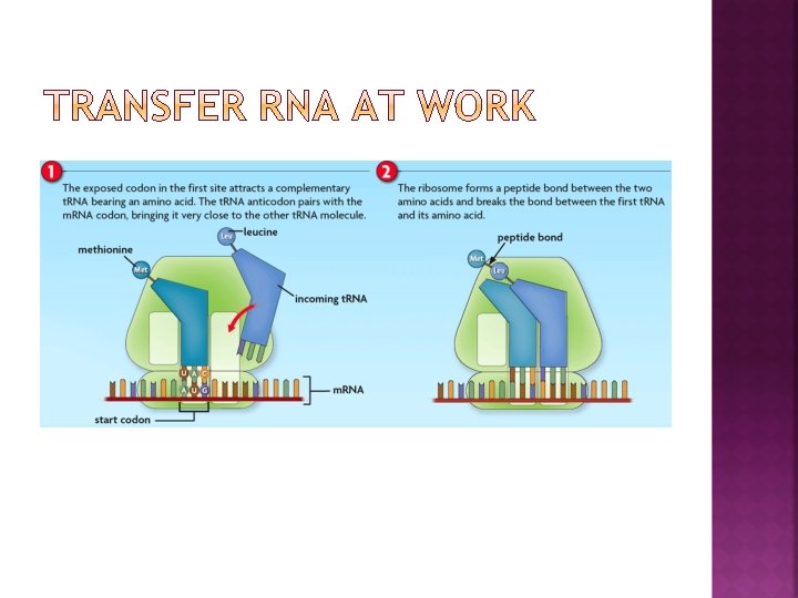
For each codon of m. RNA, there is a transfer RNA (t. RNA) molecule that has the anticodon An anticodon is a set of three nucleotides that is complementary to the m. RNA codon Needed for bonding to the m. RNA to make a “drop off” The transfer RNA also carries ONE amino acid The transfer RNA bonds temporarily to its m. RNA complement codon A chemical reaction occurs that causes the t. RNA to let go of its amino acid
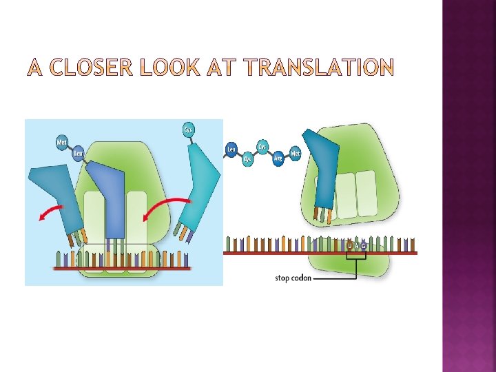
The amino acids that are dropped off by the t. RNA are linked together by a peptide bond This continues until a polypeptide chain or a protein is made based on the instructions originally provided by the DNA.
How protein synthesis is altered when there is a change in the DNA
Any change in the DNA nucleotide sequence The DNA is checked twice for this during Interphase of the cell cycle After G 1: The existing DNA is checked After G 2: The replicated DNA is checked to make sure no mutations occurred in the replication of DNA Causes of mutations DNA Replication errors Caused by mutagens: UV light, chemicals, pollutants, changes in the environment etc.
How mutations can effect protein synthesis No effect: The same amino acid sequence is created regardless Good result: A new adaptive trait comes about as a result (evolution) Bad result: Controls over the cell cycle are lost and rapid cell division (cancer) results.
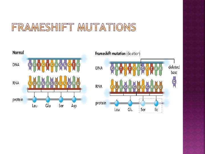
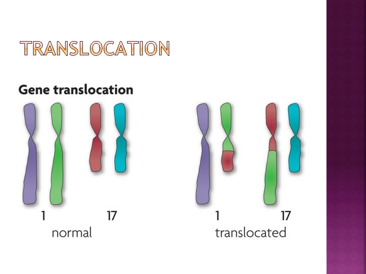
The overall length of the DNA does NOT change Substitution: Another nucleotide replaces an existing one Inversion: Two, or more, nucleotides switch locations Point Mutations The overall length of the DNA is changed Deletion: One or more nucleotides are removed Insertion: One or more extra nucleotides are added Translocation: A part of one DNA moves to another strand of DNA Frameshift Mutations
mutate d base
Yes If the mutation occurs in the germ cells that divide through meiosis to create a sperm or an egg These remain “hidden” until an offspring is created using either the sperm or the egg with the mutation No If the mutation occurs in the somatic cells that divide through mitosis These will only affect the individual, not their offspring