The Child with Cardiovascular Dysfunction Lecture 2 1






- Slides: 69
The Child with Cardiovascular Dysfunction Lecture 2 1
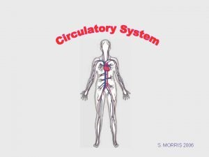
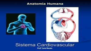
Cardiovascular disorders in children are divided into two major groups Congenital cardiac defects and acquired heart disorders. • Acquired cardiac disorders refer to disease processes or abnormalities that occur after birth. They result from various factors, including infection, autoimmune responses, environmental factors, and familial tendencies.
Congenital Heart Disease • Are anatomic abnormalities of the heart that are present at the birth and impair cardiac function. • Two main clinical manifestation – HF – Hypoxia 3
Embryologic Development • The heart and other components of the circulatory system (blood, blood vessels, lymph) begin to develop from the mesoderm during the fourth week of gestation and are completely formed by the eighth week. • Page 1252
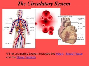
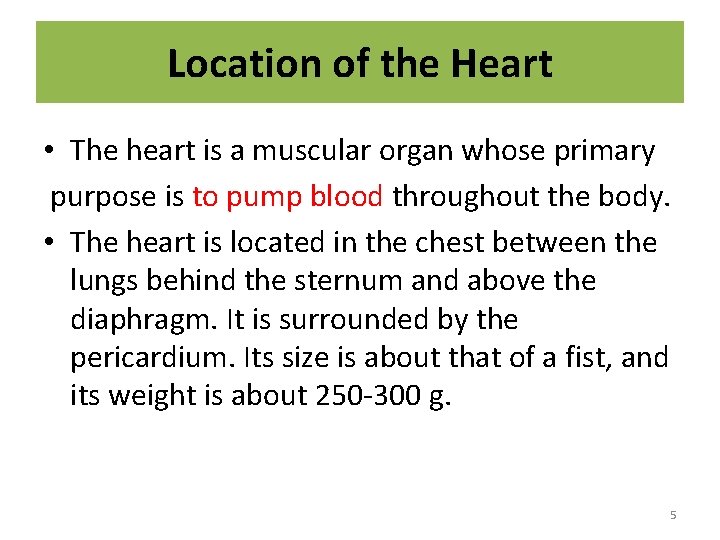
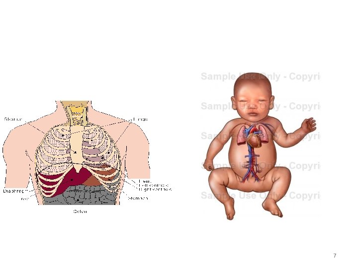
Location of the Heart • The heart is a muscular organ whose primary purpose is to pump blood throughout the body. • The heart is located in the chest between the lungs behind the sternum and above the diaphragm. It is surrounded by the pericardium. Its size is about that of a fist, and its weight is about 250 -300 g. 5
7
8
Pediatric differences • Cardiac functioning: – Infants have a great risk of heart failure than older child because the immature heart is more sensitive to volume or pressure overload. – Heart muscle fully developed at five year. – Metabolic rate and oxygen requirements double at birth so the HR is high 9
Congenital Heart Disease • Incidence: 5 -8 per 1000 live births – About 2 -3 of these are symptomatic in first year of life. – Major cause of death in first year of life (after prematurity) – Most common anomaly is VSD – 85% with newborn are expected to survive. • The exact cause of most congenital cardiac defects is unknown 10
Risk factors for CHD 1. 2. 3. 4. 5. Maternal rubella during pregnancy. Maternal alcoholism, drug use. Maternal age Maternal type one diabetes. Several genetics factors, down syndrome , turner syndrome 11
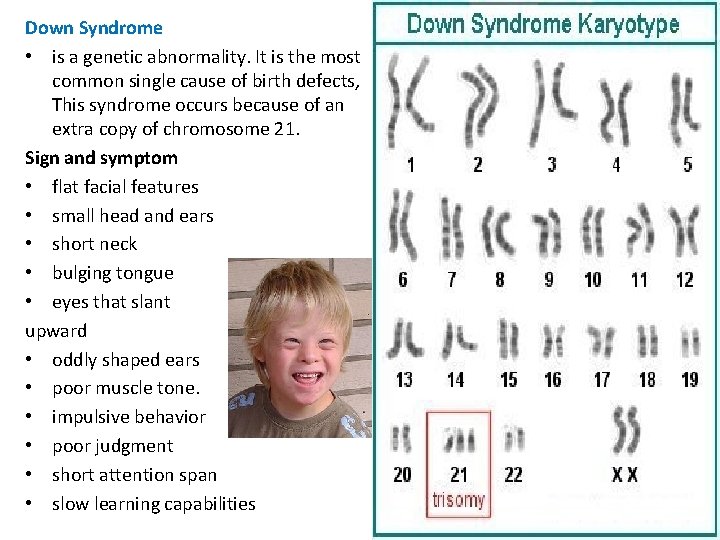
Down Syndrome • is a genetic abnormality. It is the most common single cause of birth defects, This syndrome occurs because of an extra copy of chromosome 21. Sign and symptom • flat facial features • small head and ears • short neck • bulging tongue • eyes that slant upward • oddly shaped ears • poor muscle tone. • impulsive behavior • poor judgment • short attention span • slow learning capabilities 12
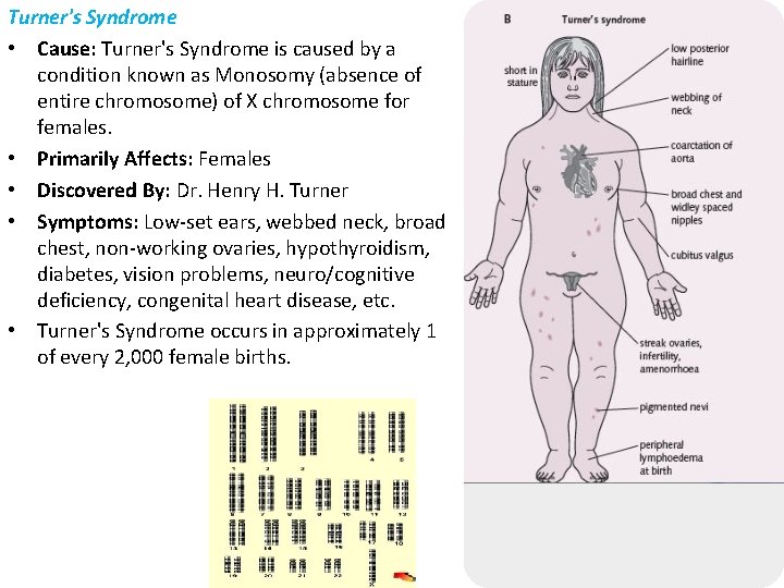
Turner's Syndrome • Cause: Turner's Syndrome is caused by a condition known as Monosomy (absence of entire chromosome) of X chromosome for females. • Primarily Affects: Females • Discovered By: Dr. Henry H. Turner • Symptoms: Low-set ears, webbed neck, broad chest, non-working ovaries, hypothyroidism, diabetes, vision problems, neuro/cognitive deficiency, congenital heart disease, etc. • Turner's Syndrome occurs in approximately 1 of every 2, 000 female births. 13
Changes at Birth 1. Foramen ovale 2. Ductus arteriosus 3. Ductus venosus 14
15
Pediatric Indicators of Cardiac Dysfunction 1. Poor feeding 2. Tachypnea/ tachycardia 3. Failure to thrive/poor weight gain/activity intolerance 4. Developmental delays 5. + Prenatal history 6. + Family history of cardiac disease 16
Fetal circulation • http: //www. indiana. edu/~anat 550/cvanim/fe tcirc/fetcirc. html 17
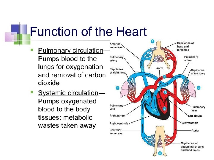
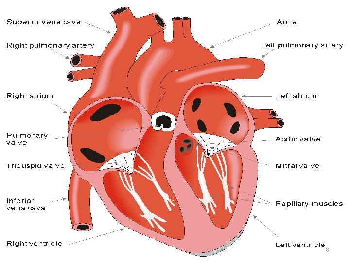
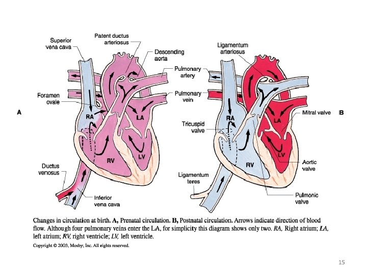
Fetal Circulation • During pregnancy blood is transferred from placenta through umbilical vein into the ductus venosus (opining into the inferior vena cava). • Then blood is carried to the right atrium in which is forced into the left atrium by an opining between the two atriums called foramen ovale. • Blood is transferred into the left ventricles and to the whole body through aorta. • Some blood pass into the right ventricle, in which it moved to the lungs by the pulmonary artery. • But, the majority of this blood pass into the aorta by an opining between aorta & pulmonary artery called ductus atreriousus
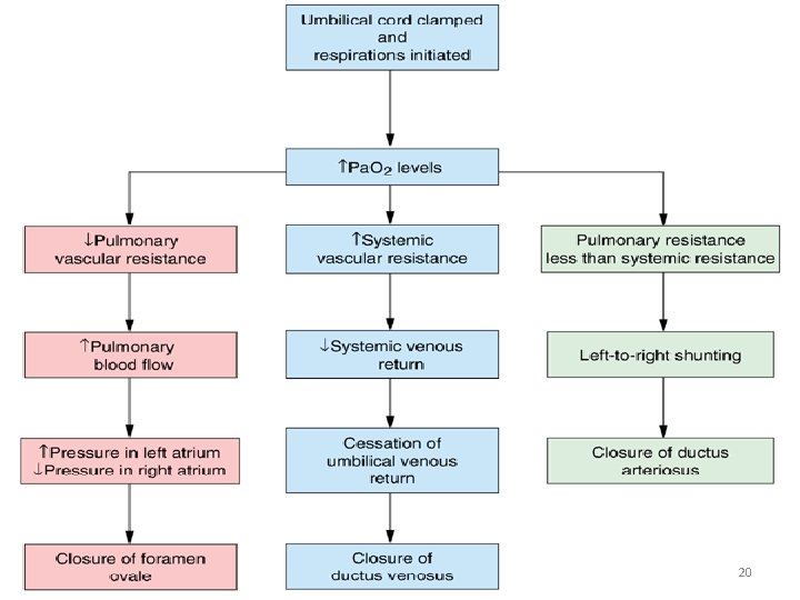
Fetal Circulation • After birth, the newborn start breathing which increase the lung expansion, helping in shifting of blood into the lungs by the pulmonary artery not into the aorta by the ductus arteriosus as during pregnancy. • This will lead to increase pulmonary blood flow & decrease pulmonary vascular resistance. • Also leading to increase blood returned to the left atrium through pulmonary veins & this leads to increase pressure in the left atrium. • The increasing pressure stimulate the closure of the foramen ovale and. Ductus arteriosus close in response to increase O 2 saturation after 10 -12 hours and permanently after 10 -21 days.
20
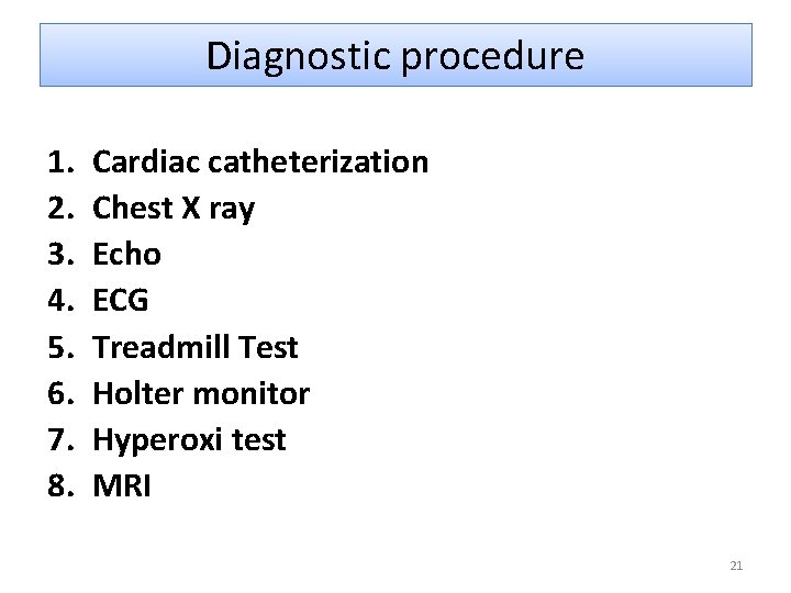
Diagnostic procedure 1. 2. 3. 4. 5. 6. 7. 8. Cardiac catheterization Chest X ray Echo ECG Treadmill Test Holter monitor Hyperoxi test MRI 21
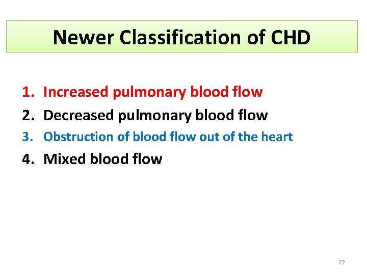
Newer Classification of CHD 1. Increased pulmonary blood flow 2. Decreased pulmonary blood flow 3. Obstruction of blood flow out of the heart 4. Mixed blood flow 22
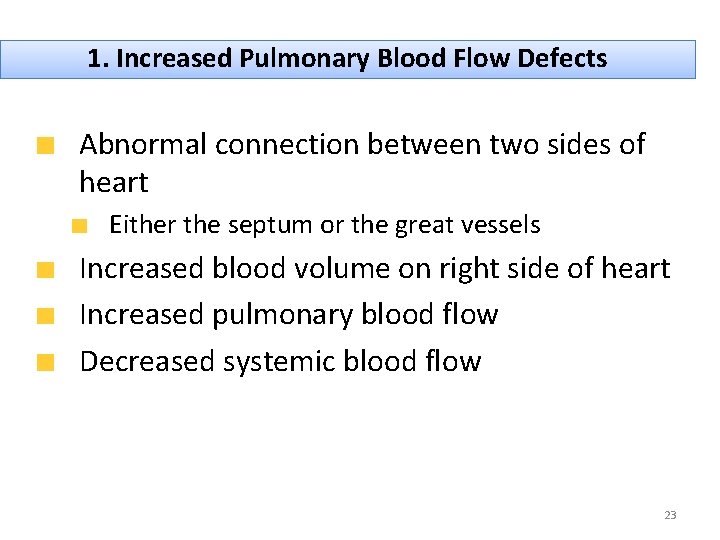
1. Increased Pulmonary Blood Flow Defects Abnormal connection between two sides of heart Either the septum or the great vessels Increased blood volume on right side of heart Increased pulmonary blood flow Decreased systemic blood flow 23
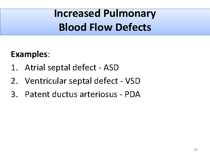
Increased Pulmonary Blood Flow Defects Examples: 1. Atrial septal defect - ASD 2. Ventricular septal defect - VSD 3. Patent ductus arteriosus - PDA 24
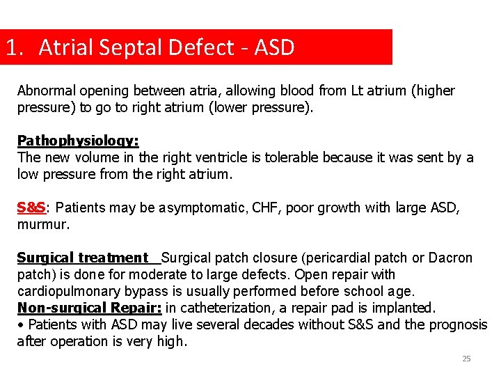
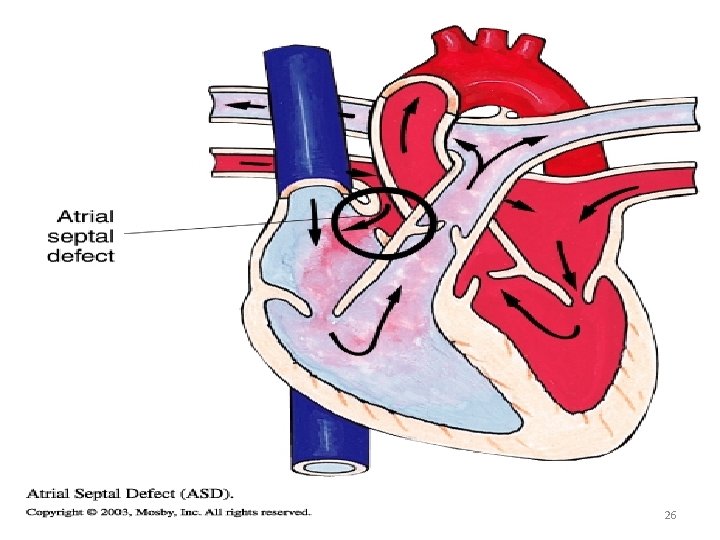
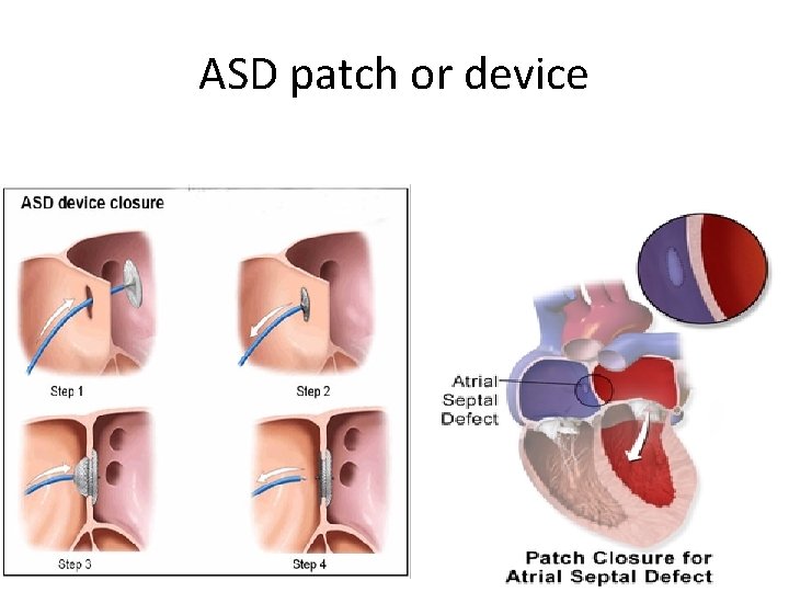
1. Atrial Septal Defect - ASD Abnormal opening between atria, allowing blood from Lt atrium (higher pressure) to go to right atrium (lower pressure). Pathophysiology: The new volume in the right ventricle is tolerable because it was sent by a low pressure from the right atrium. S&S: Patients may be asymptomatic, CHF, poor growth with large ASD, murmur. Surgical treatment Surgical patch closure (pericardial patch or Dacron patch) is done for moderate to large defects. Open repair with cardiopulmonary bypass is usually performed before school age. Non-surgical Repair: in catheterization, a repair pad is implanted. • Patients with ASD may live several decades without S&S and the prognosis after operation is very high. 25
26
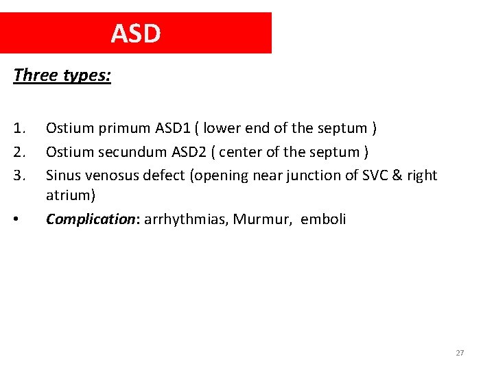
ASD Three types: 1. 2. 3. • Ostium primum ASD 1 ( lower end of the septum ) Ostium secundum ASD 2 ( center of the septum ) Sinus venosus defect (opening near junction of SVC & right atrium) Complication: arrhythmias, Murmur, emboli 27
ASD patch or device 28
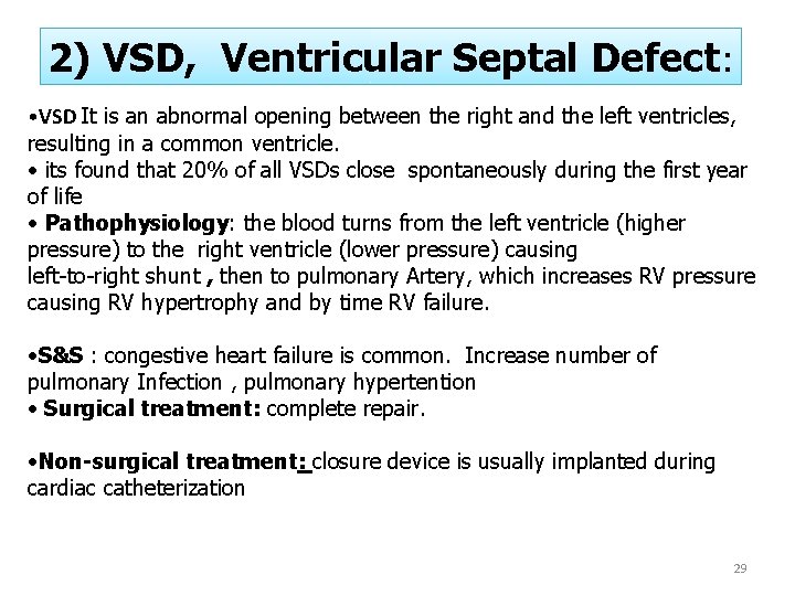
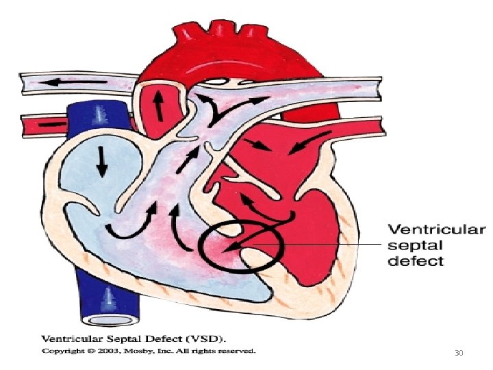
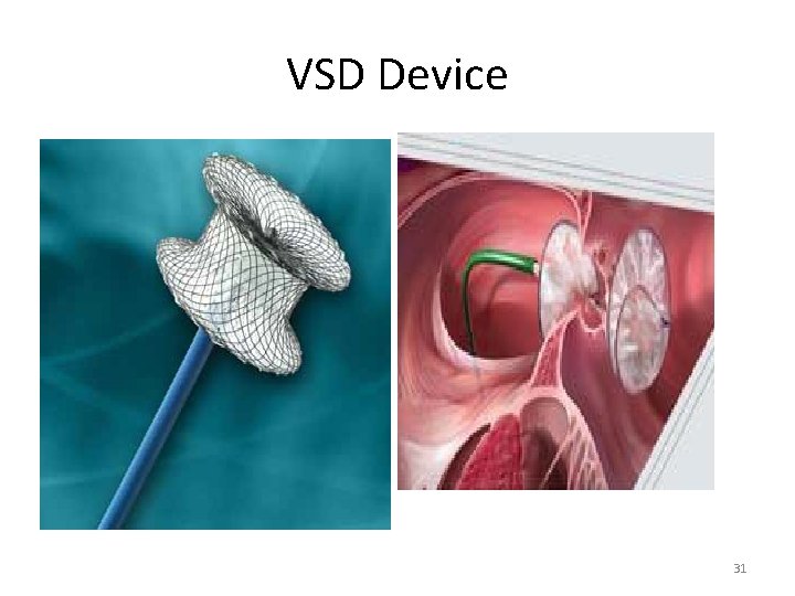
2) VSD, Ventricular Septal Defect: • VSD It is an abnormal opening between the right and the left ventricles, resulting in a common ventricle. • its found that 20% of all VSDs close spontaneously during the first year of life • Pathophysiology: the blood turns from the left ventricle (higher pressure) to the right ventricle (lower pressure) causing left-to-right shunt , then to pulmonary Artery, which increases RV pressure causing RV hypertrophy and by time RV failure. • S&S : congestive heart failure is common. Increase number of pulmonary Infection , pulmonary hypertention • Surgical treatment: complete repair. • Non-surgical treatment: closure device is usually implanted during cardiac catheterization 29
30
VSD Device 31
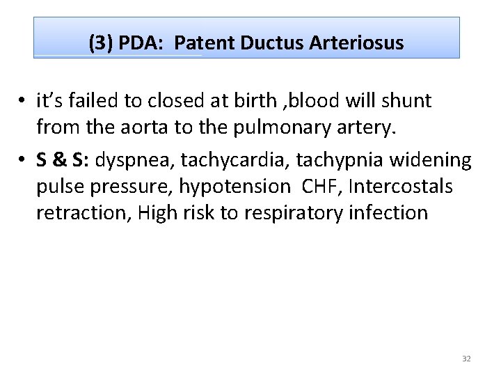
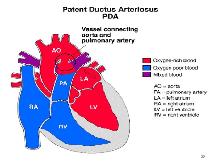
(3) PDA: Patent Ductus Arteriosus • it’s failed to closed at birth , blood will shunt from the aorta to the pulmonary artery. • S & S: dyspnea, tachycardia, tachypnia widening pulse pressure, hypotension CHF, Intercostals retraction, High risk to respiratory infection 32
33
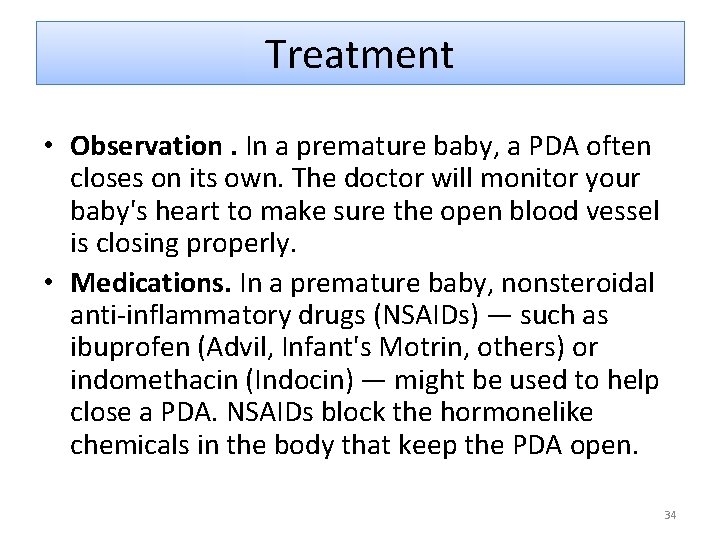
Treatment • Observation. In a premature baby, a PDA often closes on its own. The doctor will monitor your baby's heart to make sure the open blood vessel is closing properly. • Medications. In a premature baby, nonsteroidal anti-inflammatory drugs (NSAIDs) — such as ibuprofen (Advil, Infant's Motrin, others) or indomethacin (Indocin) — might be used to help close a PDA. NSAIDs block the hormonelike chemicals in the body that keep the PDA open. 34
Treatment • Open-heart surgery. . A surgeon makes a small cut between child's ribs to reach child's heart and repair the open duct using stitches or clips called a left thoracotomy.
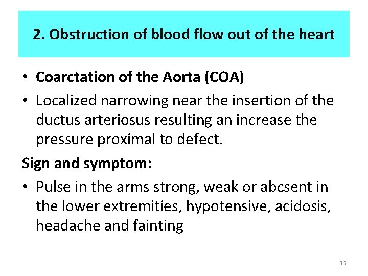
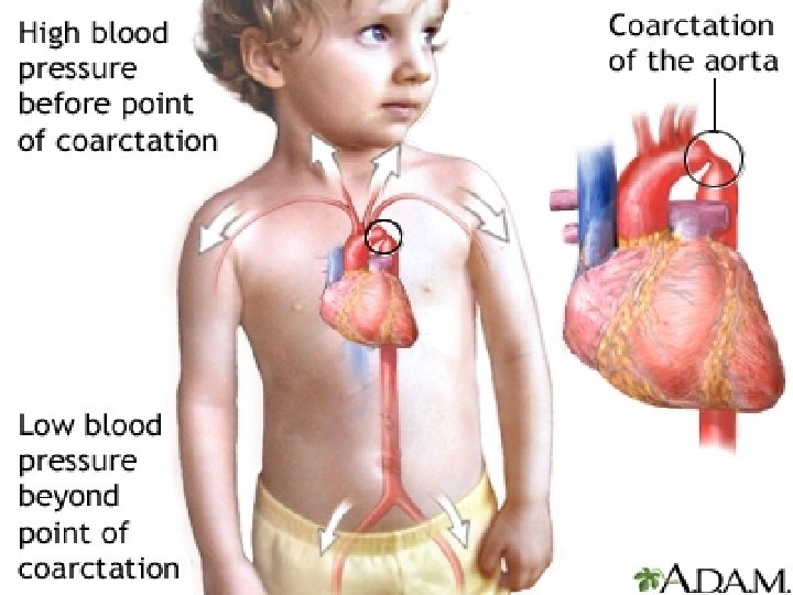
2. Obstruction of blood flow out of the heart • Coarctation of the Aorta (COA) • Localized narrowing near the insertion of the ductus arteriosus resulting an increase the pressure proximal to defect. Sign and symptom: • Pulse in the arms strong, weak or abcsent in the lower extremities, hypotensive, acidosis, headache and fainting 36
COA 37
Treatment 1. Resection with end-to-end anastomosis: • This method involves removing the narrowed segment of the aorta (resection) followed by connecting the two ends of the aorta together (anastomosis). 2. Bypass graft repair. This technique involves bypassing the narrowed area by inserting a plastic tube called a graft between the portions of the aorta. 3. Patch aortoplasty. treat coarctation by cutting across the narrowed area of the aorta and then attaching a patch of synthetic material to widen the blood vessel. Patch aortoplasty is useful if the coarctation involves a long segment of the aorta. 38
Treatment cont … 4. Balloon angioplasty and stenting • Balloon angioplasty is an option for initially treating aortic coarctation or for treating re-narrowing (re-coarctation) that has occurred after surgery. • During this procedure, the doctor inserts a thin, flexible tube (catheter) into an artery in your groin and threads it through your blood vessels to your heart using X-ray imaging. • doctor places an uninflated balloon through the opening of the narrowed aorta. When the balloon is inflated, the aorta widens and blood flows more easily. 39
3. Decreased pulmonary blood flow • Obstruct the pulmonary blood flow result in little or no blood reaching the lung to get oxygenated. 40
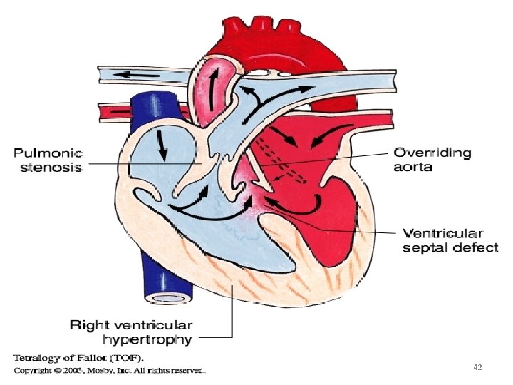
3. Decreased pulmonary blood flow • Tetralogy Of Fallot (TOF): The classic form includes: • Ventricular septal defect. • Pulmonary stenosis. • Overriding aorta. • Right venticular hypertrophy. 41
42
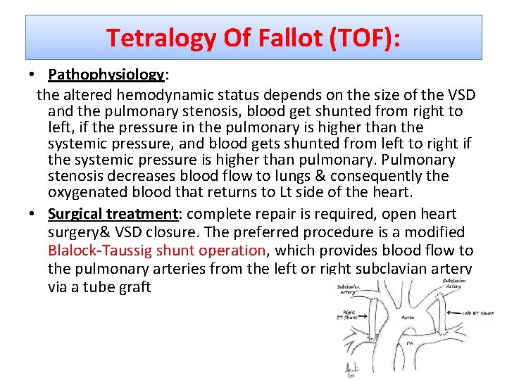
Tetralogy Of Fallot (TOF): • Pathophysiology: the altered hemodynamic status depends on the size of the VSD and the pulmonary stenosis, blood get shunted from right to left, if the pressure in the pulmonary is higher than the systemic pressure, and blood gets shunted from left to right if the systemic pressure is higher than pulmonary. Pulmonary stenosis decreases blood flow to lungs & consequently the oxygenated blood that returns to Lt side of the heart. • Surgical treatment: complete repair is required, open heart surgery& VSD closure. The preferred procedure is a modified Blalock-Taussig shunt operation, which provides blood flow to the pulmonary arteries from the left or right subclavian artery via a tube graft 43
Tetralogy Of Fallot (TOF): • Sign and symptom: cyanosis, murmur, hypoxia, clubbing of fingers, poor growth • Patient at risk of: emboli, CVA, seizures and sudden death. • Surgical intervention: complete repair in the first year of the life. 44
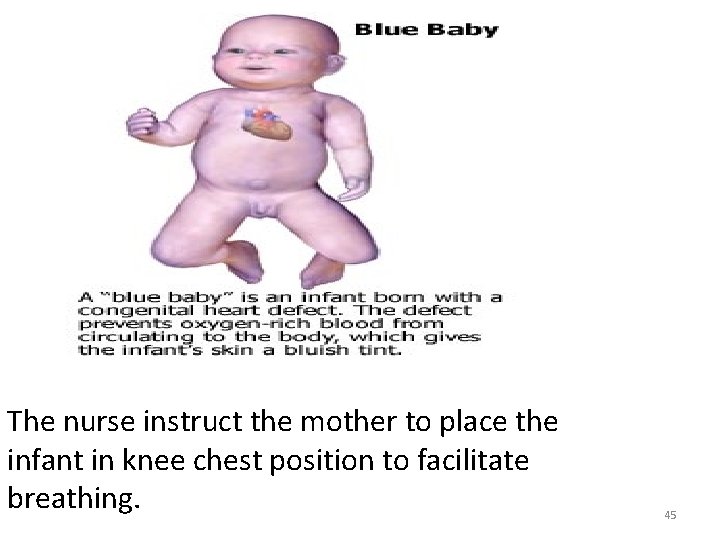
The nurse instruct the mother to place the infant in knee chest position to facilitate breathing. 45
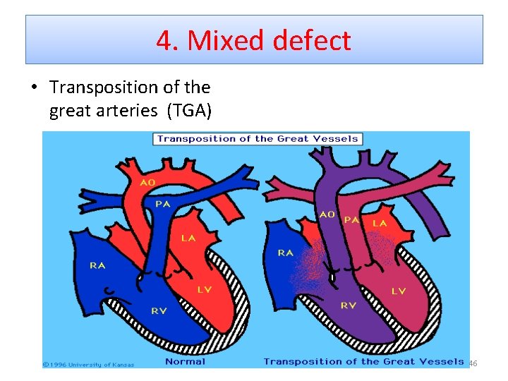
4. Mixed defect • Transposition of the great arteries (TGA) 46
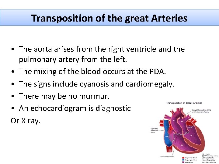
Transposition of the great Arteries • The aorta arises from the right ventricle and the pulmonary artery from the left. • The mixing of the blood occurs at the PDA. • The signs include cyanosis and cardiomegaly. • There may be no murmur. • An echocardiogram is diagnostic Or X ray. 47 47
What are the symptoms of TGA? 1. 2. 3. 4. 5. 6. Cyanosis is noted in the first hours of life Rapid breathing Hypoxia, CHF developed labored breathing Rapid heart rate Cool, clammy skin 48
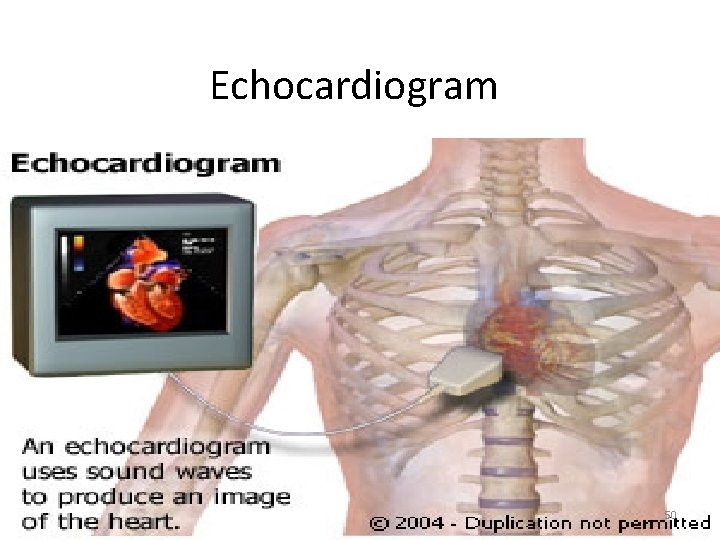
Diagnostic test 1. 2. 3. 4. Chest x-ray Electrocardiogram Echocardiogram (echo) Cardiac catheterization 49
Echocardiogram 50
Treatment • Prostaglandin is ordered to maintain a patent ductus arteriosus. • Oxygen administered • Surgery before 1 week. • Survival with out surgery is impossible. 51
Nursing Process Overview for the Child with CHD • Assessment – Health History – pregnancy, feeding problems, siblings , respiratory difficulty and family history. – Physical Examination – Sign and symptom (respiratory distress and poor feeding. – Growth problems. – Edema – Cyanosis, murmur 52 52
Assess for sign and symptoms of hypoxemia • Polycyethemia • Clubbing • Cyanosis • Assess the tongue and buccal membrane • Assessment (cont’d) Laboratory and Diagnostic Testing Cardiac Catheterization 53
• Nursing Diagnosis: • Activity intolerance related to decreased tissue oxygenation. • Anxiety related to hypoxia • Decrease cardiac out put related to increased cardiac workload. • Delayed growth and development related to hypoxia. • Risk for infection related pulmonary stasis. 54
Nursing intervention of the Child with CHD • Improving Oxygenation. • Promoting Adequate Nutrition. – Offer frequent high calorie every three hrs. Provide iron supplements Maintain hydration Blood transfusion Assisting the Child and Family to Cope. Preventing Infection. Providing Care for the Child Undergoing Cardiac Surgery – Providing Preoperative Care – Providing Postoperative Care. • Providing Patient and Family Education. • • • 55 55
Congestive Heart Failure (CHF) • CHD Most Common Cause of CHF in Infants • S/S: – Tachycardia, Diaphoresis, Tachypnea, Feeding problem, Crackles & Respiratory Distress, Edema, weight gain, Hepatomegaly, Peripheral edema, especially periorbital, Ascites , Neck vein distention (children) • CXR Shows Large Heart. • Echocardiogram is Diagnostic. 56
Etiologies of Heart failure in Child or Adolescent • • • Anemia Dysrhythmias Cardiomyopathy Hypertension Inherited Kawasaki disease Myocarditis Renal failure Other noncardiac diseases (e. g. , muscular dystrophy) Electrolyte imbalances Endocrinopathies include hyperthyroidism and hypothyroidism
Therapeutic Management (1) Improve cardiac function (increase contractility and decrease afterload), (2) Remove accumulated fluid and sodium (decrease preload). (3) Decrease cardiac demands. (4) Improve tissue oxygenation and decrease oxygen consumption.
Congestive Heart Failure (CHF) • Medical Management: Digoxin To Make Heart Work More Efficiently Not given if HR less than 90 for infant. Lasix/Diuretics To Remove Excess Fluid Oxygen: Potent Vasodilator which decreases pulmonary vascular resistance. – Rest, a neutral thermal environment, semi-Fowler position, cluster care to promote uninterrupted rest – – 59
• Nursing care CHF – Monitor physiologic functions: BP, HR, RR – Assess crackels – Prevent infection; Group care; Semi-Fowler position. – Adequate Nutrition: Feeding Techniques: 45 Degree Angle; Rest Frequently. – Promote Development: Play, Age Appropriate Toys, Physical Activities With Rest Periods – Emotional Support: Prevent Hypoxia From Agitation or Distress; Consistency of Caregiver for Patient; Refer-Parent-to-Parent Support Groups. page 1268 60
Rheumatic fever is an inflammatory disease that may develop after an infection with group A Streptococcus bacteria (such as strep throat). The disease can affect the heart, joints, skin, and brain • Rheumatic fever mainly affects children ages 5 15, and occurs approximately 14 -28 days after strep throat. 61
Signs and symptoms 1. Abdominal pain 2. Fever 3. Heart (cardiac) problems, which may not have symptoms, or may result in shortness of breath and chest pain 4. Joint pain, arthritis(mainly in the knees, elbows, ankles, and wrists) 5. Joint swelling; redness or warmth 6. Nosebleeds (epistaxis) 7. Skin nodules 8. Skin rash (erythema) 62
The Major criteria for diagnosis include: 1. Arthritis in several large joints (polyarthritis) 2. Heart inflammation (carditis) 3. Nodules under the skin (subcutaneous skin nodules) 4. Skin rash (erythema marginatum) 63
Treatment 1. antibiotics such as penicillin, sulfadiazine, or erythromycin) over the long term to prevent strep throat from returning 2. Anti-inflammatory medications such as aspirin or corticosteroids reduce inflammation to help manage acute rheumatic fever. 64
KAWASAKI DISEASE • is an acute systemic vasculitis of unknown cause. The illness is self-limiting and resolves in 6 to 8 weeks. Without treatment. • Damage to the coronary arteries (the blood vessels that supply the heart muscle) is the most common sequelae and involves dilation of the coronary arteries and/or coronary artery aneurysm formation. • The incidence rate is estimated at 112 cases per 100, 000 children less than 5 years of age.
Clinical Manifestations • high fever that is unresponsive to antibiotics and antipyretics. • bulbar conjunctivae of the eyes become reddened, with clearing around the iris. • cracked lips and the characteristic “strawberry tongue” • the child’s hands and feet become edematous, and the palms and soles become erythematous. • Cardiac manifestations during this period include • myocarditis, decreased LV function, pericardial effusion, and • mitral regurgitation
Therapeutic management • The administration of IV immune globulin (IVIG). • Aspirin is given initially in an anti-inflammatory dosage (80 to 100 mg/kg/day in divided doses every 6 hours) to control • fever and symptoms of inflammation.
Nursing management • the nurse must monitor the child’s cardiac status carefully. Intake and output and daily weight measurements are recorded. • The child should be assessed frequently • for signs of HF, including decreased urinary output, tachycardia, and respiratory distress • Nursing care focuses primarily on symptomatic relief.
GOOD LUCK !!!!!!! THANK YOU 69 69