RENAL SYSTEM Osmoregulation Extracellular vs Intracellular Water Osmotic

")


 150 ml")


 Urinary")
 -Stretch receptors spinal")

 Afferent Arteriole (takes")


 -proximal convoluted tubule (in cortex) -tubes straighten as")


")
 Proximal Convoluted Tubule (PCT) -located")
 Loop of Henle (2 main")
 Descending limb of the loop")
 Ascending limb of the loop")
 Distal Convoluted Tubule (DCT) -reabsorption")
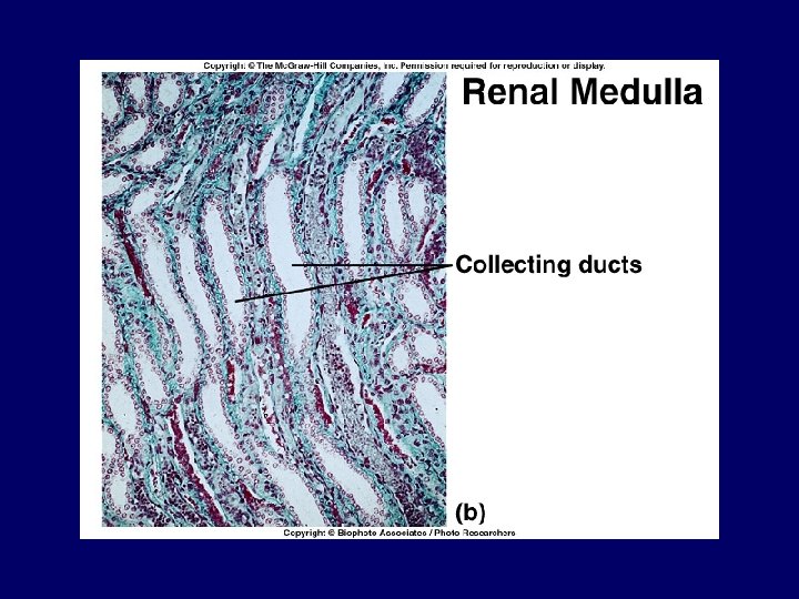
 Collecting Duct")
 Hypotonic Filtrate Excreted Reduction of Body")
 ADH released from posterior pituitary Collecting")

 Other Functions of the Urinary System: Focusing on Acid Base")
 +")

![C. Erythropoietin *Monitors Oxygen Tension *Released by kidney as [O 2] in plasma drops](https://slidetodoc.com/presentation_image/76f24175131e77036a831f4d5a0bed85/image-45.jpg "C. Erythropoietin *Monitors Oxygen Tension *Released by kidney as [O 2] in plasma drops")


")


 -depression of")
 -over-excitability of")


 Respiratory Acidosis = Increased p. CO 2 LOW")
 Respiratory Alkalosis = Decreased p. CO 2, ↑")
 Metabolic Acidosis = ↓ bicarbonate, ↓ p. H")
 Metabolic Alkalosis = ↑ bicarbonate, ↑ p. H")

- Slides: 67
RENAL SYSTEM Osmoregulation Extracellular vs Intracellular Water Osmotic Equilibrium & Water Balance Kidney Anatomy: structure & blood flow Kidney Physiology: Filtration & Glomerular Filtration Rate Reabsorption (convoluted tubules, loop of Henle, col. duct) Secretion and Excretion (collecting duct) Reading: Chapters 17 & 18
Osmoregulation Maintaining osmotic balance (non-nutritional homeostasis)
Distribution of body fluids Intracellular Fluid Extracellular Fluid
Fluid Distribution in a 67 kg individual Water = 60% of body weight (0. 6) x 67 kg = 40 liters of water Water is divided into 2 compartments: ICF = 60% ECF = 40%
Fluid Distribution in a 67 kg individual ICF ECF 60% of body water 40% of body water 25 L (60% of 40 L) 15 L (40% of 40 L) Cytoplasm Plasma (3 L) Interstitial Fluid (12 L) More Proteins High in K+ High in Na + and Cl- Ions & nutrients Ions, nutrients, wastes 300 m. Osm ** Osmotic Equilibrium **
What is osmosis? The diffusion of water. What is Diffusion? Movement of water from an area of higher concentration of water to a lower concentration of water.
Non-nutritional homeostasis Involves: -ion concentration of body fluids -volume of body fluids -why is this important? There are many strategies for maintaining osmotic equilibrium
Mammals Balance Water Intake with Water Loss 850 ml (skin & lungs) 150 ml (feces) 1500 ml (urine) 2500 ml (total) Water Intake 750 ml (moist foods) 1500 ml (liquids) 250 ml (metabolism) 2500 ml (total)
Sites of Regulation 1. THIRST: -variable sensitivity in humans 2. Osmoregulation in KIDNEYS (urine production) -principle regulation of water loss -wide variety of consumption: *we get 400 to 2500 ml of water/day *we get 10 to 25 000 mg of Na. Cl/day BUT, Osmolality of body fluids changes by <1% ! There is an obligatory water loss of ~400 ml (why? )
Mammalian Kidney Three Main Functions: 1. Remove wastes 2. Regulate fluid volume 3. Regulate ion concentrations 4. Are there any other functions?
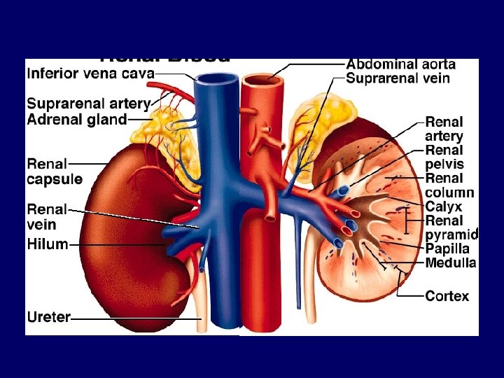
Kidney Anatomy Location -lateral to vertebral column -by 11 th & 12 th ribs
Kidney Anatomy Landmarks: -Hilum = point of exit/entry for blood vessels & ureter -Capsule = protective fibrous layer -Outer Layer = Cortex (granular) Bowman’s Capsules -Inner Layer = Medulla (striated) Renal Pyramids Loops of Henle & Collecting Ducts
Flow of Filtrate Collecting Duct Minor Calyx Major Calyx Renal Pelvis Ureter (peristalsis) Urinary Bladder Urethra
Urinary Bladder -Stores Urine (200 -300 ml = ~ 1 cup) -Stretch receptors spinal cord internal sphincter relaxes (smooth) -External sphincter = skeletal muscle (relaxed when appropriate) -Epithelial type?
Urethra -Tube that carries urine out of the body -1. 5” in females, separate from reproductive system -8” in males, it has both urinary & reproductive functions Cystitis = inflammation of urethra & bladder 10 x more common in females? Why? Treatment?
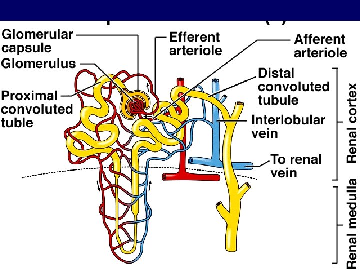
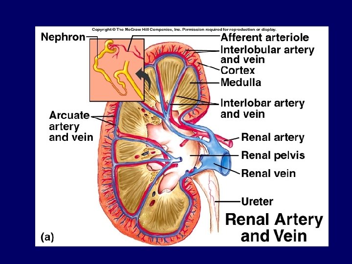
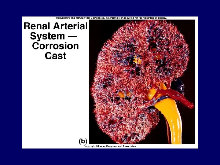
Blood Supply Aorta Renal Artery Peripheral Branches (interlobar, arcuate, interlobular arteries) Afferent Arteriole (takes blood to glomerulus) Glomerulus (ball of capillaries) Efferent Arteriole (takes blood away) Peritubular capillaries (vasa recta) Peripheral Branches (interlobular, arcuate, interlobar veins) Renal Vein Inferior Vena Cava
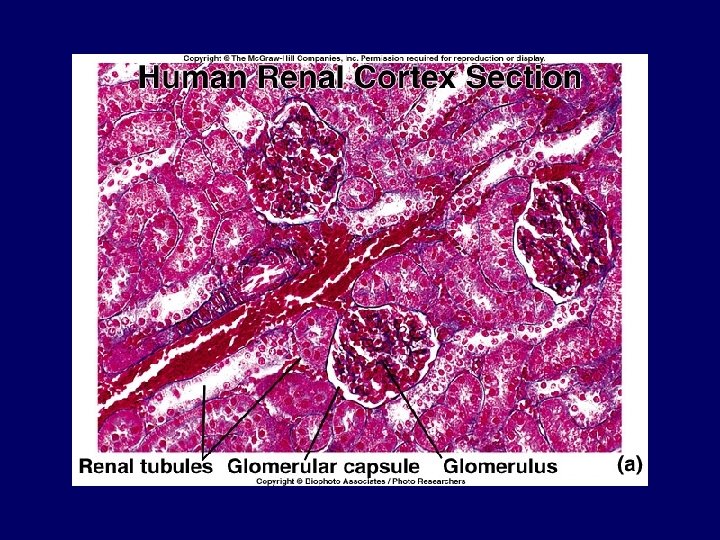
Glomerulus -ball of leaky capillaries -afferent arteriole = large -efferent arteriole = small -high hydrostatic pressure inside the glomerulus -surrounded by Bowman’s capsule (= glomerular capsule) -1 million per kidney
Nephron -series of small tubes -surrounded by peritubular capillaries -1 million per kidney -functional unit of kidney -process blood into filtrate (urine)
Nephron -Bowman’s/glomerular capsule (funnel like opening) -proximal convoluted tubule (in cortex) -tubes straighten as they enter medulla -loop of Henle (descending and ascending limbs) -distal convoluted tubule (cortex) -collecting duct (medulla)
Three Primary Functions 1. Remove Cellular Wastes 2. Regulate Body Fluid Volume 3. Regulate Ion Concentration
These 3 tasks are achieved because the kidney is capable of: Filtration: substance moves from blood to the glomerular filtrate Reabsorption: substance moves from filtrate back to blood Secretion: substance moves from blood to the filtrate (urine) Excretion: elimination of a waste product from body
Filtering Plasma Fluid is pushed OUT of plasma in glomerulus into Bowman’s Capsule -High hydrostatic pressure at glomerulus: Why? The efferent artery is small, relative to afferent artery FILTRATE =water, ions, glucose, a. a. ’s, hormones, vitamin C, waste but, NO CELLS!
Glomerular Filtration Rate -volume of filtrate made by both kidneys = 115 ml/min (women) 125 ml/min (men) -about 7. 5 L of filtrate made every hour or 180 L/day (45 gallons!) REMEMBER: -180 L of filtrate made every day -1. 5 L of urine lost every day -most of the filtrate is reabsorbed
Tubular Reabsorption (PCT + loop of Henle + DCT) Proximal Convoluted Tubule (PCT) -located mainly in cortex -microvilli what does this tell you about function? -most of the filtrate is reabsorbed here (~87%) -volume of filtrate is reduced but it’s isosmotic w/ plasma (300 m. Osm) -active transport of Na+ from filtrate back to blood -water & Cl- follow passively -glucose moves to capillaries by facilitated diffusion & active transport -NO hormonal control here
Tubular Reabsorption (PCT + loop of Henle + DCT) Loop of Henle (2 main functions) 1. If excess water is consumed = 2. If there is dehydration = -lots of dilute urine made -helps decrease body fluid volume -sml. amount of concentrated urine made -help increase body fluid volume
Tubular Reabsorption (PCT + loop of Henle + DCT) Descending limb of the loop -permeable to water -impermeable to salt (probably) -interstitial space = very salty (due to activity of ascending limb) -water moves out -filtrate gets more concentrated -bottom of loop = filtrate 1200 m. Osm (hypertonic relative to plasma)
Tubular Reabsorption (PCT + loop of Henle + DCT) Ascending limb of the loop -permeable to salt -impermeable to water -Na. Cl diffuses OUT -Na is actively transported OUT -Cl follows passively -filtrates becomes more dilute -interstitial space get very salty -top of loop = filtrate 100 m. Osm (hypotonic relative to plasma)
Tubular Reabsorption (PCT + loop of Henle + DCT) Distal Convoluted Tubule (DCT) -reabsorption of any remaining nutrients -sensitive to ALDOSTERONE -Adrenal Cortex senses Na+/K+: -if low Na+ aldosterone Na+ reabsorbed -if high K+ aldosterone K+ secreted
Regulation of Body Fluid Volume (Collecting Duct along with loop of Henle) Collecting Duct -hypotonic filtrate enters collecting duct (CD) -CD is impermeable to water unless ADH is present -ADH increases the permeability of the CD to water
Overhydration Collecting Duct Impermeable (water stays in CD) Hypotonic Filtrate Excreted Reduction of Body Fluid Volume
Dehydration Hypothalamus Senses Reduction in Fluid Volume (osmoreceptors) ADH released from posterior pituitary Collecting Duct More Permeable Water moves OUT of CD passively (gradient formed by loop of Henle) Hypertonic Filtrate Excreted
Three Primary Functions 1. Remove Cellular Wastes -by filtration in glomerulus & by excretion in calyx & beyond 2. Regulate Body Fluid Volume -by reabsorption in loop of henle (Na. Cl gradient) & CD (ADH) 3. Regulate Ion Concentration -by secretion and reabsorption in DCT (aldosterone)
Urinary System (part II) Other Functions of the Urinary System: Focusing on Acid Base Balance Reading: Chapters 17 & 18
Renal Control of Blood Pressure Juxtaglomerular Apparatus = macula densa (cells of DCT) + afferent arteriole
A. Low Blood Pressure B. High Blood Pressure
C. Erythropoietin *Monitors Oxygen Tension *Released by kidney as [O 2] in plasma drops *Acts on _________, stimulating _______ production
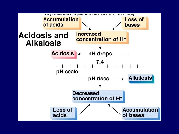
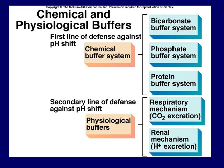
Regulation of p. H Normal Blood p. H = 7. 35 -7. 45 Maintained by chemical buffers AND physiological buffer systems
1. 2. 3. Blood p. H = 7. 35 -7. 45
Where does H + come from? 1. Aerobic Respiration of Glucose C 6 H 12 O 6 + 6 O 2 6 CO 2 + 6 H 2 O + energy CO 2 +H 2 O --> H+ + HCO 3 -
Where does H + come from? 2. Anaerobic Respiration of Glucose C 6 H 12 O 6 + O 2 energy + lactic acid (H+ )
Where does H + come from? 3. Oxidation of sulfur containing a. a. H 2 SO 4 2 - + 2 H+ Despite these 3 sites of acid (H+) production… blood p. H is close to neutral (7. 35 -7. 45)
Regulation of blood p. H CHEMICAL BUFFERS = 1 st line of defense ACID: Dissociates to release 1 or more H + when in H O 2 BASE: Accepts H+ when in H 2 O
Three Chemical Buffer Systems 1. Protein Buffer Systems: 2 HN --- aa --- COOH HN COO 3 amino group acts like base when p. H is too low carboxyl group acts like acid when p. H is too high e. g. Hb (most of the H+ yielded in CO 2 transport is carried by Hb)
Three Chemical Buffer Systems 2. Carbonic Acid Bicarbonate Buffer System: Bicarbonate (HCO 3 -) can act a weak base (pick up H+) Carbonic Acid (H 2 CO 3) can act as a weak acid (release H+) H+ + HCO 3 - --> H 2 CO 3
Three Chemical Buffer Systems 3. Phosphate Buffer System: PO 44 - ions = a major intracellular ion H 2 PO 4 - H+ + HPO 42 Dihydrogen P = weak acid, can buffer strong bases HPO 42 - + H+ H 2 PO 4 Monohydrogen P = -weak base, can buffer strong acid -also in urine to buffer acids
Chemicals buffers try to keep p. H between 7. 34 and 7. 45…but under extreme conditions the chemical buffers can’t keep up!
What happens if p. H gets out of range? ACIDOSIS (<7. 35) -depression of CNS by inhibiting synaptic transmission -if less than 7. 0 low nerve function disorientation, coma, death
What happens if p. H gets out of range? ALKALOSIS (>7. 45) -over-excitability of CNS & PNS -nerves send impulses repetitively, even w/out normal stimulus -nervousness, muscle spasms, convulsions, death 1. p. H also affects enzyme function 2. We can only live ~ 1 min at p. H extremes So, p. H is closely monitored!
Regulation of blood p. H PHYSIOLOGICAL BUFFERS = 2 nd line of defense These buffer systems work when p. H changes lead to alkalosis or acidosis
Physiological Buffer Systems *Metabolic Imbalance Respiratory Compensation Exhale CO 2 Begins within minutes Finished within 2 hours *Respiratory Imbalance Metabolic Compensation Mainly in kidney Secretion of H+ Reabsorption of HCO 3 Begins within minutes Takes days to finish
H 2 O + CO 2 H 2 CO 3 H+ + HCO 3 -
ACIDOSIS & ALKALOSIS (causes & compensations) Respiratory Acidosis = Increased p. CO 2 LOW p. H Cause = -hypoventilation -emphysema, COPD, pulmonary edema, trauma to respiratory center, airway obstruction, … Compensation = metabolic (kidney) inc. secretion of H+ (PCT) inc. reabsorption of HCO 3 - (PCT, DCT, CD) H 2 O + CO 2 <--> H 2 CO 3 <--> H+ + HCO 3 -
ACIDOSIS & ALKALOSIS (causes & compensations) Respiratory Alkalosis = Decreased p. CO 2, ↑ p. H Cause = hyperventilation O 2 deficit, altitude, anxiety, pulmonary disease Compensation = metabolic reduced secretion of H+ reduced reabsorption of HCO 3 - what else could you do as a treatment? H 2 O + CO 2 <--> H 2 CO 3 <--> H+ + HCO 3 -
ACIDOSIS & ALKALOSIS (causes & compensations) Metabolic Acidosis = ↓ bicarbonate, ↓ p. H Cause = -Loss of HCO 3 - -Due to diarrhea with lower GI losses, -Due to kidney dysfunction Compensation = Respiratory Hyperventilation to blow off CO 2 H 2 O + CO 2 <--> H 2 CO 3 <--> H+ + HCO 3 -
ACIDOSIS & ALKALOSIS (causes & compensations) Metabolic Alkalosis = ↑ bicarbonate, ↑ p. H Cause = Loss of acid (vomiting, gastric suctioning) Excessive intake of alkaline drugs Compensation = Respiratory Hypoventilation, slows CO 2 loss H 2 O + CO 2 <--> H 2 CO 3 <--> H+ + HCO 3 -
END