
Osmoregulation Chapter 44 Osmoregulation Balance of water solute
Osmoregulation Chapter 44
Osmoregulation Balance of water & solute concentration Intracellular & extracellular Ions (solutes) Na+1, Cl-1
Osmolarity Hyperosmotic Hypoosmotic Isoosmotic Normal saline (0. 9%) 5% dextrose
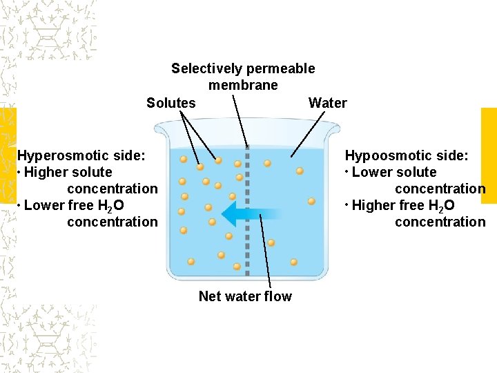
Selectively permeable membrane Solutes Water Hypoosmotic side: • Lower solute concentration • Higher free H O 2 concentration Hyperosmotic side: • Higher solute concentration • Lower free H O 2 concentration Net water flow
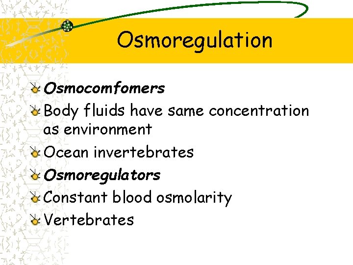
Osmoregulation Osmocomfomers Body fluids have same concentration as environment Ocean invertebrates Osmoregulators Constant blood osmolarity Vertebrates
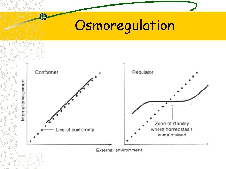
Osmoregulation
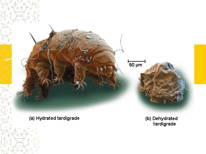
50 µm (a) Hydrated tardigrade (b) Dehydrated tardigrade
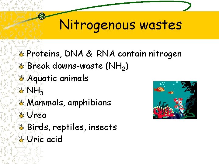
Nitrogenous wastes Proteins, DNA & RNA contain nitrogen Break downs-waste (NH 2) Aquatic animals NH 3 Mammals, amphibians Urea Birds, reptiles, insects Uric acid
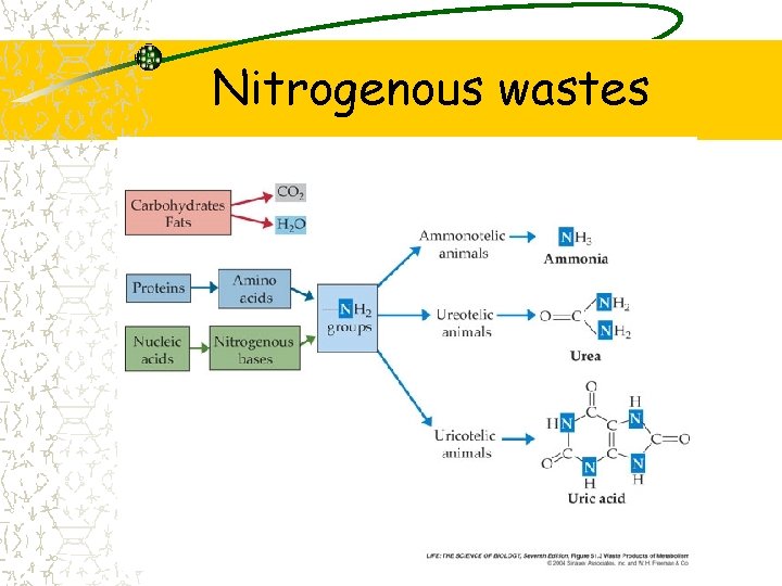
Nitrogenous wastes
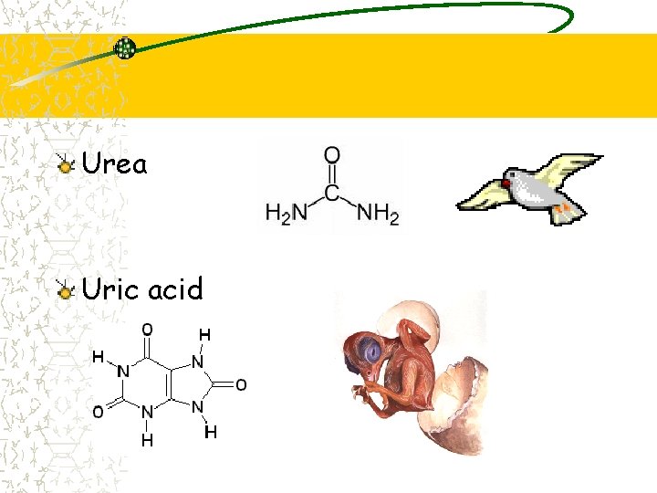
Urea Uric acid
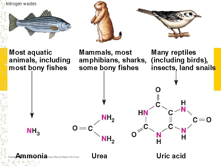
Nitrogen wastes Most aquatic animals, including most bony fishes Ammonia Many reptiles Mammals, most amphibians, sharks, (including birds), insects, land snails some bony fishes Urea Uric acid
Excretory process Excretory systems Regulate solute concentrations Produce urine Refine filtrate from body fluids
Excretory process Filtration Blood (body fluids) is filtered Forms filtrate (urine) Reabsorption Ions & nutrients are recovered Returned to body from filtrate
Excretory process Secretion Wastes/water from capillaries to filtrate (urine) Excretion Release filtrate (urine) from body Release nitrogenous metabolites
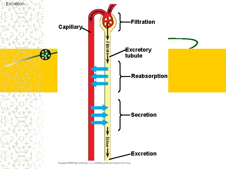
Excretion Filtration Capillary Filtrate Excretory tubule Reabsorption Secretion Urine Excretion
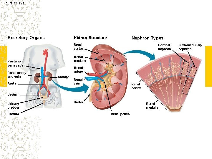
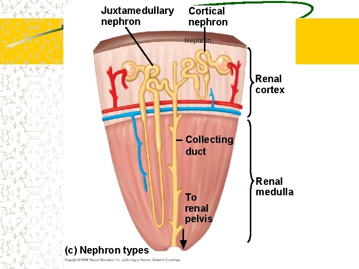
Figure 44. 12 a Excretory Organs Kidney Structure Nephron Types Renal cortex Renal medulla Posterior vena cava Renal artery and vein Aorta Cortical nephron Renal artery Kidney Renal vein Renal cortex Ureter Urinary bladder Urethra Ureter Renal medulla Renal pelvis Juxtamedullary nephron
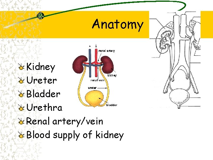
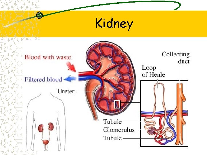
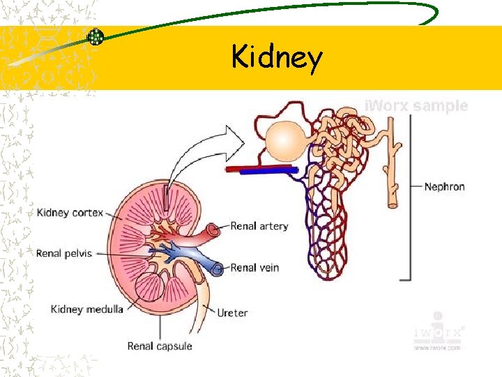
Anatomy Kidney Ureter Bladder Urethra Renal artery/vein Blood supply of kidney
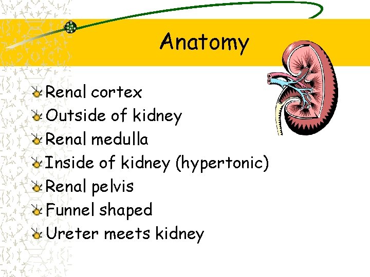
Anatomy Renal cortex Outside of kidney Renal medulla Inside of kidney (hypertonic) Renal pelvis Funnel shaped Ureter meets kidney
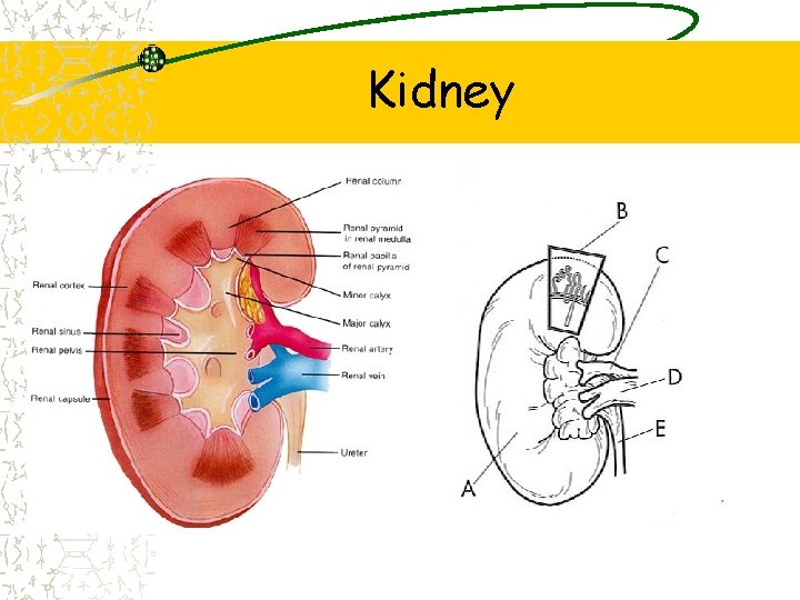
Kidney
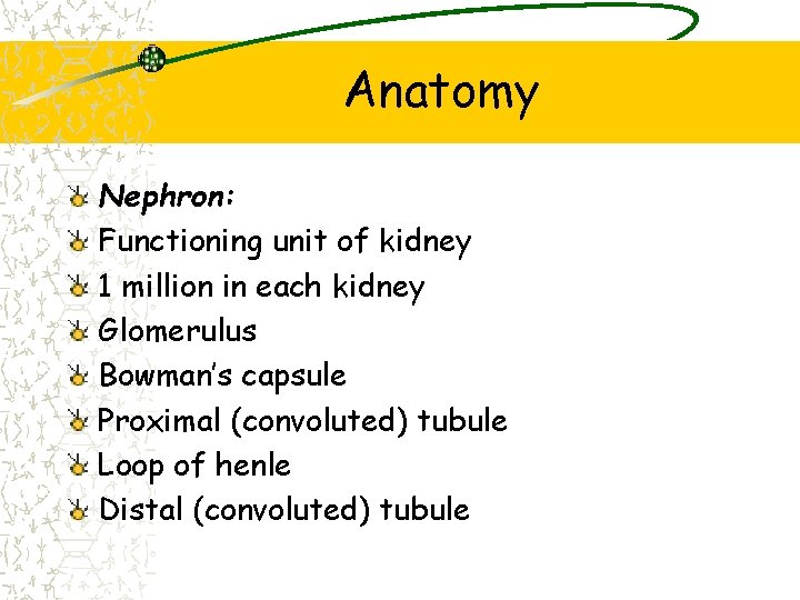
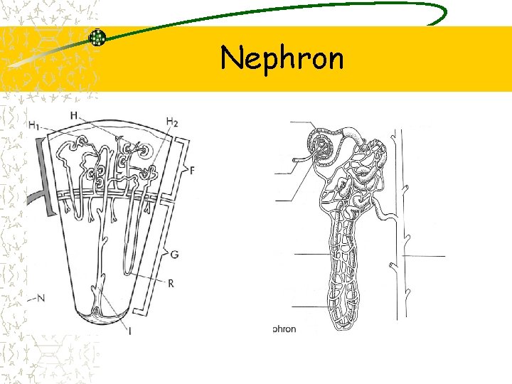
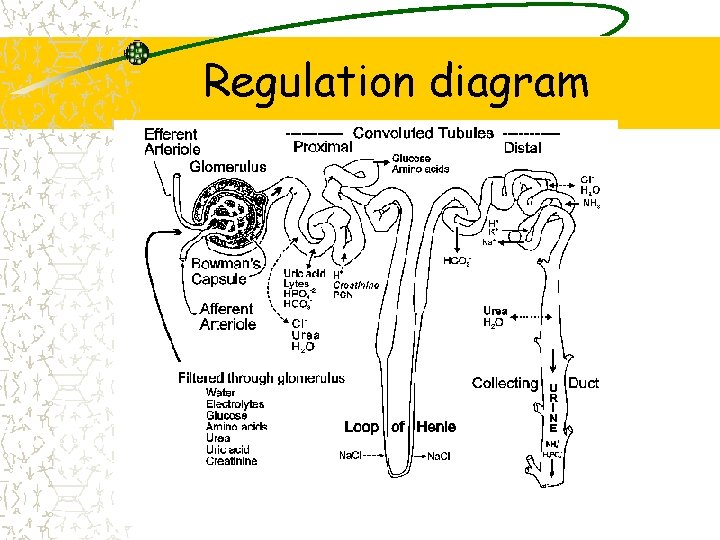
Anatomy Nephron: Functioning unit of kidney 1 million in each kidney Glomerulus Bowman’s capsule Proximal (convoluted) tubule Loop of henle Distal (convoluted) tubule
Nephron
Kidney
Kidney
Pathway
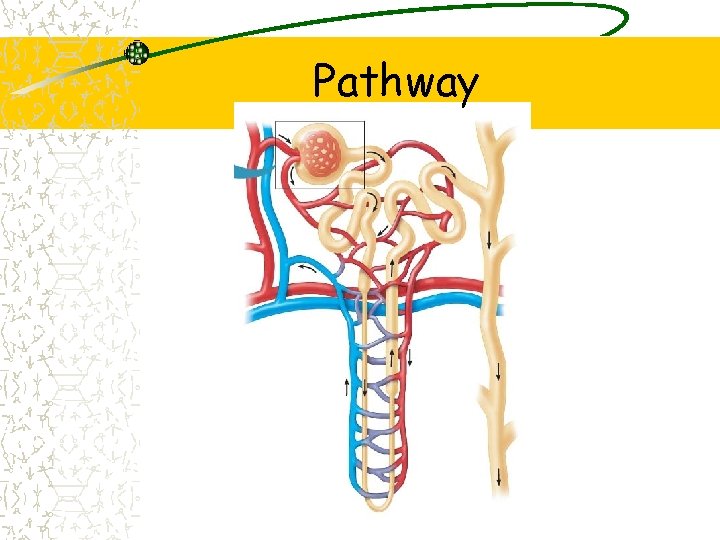
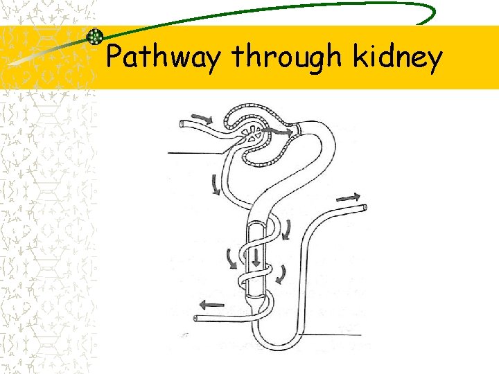
Pathway through kidney Blood forced into glomerulus Bed of capillaries Blood cells & proteins are too large Plasma goes through with minerals & water Filtrate Enters Bowman’s capsule Proximal tubule Located in renal cortex
Pathway through kidney Enters loop of Henle In the loop goes deep in the renal medulla Fluid returns Distal tubule Drains into collecting duct Goes into renal pelvis (urine) Ureter
Pathway through kidney
Juxtamedullary nephron Cortical nephron Nephron Renal cortex Collecting duct To renal pelvis (c) Nephron types Renal medulla
Nephron
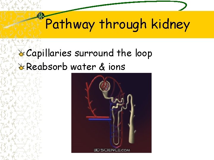
Pathway through kidney Capillaries surround the loop Reabsorb water & ions
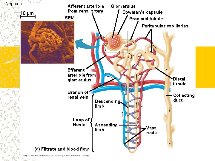
Nephron 10 µm Afferent arteriole from renal artery SEM Glomerulus Bowman’s capsule Proximal tubule Peritubular capillaries Efferent arteriole from glomerulus Branch of renal vein Loop of Henle (d) Filtrate and blood flow Distal tubule Collecting duct Descending limb Ascending limb Vasa recta
Pathway through kidney 1000 -2000 L of blood flow through a day 180 L of water a day Urinate only 1. 5 L per day Reabsorb glucose, aa, vitamins & water Excrete wastes, harmful substances, H+ & water Maintain homeostasis (blood volume, p. H electrolytes & BP)
Regulation diagram
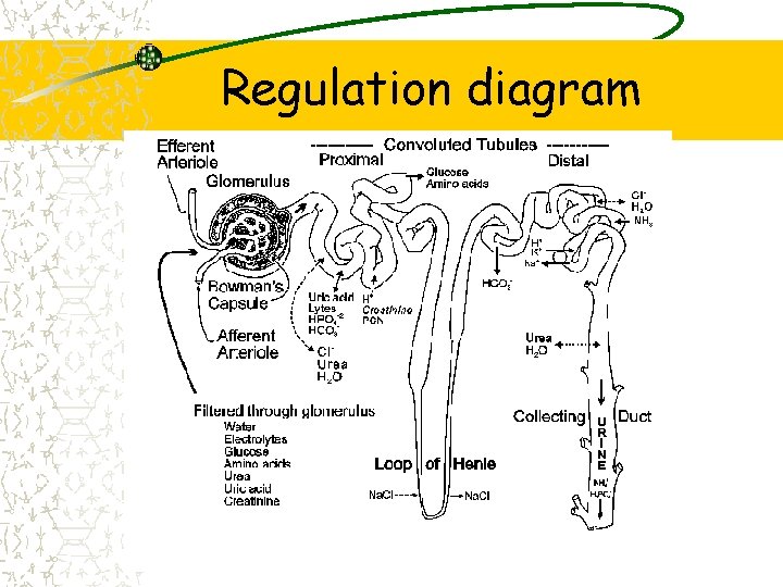
Closer look Proximal tubule Most nutrients reabsorbed to blood Na. Cl, H 2 O, HCO 3 -1 reabsorbed to blood NH 3, H+1 secreted into the tubule from blood
Bowman’s Capsule and Proximal Tubule
Closer look Loop of Henle Descending loop water is reabsorbed Ascending loop Na. Cl is reabsorbed Longer loop-more Na. Cl is absorbed
Closer look Distal tubule Regulation of K+1 & Na. Cl here Depends on body’s needs K+ is secreted into tubule Na. Cl reabsorbed into tubule

Loop of Henle and Distal Tubule
Closer look Collecting duct Na. Cl can be reabsorbed here Some urea diffuses out of tube Remaining filtrate passes as urine
Collecting Duct
Regulation diagram
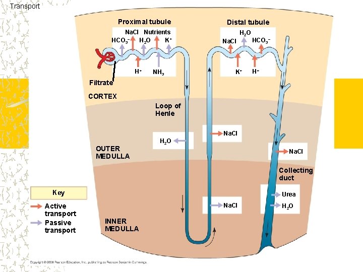
Transport Proximal tubule Na. Cl Nutrients HCO 3– H 2 O K+ H+ NH 3 Distal tubule H 2 O Na. Cl K+ HCO 3– H+ Filtrate CORTEX Loop of Henle Na. Cl OUTER MEDULLA H 2 O Na. Cl Collecting duct Key Active transport Passive transport Urea Na. Cl INNER MEDULLA H 2 O
Regulation of kidney ADH (antidiurectic hormone) Vasopressin Released by posterior pituitary gland Released in response to increased solute concentration in plasma Dehydrated or eating salty foods Distal tubule/collecting duct become more permeable to water Decreases the amount of water in urine Reabsorbs more water
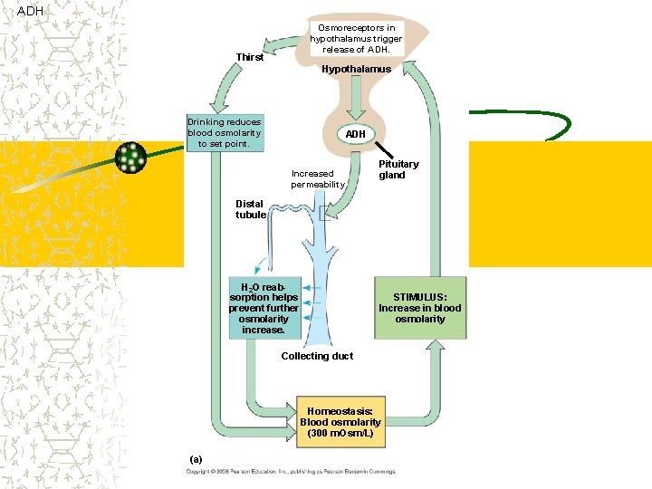
ADH Osmoreceptors in hypothalamus trigger release of ADH. Thirst Hypothalamus Drinking reduces blood osmolarity to set point. ADH Increased permeability Pituitary gland Distal tubule H 2 O reabsorption helps prevent further osmolarity increase. STIMULUS: Increase in blood osmolarity Collecting duct Homeostasis: Blood osmolarity (300 m. Osm/L) (a)
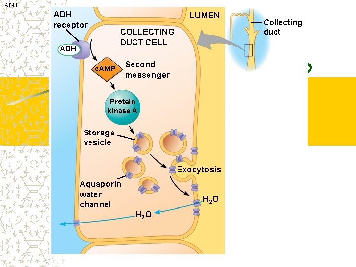
ADH receptor LUMEN COLLECTING DUCT CELL ADH c. AMP Second messenger Protein kinase A Storage vesicle Exocytosis Aquaporin water channel H 2 O Collecting duct
Effect of ADH
Regulation of kidney Low blood volume Juxtaglomerular apparatus Tissue located near artery supplies blood to glomerulus Secretes renin (enzyme) Angiotensinogen to angiotensin I Angiotensin I to angiotension II Causes blood vessels to constrict Causes release of aldosterone
Regulation of kidney Aldosterone Released by the adrenal cortex Due to low sodium levels Due to low blood volume Stimulates distal tubules/collecting ducts to reabsorb sodium Chlorine & water follow Stimulates potassium to be excreted
Regulation of kidney Aldosterone Increases serum sodium levels Increases water (blood volume) Decreases serum potassium levels ACE inhibitor Blood pressure medication Blocks the affects of aldosterone
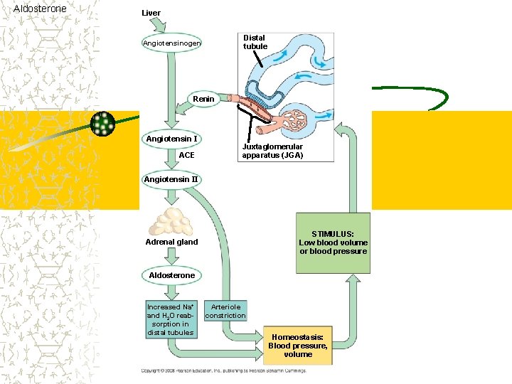
Aldosterone Liver Distal tubule Angiotensinogen Renin Angiotensin I ACE Juxtaglomerular apparatus (JGA) Angiotensin II STIMULUS: Low blood volume or blood pressure Adrenal gland Aldosterone Increased Na+ and H 2 O reabsorption in distal tubules Arteriole constriction Homeostasis: Blood pressure, volume

Regulation of kidney ANH (atrial natriuretic homone) Secreted by right atrium Due to increased blood volume Causes sodium/water to be released Lowers blood volume Opposes aldosterone
Problems Diabetes insipidus Lacks ADH Pituitary problem Increased urination Leads to dehydration & low BP Alcohol consumption Blocks ADH
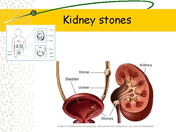
Kidney stones
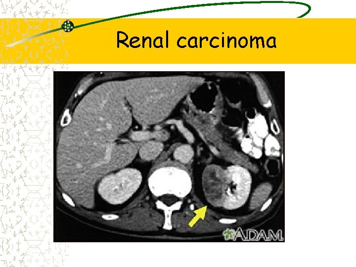
Renal carcinoma
- Slides: 54