
Lecture 4 Intracellular Accumulations By Prof Dr H
Lecture 4 Intracellular Accumulations By Prof. Dr. H. Lotfy El-Gammal 2019
• Definition: Intracellular accumulation of substances in abnormal amounts can occur within the cytoplasm (especially lysosomes) or nucleus of the cell. • This phenomenon was previously referred to as infiltration, meaning thereby that some thing unusual has infiltrated the cell from outside which is not always the case. • Intracellular accumulation of the substance in mild degree causes reversible cell injury while more severe damage results in irreversible cell injury.

Abnormal intracellular accumulations can be divided into 3 groups: i) Accumulation of constituents of normal cell metabolism produced in excess e. g. accumulations of lipids (fatty change, cholesterol deposits), proteins and carbohydrates. ii) Accumulation of abnormal substances produced as a result of abnormal metabolism due to lack of some enzymes e. g. storage diseases or inborn errors of metabolism. iii) Accumulation of pigments e. g. endogenous pigments under special circumstances, and exogenous pigments due to lack of enzymatic mechanisms to degrade the substances or transport them to other sites.

• Fatty Change (Steatosis) • Definition: Fatty change, steatosis or fatty metamorphosis is the intracellular accumulation of neutral fat within parenchymal cells. • It includes the older, now abandoned, terms of fatty degeneration and fatty infiltration because fatty change neither necessarily involves degeneration nor an infiltration. • The deposit is in the cytosol and represents an absolute increase in the intra cellular lipids. • Fatty change is particularly common in the liver but may occur in other non-fatty tissues as well e. g. in the heart, skeletal muscle, kidneys (lipoid nephrosis or minimum change disease) and other organs.
• Fatty Liver • Liver is the commonest site for accumulation of fat because it plays central role in fat metabolism. • Depending upon the cause and amount of accumulation, fatty change may be mild and reversible, or severe producing irreversible cell injury and cell death. • Etiology Fatty change in the liver may result from one of the two types of causes: 1. Conditions with excess fat • These are conditions in which the capacity of the liver to metabolise fat is exceeded e. g. i) Obesity. ii) Diabetes mellitus. iii) Congenital hyperlipidaemia.
2. Liver cell damage These are conditions in which fat cannot be metabolised due to liver cell injury e. g. i) Alcoholic liver disease (most common). ii) Starvation. iii) Protein calorie malnutrition. iv) Chronic illnesses (e. g. tuberculosis). v) Acute fatty liver in late pregnancy. vi) Hypoxia (e. g. anaemia, cardiac failure). vii) Hepatotoxins (e. g. carbon tetrachloride, chloroform, ether, aflatoxins and other poisons). viii) Drug-induced liver cell injury (e. g. administration of methotrexate, steroids, CCl 4, halothane anaesthetic, tetracycline etc). ix) Reye’s syndrome.
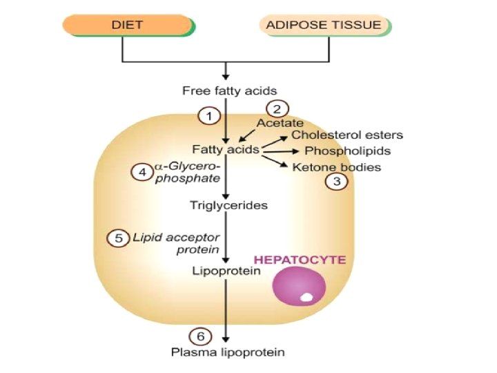
In fatty liver, intracellular accumulation of triglycerides occurs due to defect at one or more of the following 6 steps in the normal fat metabolism shown in Figure 63: 1. Increased entry of free fatty acids into the liver. 2. Increased synthesis of fatty acids by the liver. 3. Decreased conversion of fatty acids into ketone bodies resulting in increased esterification of fatty acids to triglycerides. 4. Increased α-glycerophosphate causing increased esterification of fatty acids to triglycerides. 5. Decreased synthesis of ‘lipid acceptor protein’ resulting in decreased formation of lipoprotein from triglycerides. 6. Block in the excretion of lipoprotein from the liver into plasma.

• Pigments • Definition: Pigments are coloured substances present in most living beings including humans. There are 2 broad categories of pigments: endogenous and exogenous. A. Endogenous Pigments • Definition: Endogenous pigments are either normal constituents of cells or accumulate under special circumstances e. g. melanin, alkaptonuria, haemoprotein-derived pigments, and lipofuscin
• Various disorders of melanin pigmentation cause generalised and localised hyperpigmentation and hypopigmentation: i) Generalised hyperpigmentation: a) In Addison’s disease, there is generalised hyper pigmentation of the skin, especially in areas exposed to light, and of buccal mucosa. b) Chloasma observed during pregnancy is the hyperpigmentation on the skin of face, nipples, and genitalia and occurs under the influence of oestrogen. A similar appear ance may be observed in women taking oral contraceptives. c) In chronic arsenical poisoning, there is characteristic raindrop pigmentation of the skin.
ii) Focal hyperpigmentation: • d) Melanotic tumours, both benign such as pigmented naevi (Figure 64), and malignant such as melanoma, are associated with increased melanogenesis.
iii) Generalised hypopigmentation • Albinism is an extreme degree of generalised hypopigmentation in which tyrosinase enzyme is genetically defective and no melanin is formed in the melanocytes. • Oculocutaneous albinos have no pigment in the skin and have blond hair, poor vision and severe photophobia. • They are highly sensitive to sunlight. Chronic sun exposure may lead to precancerous lesions and squamous and basal cell cancers of the skin in such individuals.
Haemoprotein-derived Pigments • Definition: Haemoproteins are the most important endogenous pigments derived from haemoglobin, cytochromes and their break-down products. • For an understanding of disorders of haemoproteins, it is essential to have knowledge of normal iron metabolism and its transport. • In disordered iron metabolism and transport, haemoprotein-derived pigments accumulate in the body. • These pigments are haemosiderin, acid haematin (haemozoin), bilirubin, and porphyrins.
Bilirubin • Bilirubin is the normal non-iron containing pigment present in the bile. • It is derived from porphyrin ring of the haem moiety of haemoglobin. • Normal level of bilirubin in blood is less than 1 mg/dl. • Excess of bilirubin or hyperbilirubinaemia causes an important clinical condition called jaundice. • Hyperbilirubinaemia may be unconjugated or conjugated; accordingly jaundice may appear in one of the following 3 ways: i) An increase in the rate of bilirubin production due to excessive destruction of red cells (predominantly unconjugated hyperbilirubinaemia).
ii) A defect in handling of bilirubin due to hepatocellular injury (biphasic jaundice). iii) Some defect in bilirubin transport within intrahepatic or extrahepatic biliary system (predominantly conjugated hyperbilirubinaemia). • Excessive accumulation of bilirubin pigment can be seen in different tissues and fluids of the body, especially in the hepatocytes, Kupffer cells and bile sinusoids. • Skin and sclera become distinctly yellow. • In infants, rise in unconjugated bilirubin may produce toxic brain injury called kernicterus.
Lipofuscin (Wear and Tear Pigment) • Definition: Lipofuscin or lipochrome is yellowishbrown intracellular lipid pigment (lipo = fat, fuscus = brown). • The pigment is often found in atrophied cells of old age and hence the name ‘wear and tear pigment’. • It is seen in the myocardial fibers, hepatocytes, Leydig cells of the testes and in neurons in senile dementia. • However, the pigment may, at times, accumulate rapidly in different cells in wasting diseases unrelated to ageing. • Lipofuscin granules are composed of lipid-protein complexes.
B. Exogenous Pigments • Definition: Exogenous pigments are the pigments introduced into the body from outside such as by inhalation, ingestion or inoculation. • Inhaled Pigments • The lungs of most individuals, especially of those living in urban areas due to atmospheric pollutants and of smokers, show a large number of inhaled pigmented materials. • The most commonly inhaled substances are carbon or coal dust; others are silica or stone dust, iron or iron oxide, asbestos and various other organic sub stances. • These substances may produce occupational lung diseases called pneumoconiosis. • The pigment particles after inhalation are taken up by alveolar macrophages. • Some of the pigment-laden macrophages are coughed out via bronchi, while some settle in the interstitial tissue of the lung and in the respiratory bronchioles and pass into lymphatics to be deposited in the hilar lymph nodes.
• Anthracosis (i. e. deposition of carbon particles) is seen in almost every adult lung and generally provokes no reaction of tissue injury (Figure 66). • However, extensive deposition of particulate material over many years in coal-miners’ pneumoconiosis, silicosis, asbestosis etc. provoke low grade inflammation, fibrosis and impaired respiratory function.

Ingested Pigments Chronic ingestion of certain metals may produce pigmentation. The examples are as under: i) Argyria is chronic ingestion of silver compounds and results in brownish pigmentation in the skin, bowel, and kidney. ii) Chronic lead poisoning may produce the characteristic blue lines on teeth at the gumline. iii) Melanosis coli results from prolonged ingestion of certain cathartics. iv) Carotenaemia is yellowish-red colouration of the skin caused by excessive ingestion of carrots which contain carotene.
Injected Pigments (Tattooing) • Pigments like India ink, cinnabar and carbon are introduced into the dermis in the process of tattooing where the pigment is taken up by macrophages and lies permanently in the connective tissue. • The examples of injected pigments are prolonged use of ointments containing mercury, dirt left accidentally in a wound, and tattooing (Figure 67) by pricking the skin with dyes.

- Slides: 22