Harvey Lodish Arnold Berk Paul Matsudaira Chris A

Harvey Lodish • Arnold Berk • Paul Matsudaira • Chris A. Kaiser • Monty Krieger • Matthew P. Scott • Lawrence Zipursky • James Darnell Molecular Cell Biology Fifth Edition Chapter 7: Transport of Ions and Small Molecules Across Cell Membranes Copyright © 2004 by W. H. Freeman & Company

Cell membrane Barrier to the passage of most polar molecule ·Maintain concentration of solute

Aquaporin, the water channel, consists of four identical transmembrane polypeptides

Relative permeability pf synthetic lipid bilayer to different classes of molecule

Diffusion rate depends on : 1. Concentration gradient or electrochemical gradient 2. Hydrophobicity 3. i. e. higher partition coefficient 4. 3. Particle size
 couple with")
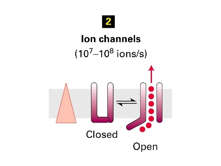
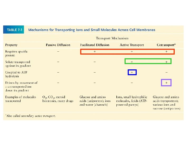
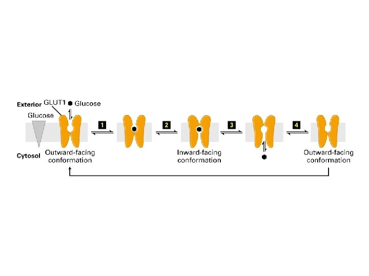
Three main class of membrane protein 1. ATP- power pump( carrier, permease) couple with energy source for active transport binding of specific solute to transporter which undergo conformation change 2. Channel protein formation of hydrophilic pore allow passive movement of small inorganic molecule

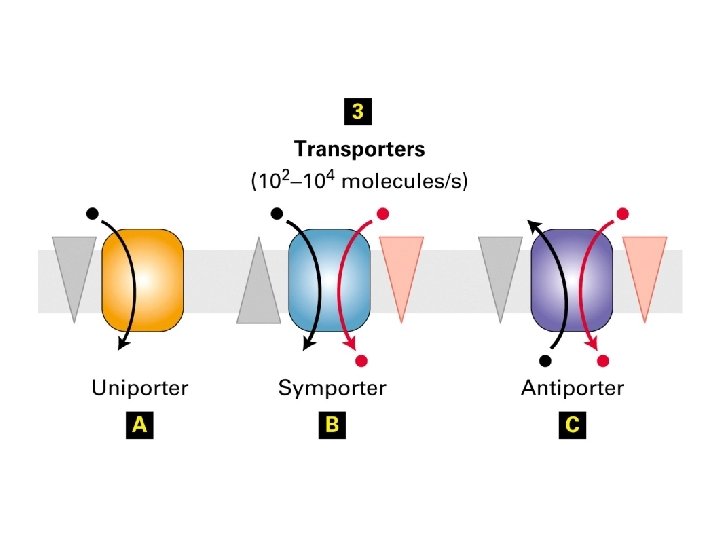
3. Transporters uniport symport antiport
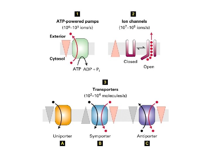

1. All transmembrane 2. proteins 2. ATP binding sites 3. Move molecules uphill against its gradient
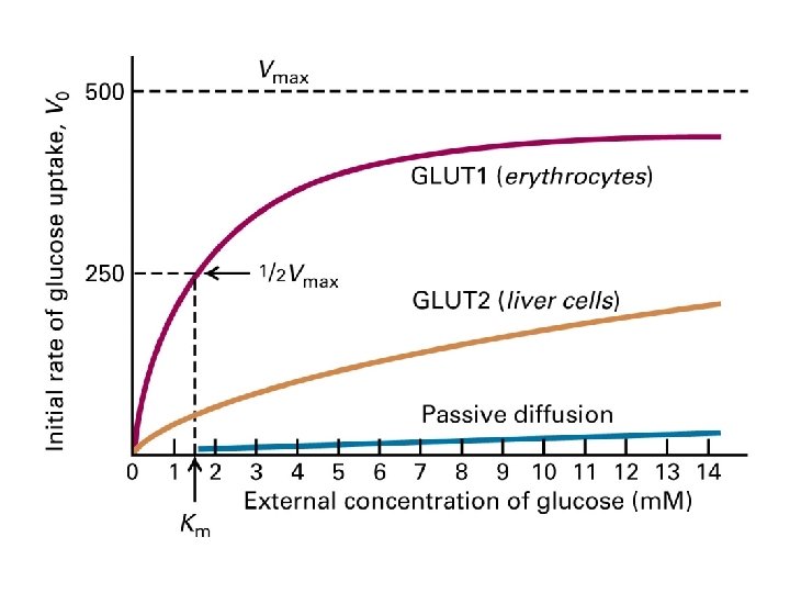

Kinetics of simple diffusion and carrier mediated diffusion

Unique features for Uniport transport: 1. Higher diffusion rate for uniport 2. Irrelevant to the partition coefficient 3. Transport rate reach Vmax when each uniport working at its maximal rate 4. Each uniport transports only a single species of molecules or single or closely related molecules

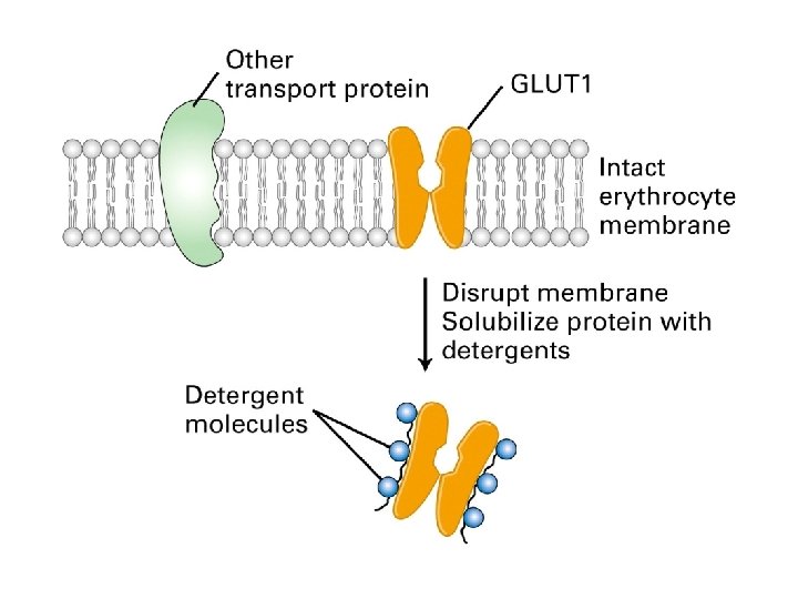
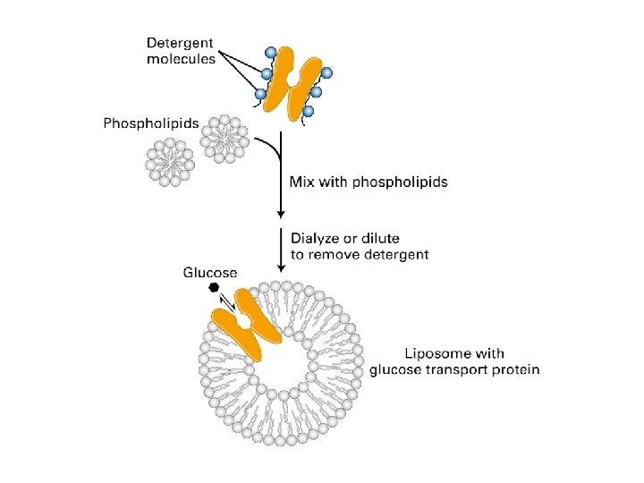
Liposome containing a single type of ytransport protein are useful in studying functional properties of transport protein
 GLUT 1 GLUT 2: express in liver cell")
Families of GLUT proteins( 1 -12) GLUT 1 GLUT 2: express in liver cell ( glucose storage) and ß cell( glucose uptake) pancrease GLUT 4: found in intracellular membrane, increase expression by insulin, lowers the blood glucose

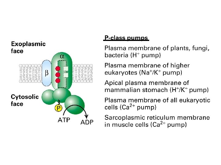
ATP powered pump 1. P- class 2. 2 , 2 subunit 3. i. e. Na+-K+ ATP ase, Ca+ATP ase, H+pump 4. 2. F-class 5. locate on bacterial membrane , chloroplast and mitochondria 6. pump proton from exoplasmic space to cytosolic for ATP synthesis 7. 3. V-class 8. maintain low p. H in plant vacuole

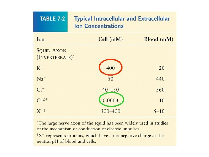
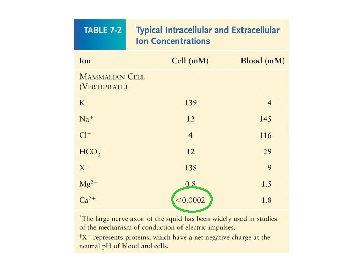

Operational model of the Ca+-ATP ase in the SR membrane of skeletal muscle cells Higher Ca+2 Lower Ca+2

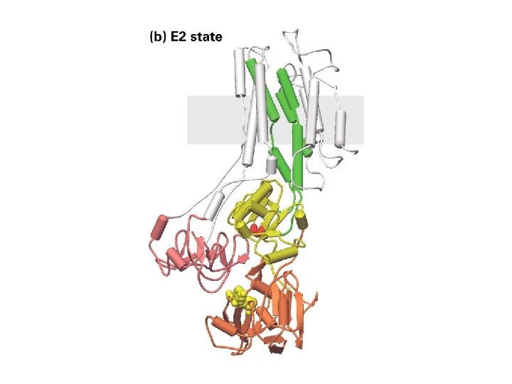
Structure of the catalytic subunit of the muscle Ca+2 ATP ase

-helix Phosphorylation site

Operational model of the Na+/K+ ATP ase in the plasma membrane Higher affinity for Na+

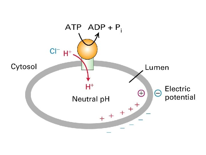
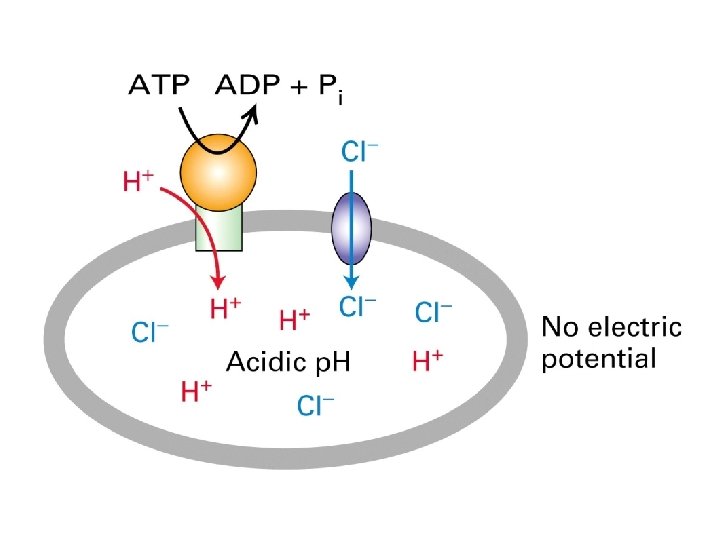
V-class H+ ATP ase pump protons across lysosomal and vacuolar membrane

Effect of proton pumping by V-class ion pumps on H+ concentration gradients and electric potential gradients across cellular membrane

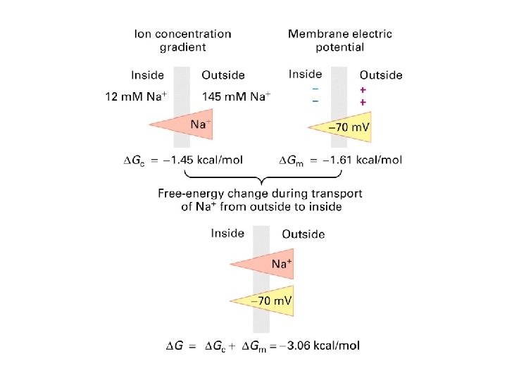
Generation of electrochemical gradient Electrochemical gradient combines the membrane potential and concentration gradient which work additively to increase the driving force
 domain 6 - helix form pathways for")
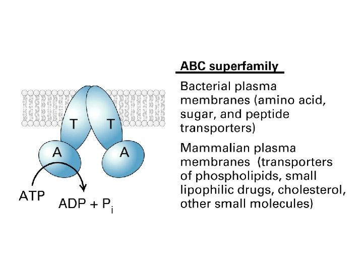
ABC transporter 2 T ( transmembrane ) domain 6 - helix form pathways for transported substance · 2 A ( ATP- binding domain) 30 -40% homology for membranes

i. e. bacterial permease use ATP hydrolysis transport a. a , sugars, vitamines, or peptides inducible, depend on the environmental condition i. e. mammalian ABC transporter ( Multi Drug Resistant) export drug from cytosol to extracellular medium mdr gene amplified by drus stimulation mostly hydrophobic for MDR proteins

Structural model for E. coli flippase 6 - helix

Flippase model of transport by MDR 1 and similar ABC proteins
 2. 3. defect in ABC")
Diseases linked with ABC proteins 1. ALD( X-link adrenoleukodestrophy) 2. 3. defect in ABC transport protein( ABCD 1) located on peroxisome, used for transport for very long fatty acid 4. 2. Tangiers disease 5. Dificiency in plasma ABCA 1 proteins, which is used for transport of phospholipis and cholesterol 6. 3. Cystic fibrosis 7. mutation of CTFR( cyctic fibrosis transmenbrane regulator; a Cl- transporter in the apical membrane of lung, sweat gland pancrease)

Ion Channel Generation of electrochemical gradient across plasma membrane i. e. Ca+ gradient regulation of signal transduction , muscle contraction and triggers secretion of digestive enzyme in to exocrine pancreastic cells i. e. Na+ gradient uptake of a. a , symport, antiport

Q: how does the electrochemical gradient formed? Selective movement of Ions Create a transmembrane electric potential difference
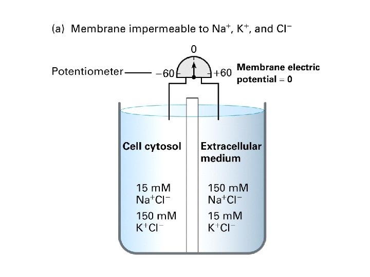
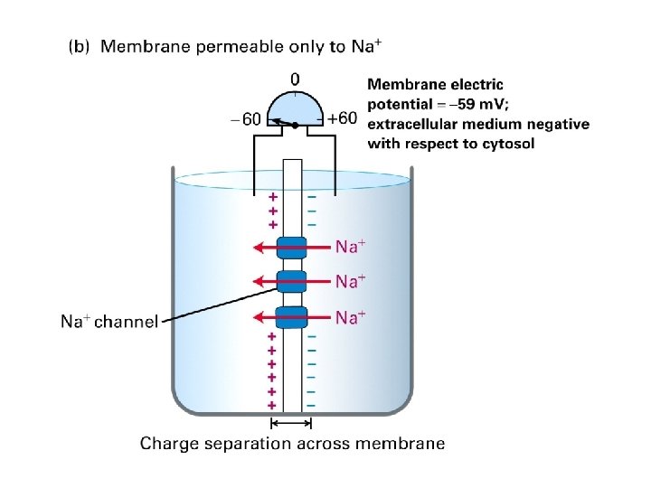
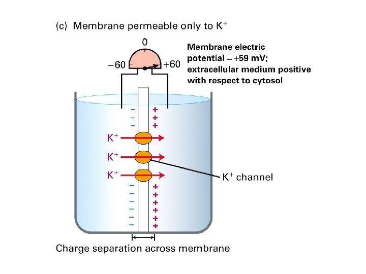

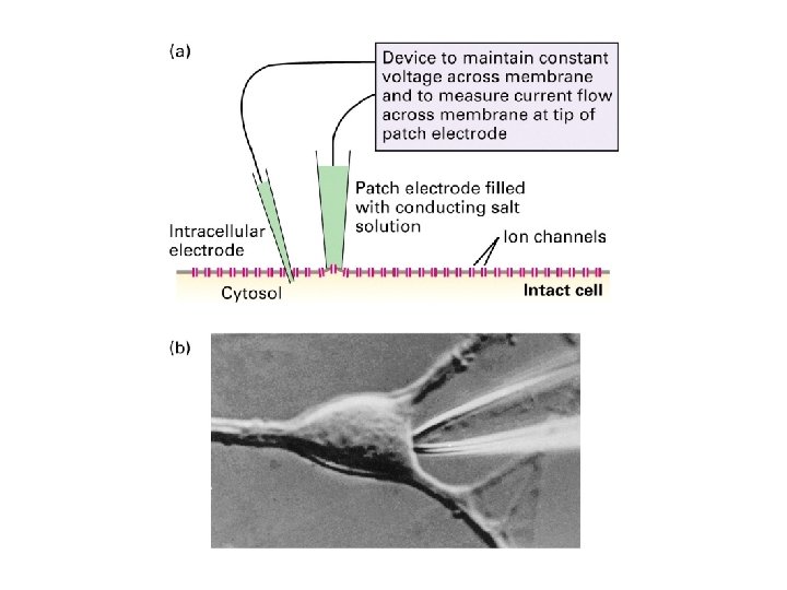
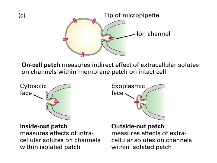
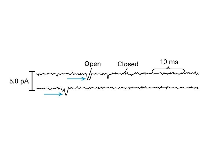
Measuring the electrochemical gradient

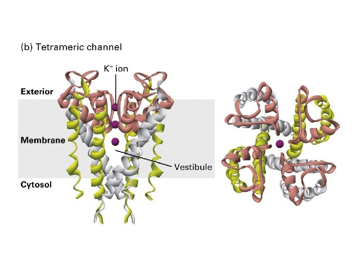
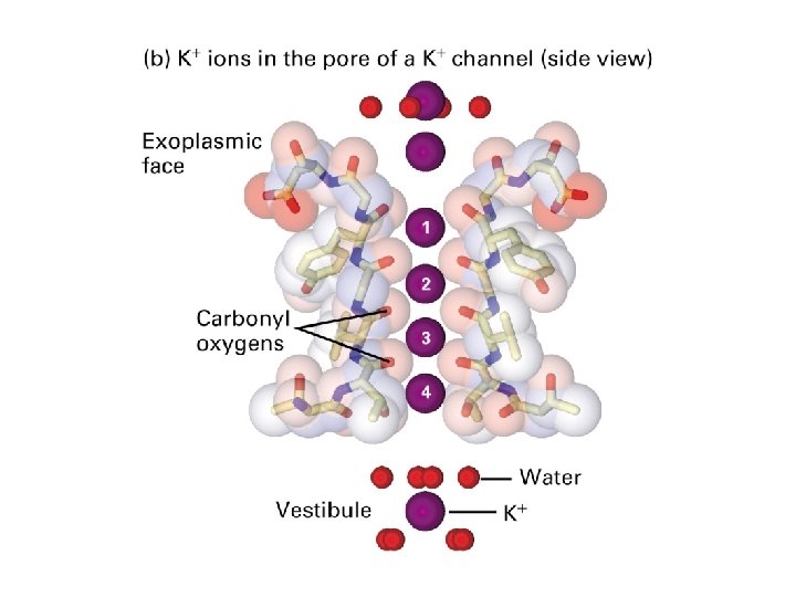
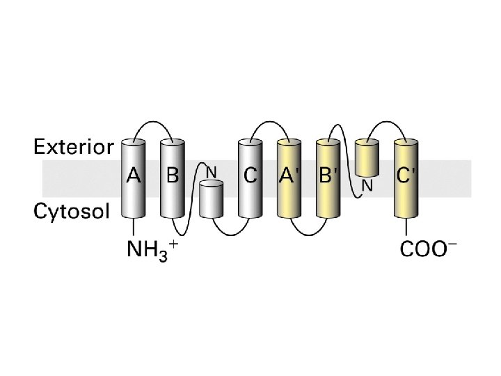
Structure of resting K+channel from the bacterium Streptomyces lividans

Important for selection

Smaller Na+ does not fit perfectly Replacement of carboxyl backbone from P segment


Oocyte expression assay is useful in comparing the function of normal and mutant forms of channel proteins

Cotransport: Use the energy stored in Na+ or H+ electrochemical gradient to power the transport of another subatance Symport: the transportd molecules and cotransported ion move in the same direction Antiport: the transported molecules move in opposited direction

Operation Model for the two-Na+/one glucose symport Glucose transport against its gradient in the epithelial cells of intestine 1 glucose in 2 Na+ in G=0

Na+ linked antiport Exports Ca+2 from cardiac Muscle Cells 3 Na+ out+ Ca+2 in 3 Na+ in+ Ca+2 out maintenance of low cytosolic Ca+2 concentration i. e. inhibition of Na+/K+ ATPase by Quabain and Digoxin raises cytosolic Na+ lowers the efficiency of Na+/Ca+2 antiport increases cytosolic Ca+2 ( used in cogestive heart failure)

Cotransporters that regulate cytosolic p. H H 2 CO 3 H+ H+ + HCO- can be neutrolized by 1. Na+/HCO 3 -/Cl- antiport 2. Cabonic anhydrase HCO 33. Na+/H+ antiport CO 2+OH-

The activity of membrane transport proteins that regulate the cytosolic p. H of mammalian cells changes with p. H

Plant vacuole membrane p. H 3— 6 Low acidity maintained by V-class ATP-powered pump PPi -powered pump

Concentration of ions and sucrose by the plant vacuole

Movement of water Osmosis: movement of water across semipermeable membrane Osmotic pressure: hydrostatic pressure uses to stop the net flow of water
 B A")
Osmotic pressure =RT( C -C ) B A

Expression of aquaporin by frog oocytes increases their permeability Aquaporin 1 erythrocyte Aquaporin 2 kidney cells
")
Water channel pprotein( aquaporin)

tetrameric · 6 -helices for each subunit

· 2 -nm-long water selective gate 0. 28 nm gate width ·Highly conserved arginine and histidine in the gate H 2 O for HO bonding with cystein

Transepithelial transport Import of molecules on the lumen side of intestinal epithelial cells and their export on the blood facing sides

Transcellular transport of glucose from the intestinal lumen into the blood Cholera toxin activated by Cl-

Acidification of the stomach lumen by parietal cells in the gastric lining

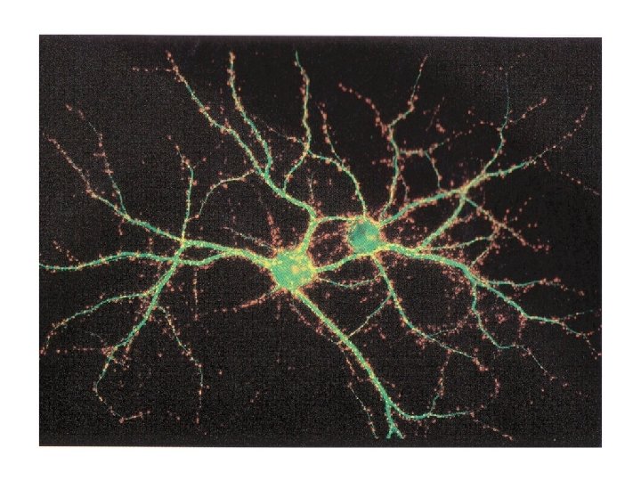
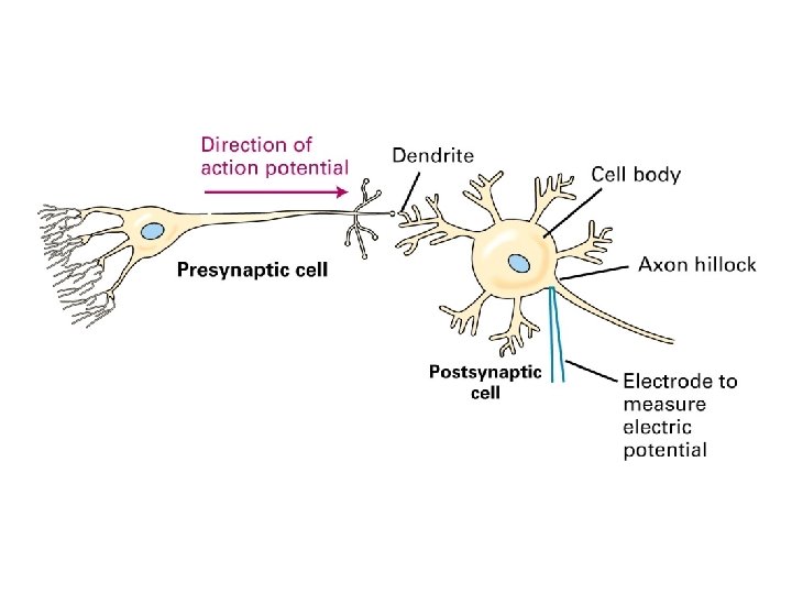
Typical morphology of two types of mammalian neurons 100 m/sec

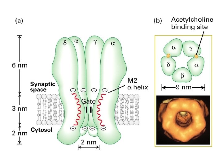
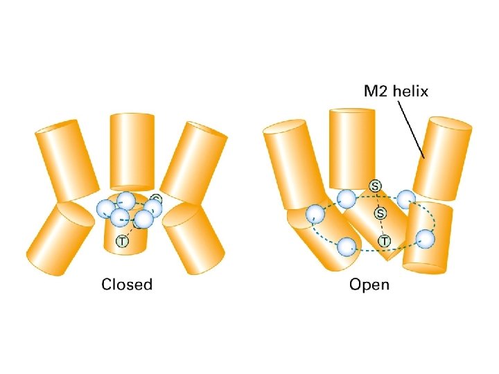
Neurotransmitters Receptors 1. Ligand gated ion channels 2. G-protein coupled receptors Synaptic vesicle: Storage of neurotransmitter. Low p. H of vesicle lumen powers entry of neuritransmitter into lumen by H+/protein antipoter

Structures of small molecules function as neurotransmitters

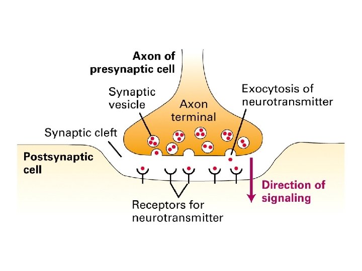
Exocytosis of synaptic vesicle 1. Action potential 2. Influx of Ca+2 triggers release of neurotransmitter

Cycling of nuerotransmitters and of synaptic vesicles in axon terminals H+/protein antiport

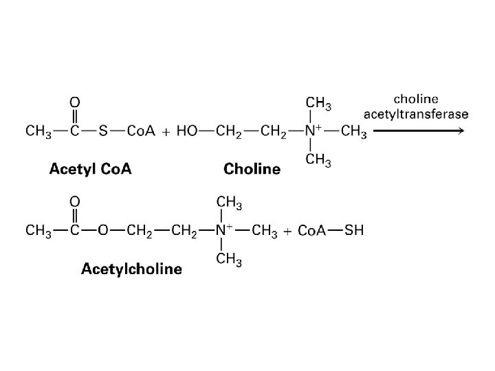
Signaling at synapse id terminated by degradation or reuptake of neurotransmitter 1. degradation i. e. acetyocholine hydrolyzed by acetyocholineaterase 2. reuptake i. e. transport into axon terminals by Na+/linked symport transporters for GABA, norepinephrine, dopamine, and serotonin

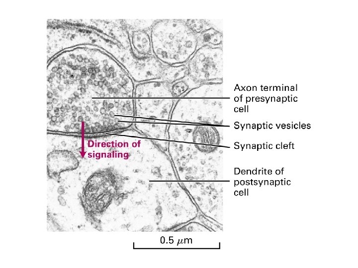
Synaptic vesicles in the axon terminal near the region where neurotransmitter release

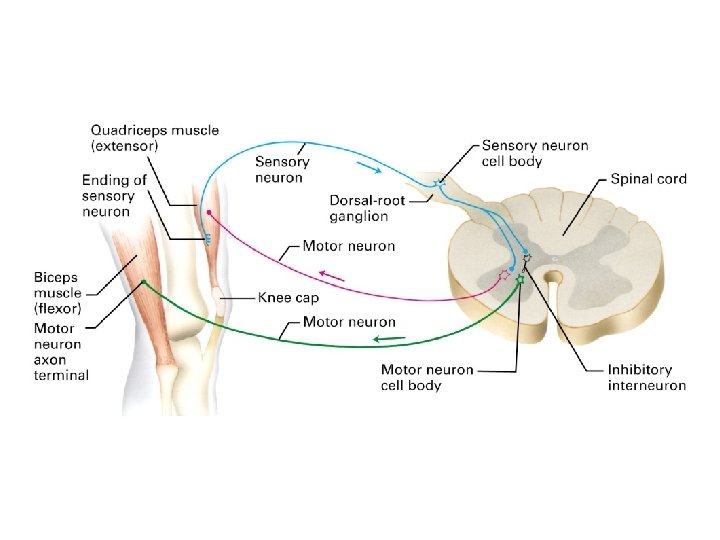
Sequential activation of gated ion channels at a neurotransmuscular junction

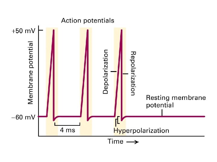
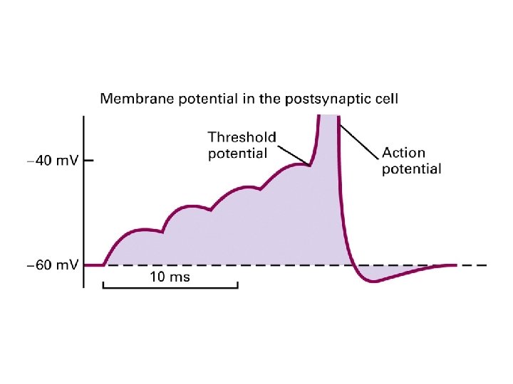
Incoming signals must reach the threshold potential to trigger an action potential in post synaptic cells
- Slides: 94