Osmotic effect Dr Haya AlGhazali l Osmotic pressure


")


 *")

- Slides: 20
Osmotic effect Dr. Haya Al-Ghazali
l Osmotic pressure: is the pressure applied by a solution to prevent the inward flow of water across a semipermeable membrane
l A semipermeable membrane: also termed (a selectively-permeable membrane, a partially-permeable membrane or a differentially-permeable membrane), is a membrane that will allow certain molecules or ions to pass through it by diffusion and occasionally specialized "facilitated diffusion"
Diffusion: describes the spread of particles through random motion from regions of higher concentration to regions of lower concentration l Active transport l
l Cytolysis, or osmotic lysis: occurs when a cell bursts due to an osmotic imbalance that has caused excess water to move into the cell. It occurs in a hypotonic environment, where water diffuses into the cell and causes its volume to increase. If the volume of water exceeds the cell membrane's capacity then the cell will burst.
osmotic concentration: concentration of osmotically active particles in solutions. l osmotic fragility : the susceptibility of red blood cells to hemolyze in hypotonic saline solutions; may be used as a test of red cell fragility when a series of increasing concentrations is used. l
l l l This test is performed to detect hereditary spherocytosis and thalassemia. Hereditary spherocytosis makes red blood cells more fragile than normal. • Spherocytosis is a genetic disease that causes a defect in the red blood cell's cytoskeleton, causing the red blood cells to be small, sphereshaped, and fragile instead of donut-shaped and flexible. Some red blood cells in patients with thalassemia are more fragile than normal, but a larger number are less fragile than normal
l Osmotic fragility decreased in: Ø Thalassemia. Ø Iron deficiency anemia. Ø Sickle cell anaemia. l Osmotic fragility of red cells increased in: Ø Hereditary spherocytosis. Ø Acquired spherocytosis
Molar concentration or molarity : is most commonly in units of moles of solute per liter of solution l Molality (mol/kg, molal, or m): denotes the number of moles of solute per kilogram of solvent (not solution) l
Osmolarity: is the measure of solute concentration, defined as the number of osmoles (Osm) of solute per liter (L) of solution (osmol/L or Osm/L). l
For non-ionised substances the osmolar concentration is the same as the Molar concentration. l For substances becoming ionised in solution it is equal to molar concentration multiplied by the number of ions produced by the molecule. l
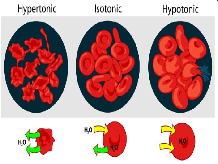
l Hypertonicity: A hypertonic solution is a solution having a greater solute concentration than the cytosol. It contains a greater concentration of impermeable solutes on the external side of the membrane. When a cell’s cytoplasm is bathed in a hypertonic solution the water will be drawn into the solution and out of the cell by osmosis. If water molecules continue to diffuse out of the cell, it will cause the cell to shrink, or crenate
l Hypotonicity: A hypotonic solution contains a lesser concentration of impermeable solutes than the solution on the external side of the membrane. When a cell’s cytoplasm is bathed in a hypotonic solution the water will be drawn out of the solution and into the cell by osmosis. If water molecules continue to diffuse into the cell, it will cause the cell to swell, up to the point that lysis (rupture) may occur
Isotonicity: l Isotonic solutions contain equal concentrations of impermeable solutes on either side of the membrane.
Test l Purpose: 1 - To aid diagnosis of hereditary spherocytosis & Thalassemia. 2 - To supplement a stained cell examination to detect morphologic RBC abnormalities. l Material: Ø Specimen: whole blood Ø Collection Medium: Na Heparin tube or Lithium Heparin tube. Ø Minimum: 5 ml whole blood. Ø Rejection Criteria: Hemolyzed specimen. Ø Methodology: Spectrophotometer.
l. Test tube l 1 l 2 l 3 l 4 l 5 l 6 l 7 l 8 l 9 l 10 l 11 l 12 l 13 l 14 Procedure l 1%Nacl(ml) l. D. W. (ml) l. Final l 10. 0 l 1. 00 l 8. 5 l 1. 5 l 0. 85 l 7. 5 l 2. 5 l 0. 75 l 6. 5 l 3. 5 l 0. 65 l 6. 0 l 4. 0 l 0. 60 l 5. 5 l 4. 0 l 0. 55 l 5. 0 l 0. 50 l 4. 5 l 5. 50 l 0. 45 l 4. 0 l 6. 0 l 0. 40 l 3. 5 l 6. 0 l 0. 35 l 3. 0 l 7. 0 l 0. 30 l 2. 0 l 8. 0 l 0. 20 l 1. 0 l 9. 0 l 0. 10 l 0. 0 l 10. 0 l 0. 00 conc. (%)
• • Then we divide every volume in 2 tubes so now we get 28 tubes. Add 50 micron of whole blood to every tube. let the tubes at R. T for 30 min at 2500 rpm. Well mixing by using the vortex. Centrifuge for 5 minutes at 2500 rpm. Now we will measure the absorbance in the tubes by using spectrophotometer (540 nm). calculate the % of hemolysis.
Result % of hemolysis = (Abs of tube / Abs of tube 14) * 100% • • Normal Range: Hemolysis begins 0. 45% and complete 0. 35%