PAPULOSQUAMOUS DISEASES Dr Saleh Al rasheed Consultant in










- Slides: 86
PAPULOSQUAMOUS DISEASES Dr. Saleh Al rasheed Consultant in Dermatology & Laser Surgery Assistant Professor - Dermatology Department College of Medicine KSU , Riyadh.
Papulosquamous diseases are those in which the primary lesions typically consist of papules with scale
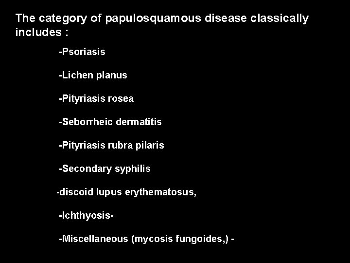
The category of papulosquamous disease classically includes : -Psoriasis -Lichen planus -Pityriasis rosea -Seborrheic dermatitis -Pityriasis rubra pilaris -Secondary syphilis -discoid lupus erythematosus, -Ichthyosis-Miscellaneous (mycosis fungoides, ) -
Psoriasis
Prevalence • Psoriasis occurs in 2%(1 -3%) of the world’s population • Equal frequency in males and females • May occur at any age from infancy to the 10 th decade of life • First signs of psoriasis – Females mean age of 27 years – Males mean age of 29 years 5
Prevalence • Two-thirds of patients have mild disease • One-third have moderate to severe disease • Early onset (prior to age 15) – Associated with more severe disease – More likely to have a positive family history • Life-long disease – Remitting and relapsing unpredictably – Spontaneous remissions of up to 5 years have been reported in approximately 5% of patients 6
Genetics and Pathogenesis • Psoriasis and the Immune System – The major histocompatibility complex (MHC) • Short arm of chromosome 6 – Histocompatibility Antigens (HLA) • HLA-Cw 6 • HLA-B 13, -B 17, -B 37, -Bw 16 – T-lymphocyte-mediated mechanism 8
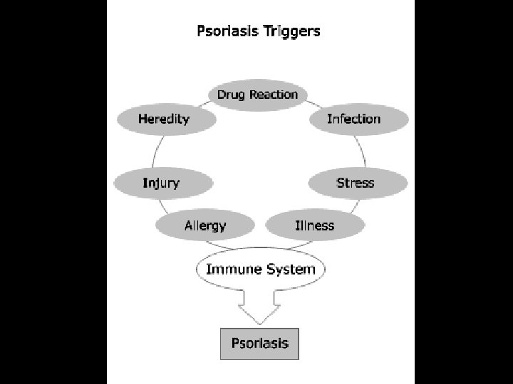
Triggering factors> • Infections- streptococcal pharyngitis/tonsillitis • Drugs- NSAID, beta blockers, lithium, antimalarials, corticosteroids • Trauma- Koebner phenomenon • Pregnancy • Stress • Alcohol • Sunlight- worsening in ~10% of patients although majority beneficial to sun exposure
Psoriasis : clinical features Well defined & circumscribed plaque • Erythematous • Silvery scaling • Symmetrical • Extensors of limbs, scalp, sacral area q. Auspitz’s sign: • light scraping of the scale with a wooden spatula produces multiple bleeding points • extreme thinning of the epidermis over the capillary laden dermal papillae •
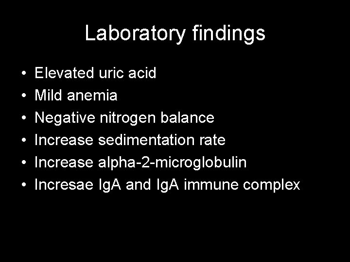
Psoriasis as a Systemic Disease • • Koebner Phenomenon Elevated ESR Increased uric acid levels → gout Mild anemia Elevated α 2 -macroglobulin Elevated Ig. A levels Increased quantities of Immune Complexes 12
Clinical Variants of Psoriasis 13
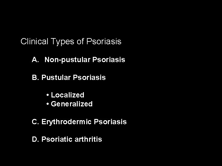
Clinical Types of Psoriasis A. Non-pustular Psoriasis B. Pustular Psoriasis • Localized • Generalized C. Erythrodermic Psoriasis D. Psoriatic arthritis
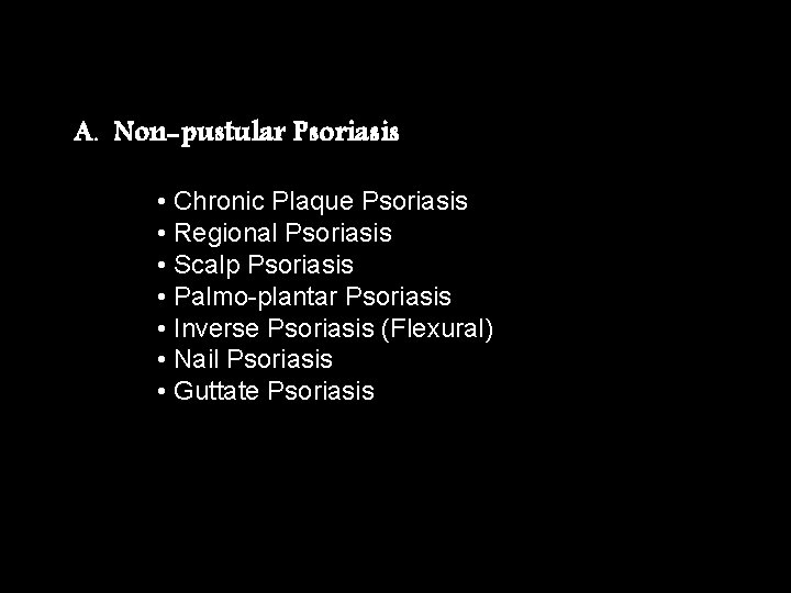
A. Non-pustular Psoriasis • Chronic Plaque Psoriasis • Regional Psoriasis • Scalp Psoriasis • Palmo-plantar Psoriasis • Inverse Psoriasis (Flexural) • Nail Psoriasis • Guttate Psoriasis
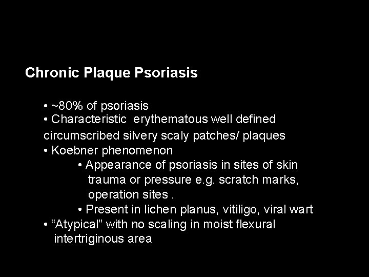
Chronic Plaque Psoriasis • ~80% of psoriasis • Characteristic erythematous well defined circumscribed silvery scaly patches/ plaques • Koebner phenomenon • Appearance of psoriasis in sites of skin trauma or pressure e. g. scratch marks, operation sites. • Present in lichen planus, vitiligo, viral wart • “Atypical” with no scaling in moist flexural intertriginous area
Chronic Plaque Psoriasis • • Most Common Variant Plaques may be as large as 20 cm Symmetrical disease Sites of Predilection – Elbows – Knees – Presacrum – Scalp – Hands and Feet 17
Chronic Plaque Psoriasis • May be widespread – up to 80% BSA • Genitalia involved in up to 30% of patients • Most patients have nail changes – Nail pitting – “Oil Spots” – Involvement of the entire nail bed • Onychodystrophy • Loss of nail plate 18
q. Auspitz’s sign: • light scraping of the scale with a wooden spatula produces multiple bleeding points • extreme thinning of the epidermis over the capillary laden dermal papillae
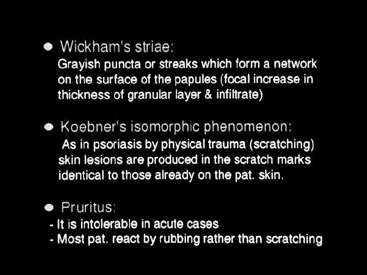
Koebner phenomenon • Appearance of psoriasis in sites of skin trauma or pressure e. g. scratch marks, operation sites. • Present in : lichen planus, vitiligo, viral wart
Scalp Psoriasis • Common • Well demarcated erythematous silvery scaly plaque with normal skin intervening • Post-auricular area commonly involved • Non scarring alopecia when severe, regrow when condition improve • DDx with seborrhoeic dermatitis by presence of typical plaque elsewhere +/- psoriatic nail changes
Palmoplantar Psoriasis • Common • Indurated heavily scaled plaque +/fissuring • Well-demarcated • DDx with foot/ hand eczema
Nail Psoriasis • ~50% of psoriasis have nail changes • Pitting • Onycholysis (separation of nail plate from nail bed) • Oil drop sign (a yellow brown, subungual spot surrounded by erythema) • Subungual hyperkeratosis • Secondary onychomycosis is common
Guttate Psoriasis • Latin “gutta” means a drop • Mainly affects children and young adults • Characterized by numerous 0. 5 to 1. 5 cm papules /plaques • Very small plaques generalized with centripetal distribution • May coalescent into larger plaques • Preceded by streptococcal tonsillitis or pharyngitis 2 weeks before onset • Spontaneous remissions in children • Often chronic in adults
Flexural Psoriasis • Psoriasis affecting axillae, perineum and umbilicus • Atypical psoriasis as friction & humidity removes the scale (diagnostic confusion) • Irritating when sweating
Life–Threatening Forms of Psoriasis • Generalized Pustular Psoriasis • Erythrodermic Psoriasis 26
Palmoplantar Pustular Psoriasis • Relatively uncommon variant • Painful, sterile pustules develop within plaque at palms and soles • Pustules resolve to leave post inflammatory hyperpigmentation • Female predominance • 20% associated with psoriasis elsewhere • Almost exclusively associated with smoking • Resistant to topical treatment
Generalized Pustular Psoriasis • Von Zumbusch’s disease • Erythematous edematous plaques studded with monomorphic sterile pustules. often after short episodes of fever of 39˚ to 40˚C • Weight loss , Muscle Weakness, Hypocalcemia Leukocytosis , Elevated ESR • Precipitated by : • withdrawal of oral steroid or widespread use of ultra potent topical steroid • pregnancy • Can be life-threatening due to fluid loss, sepsis
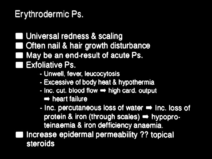
Erythrodermic Psoriasis • >90% of BSA affected • Life threatening with transcutaneous fluid loss, temperature dysregulation, sepsis, high output, cardiac failure • Other DDx of erythrodermaatopic eczema, drug eruption , cutaneous T cell lymphoma, pityriasis rubra pilaris
Erythrodermic Psoriasis • Triggering Factors – Systemic Infection – Withdrawal of high potency topical or oral steroids – Withdrawal of Methotrexate – Phototoxicity – Irritant contact dermatitis 31
Drug-provoked (Induced)psoriasis: Reported agent MOST COMMONLY ASSOCIATED AGENTS Beta blockers Lithium Antimalarial. A nti-Inflammatory Drugs Nonsteroidal Anti-Inflammatory Drugs
Psoriatic Arthritis • ~10% of chronic plaque psoriasis • Only ~15% of cases with skin and joint disease begin simultaneously • ~60% of skin disease precedes arthritis • ~25% of arthritis precedes skin disease • Probably a positive correlation between severity of skin disease and arthritis developing • Association: HLA B 27: sacro-ileitis; • B 38 and DR 7: peripheral arthritis; • B 39: all types; • DR 4: symmetrical arthritis
5 Types of Psoriatic Arthropathy • Classical distal arthropathy-distal IP joint • Seronegative RA-like polyarthritis • Oligoarticular asymmetrical arthritis • Spondyloarthropathy- Ankylosing spondylitislike • Arthritis mutilans
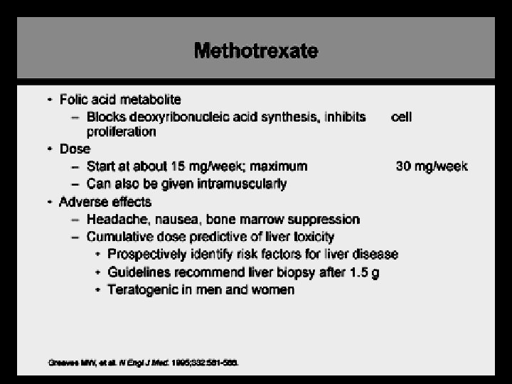
Psoriatic Arthritis treatment • NASIDs- may exacerbate psoriasis • Methotrexate • Sulphasalazine • Cyclosporine • Systemic steroid- may make the skin lesions more difficult to control • Biologics
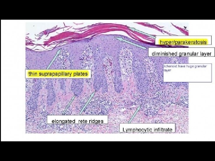
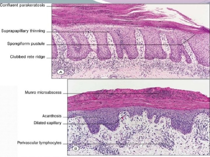
Histopathological changes • Inflammation • Epidermal keratinocyte hyperproliferation • ( parakeratosis) incomplete cornification of keratinocytes with retention of nuclei • (acanthosis), irregular thickening of the epidermis over the rete ridges but thinning over dermal papillae • (munro abscesses) epidermal polymorphonuclear leucocyte infiltrates • Vascular proliferation : dilated capillary loops in the dermal papillae
Laboratory findings • • • Elevated uric acid Mild anemia Negative nitrogen balance Increase sedimentation rate Increase alpha-2 -microglobulin Incresae Ig. A and Ig. A immune complex
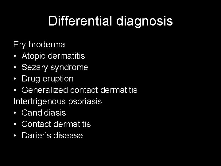
Differential diagnosis Erythroderma • Atopic dermatitis • Sezary syndrome • Drug eruption • Generalized contact dermatitis Intertrigenous psoriasis • Candidiasis • Contact dermatitis • Darier’s disease
Differential diagnosis Psoriasis vulgaris • Nummular eczema • Mycosis fungoides, plaque stage • Tinea corporis Guttate psoriasis • Pityriasis rosea • Pityriasis lichenoides et varioliformis • Syphillis • Tinea corporis
Differential diagnosis Nail psoriasis • Tinea ungium • Dyskeratosis : secondary to injury Scalp and face • Seborrheic dermatitis Genitalia • In situ squamous cell CA
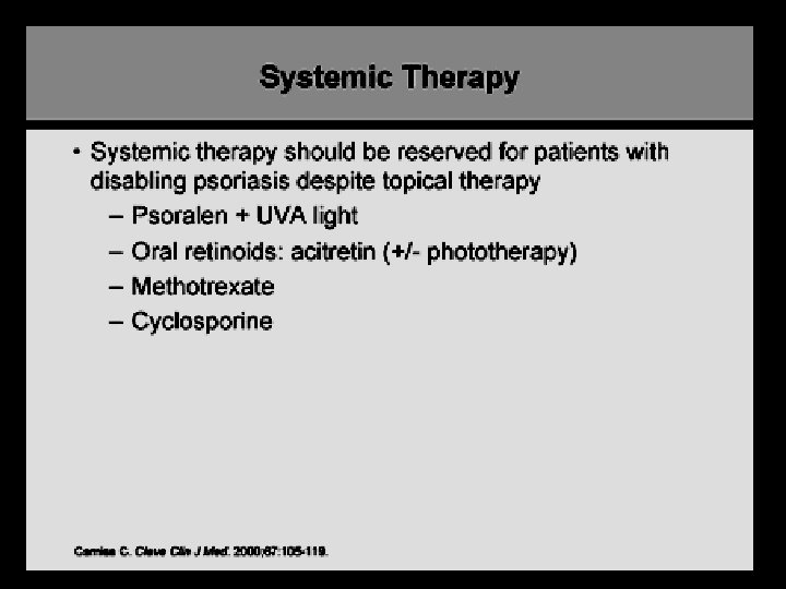
Treatment Options • Monotherapy • Combination therapy • Rotational therapy • Sequential therapy
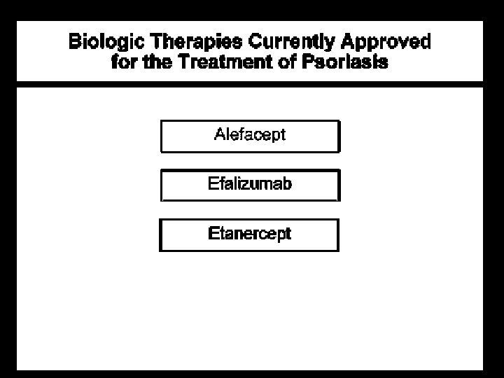
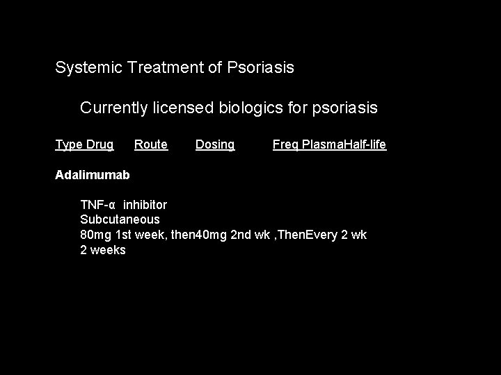
Systemic Treatment of Psoriasis Currently licensed biologics for psoriasis Type Drug Route Dosing Freq Plasma. Half-life Adalimumab TNF-α inhibitor Subcutaneous 80 mg 1 st week, then 40 mg 2 nd wk , Then. Every 2 wk 2 weeks
Etanercept Subcutaneous 50 mg (0. 8 mg/kg, max 50 mg) First 12 week: twice a wk Then once aweek 70 hours
Infliximab IL-12/23 Antibody Intravenous 5 mg/kg 0, 2, 6 wk, then every 8 wk 8 -9. 5 days
Ustekinumab Subcutaneous 45 mg for <100 kg 90 mg for >100 kg 0, 4 wk, thenevery 12 wk 15 -32 days
Absolute Contraindications: Pregnancy/breastfeeding Active (chronic) infections (including tuberculosis and active chronic hepatitis B) Congestive heart failure (NYHA grade III or IV) Relative contraindications History of recurrent infections PUVA >200 treatments (especially if followed by cyclosporin use) HIV or AIDS Hepatitis C Congestive heart failure (NYHA grade I or II) SLE, Demyelinating disease Malignancies or lymphoproliferative disorders Live vaccines
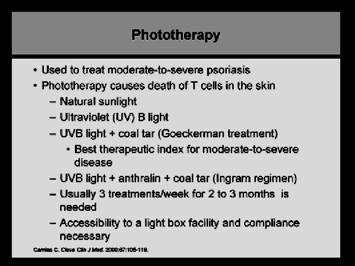
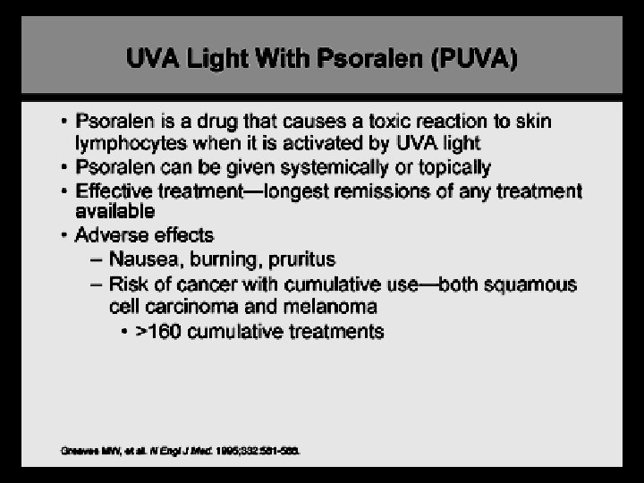
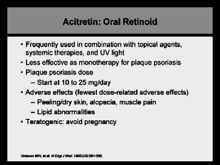
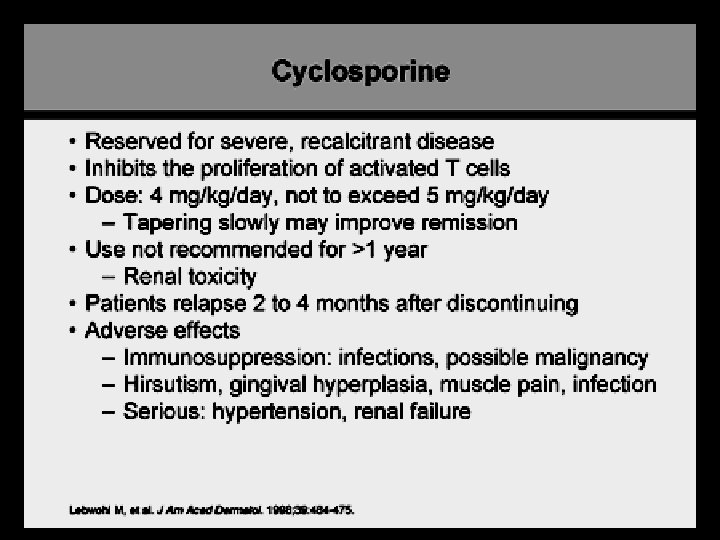
Treatment Modalities: • Combination therapy • -contraindicated in additive increase risk/ S/E, e. g. • Phototherapy +Cs. A increase risk of cutaneous cancer • Acitretin +MTX increase risk of hepatotoxicity
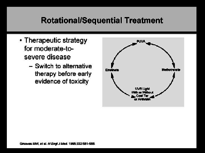
Treatment Modalities: • Rotational therapy - use of therapies for a specified period (e. g. 1 -2 year) then rotate to an alternative therapy to minimize long-term toxicity in any given therapy and decrease therapy “resistance/ tachyphylaxis
Treatment Modalities: • Sequential therapy • Induction phase: use stronger potentially more toxic agents to clear psoriasis initially • e. g. Ultrapotent topical steroid or Cs. A • Transitional phase • e. g. OM steroid+ Nocte calcipotriol or acitretin • Maintenance phase: use of a “weaker”, less toxic agent for maintenance • e. g. weekday calcipotriol +weekend steroid or acitretin +/-UVB/ PUVA
PITYRIASIS ROSEA
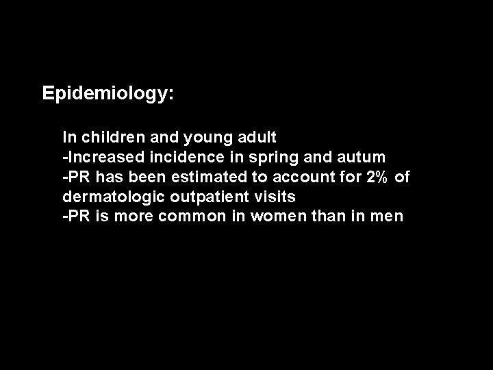
-Epidemiology: In children and young adult -Increased incidence in spring and autum -PR has been estimated to account for 2% of dermatologic outpatient visits -PR is more common in women than in men
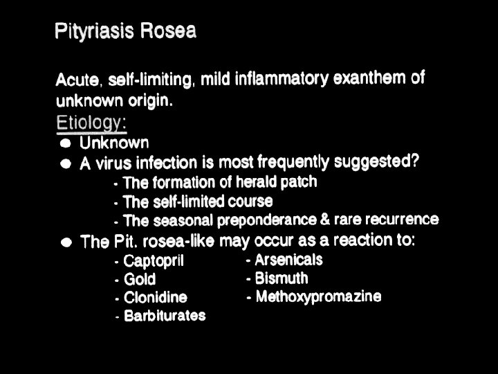
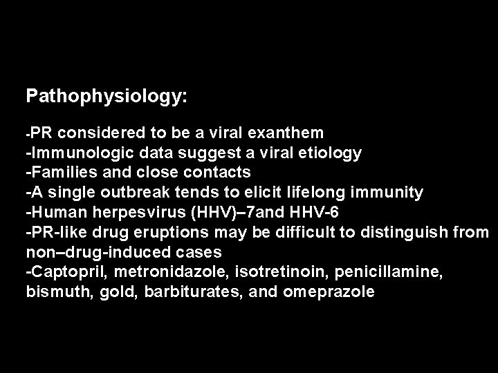
Pathophysiology: -PR considered to be a viral exanthem -Immunologic data suggest a viral etiology -Families and close contacts -A single outbreak tends to elicit lifelong immunity -Human herpesvirus (HHV)– 7 and HHV-6 -PR-like drug eruptions may be difficult to distinguish from non–drug-induced cases -Captopril, metronidazole, isotretinoin, penicillamine, bismuth, gold, barbiturates, and omeprazole
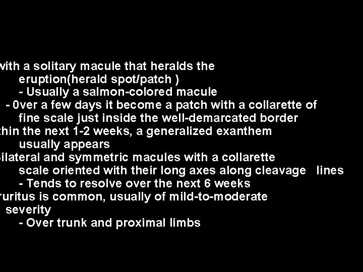
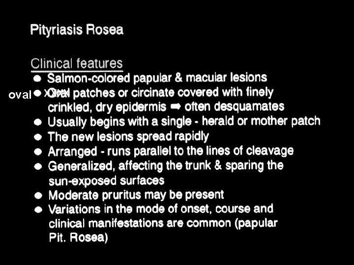
with a solitary macule that heralds the eruption(herald spot/patch ) - Usually a salmon-colored macule - 0 ver a few days it become a patch with a collarette of fine scale just inside the well-demarcated border thin the next 1 -2 weeks, a generalized exanthem usually appears Bilateral and symmetric macules with a collarette scale oriented with their long axes along cleavage lines - Tends to resolve over the next 6 weeks ruritus is common, usually of mild-to-moderate severity - Over trunk and proximal limbs
oval XXXX
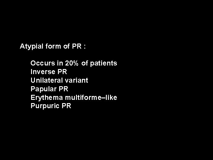
Atypial form of PR : Occurs in 20% of patients Inverse PR Unilateral variant Papular PR Erythema multiforme–like Purpuric PR
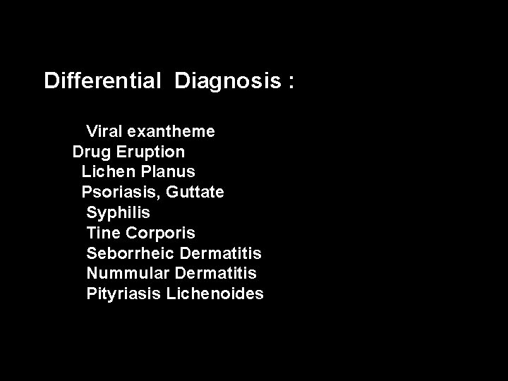
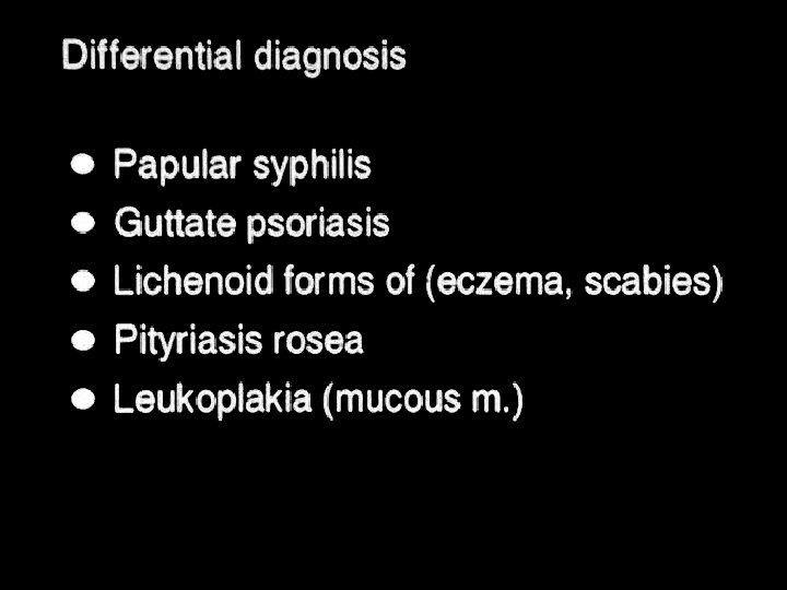
Differential Diagnosis : Viral exantheme Drug Eruption Lichen Planus Psoriasis, Guttate Syphilis Tine Corporis Seborrheic Dermatitis Nummular Dermatitis Pityriasis Lichenoides
Lichen planus (LP)
Background: -Lichen planus (LP) is a pruritic, papular eruption characterized by its violaceous color; polygonal shape; and, sometimes, fine scale -It is most commonly found on the flexor surfaces of the upper extremities, on the genitalia, and on the mucous membranes.
Epidemiology : -Approximately 1% of all new patients seen at health care clinics -Rare in children -F=M -No racial predispositions have been noted -LP can occur at any age but two thirds of patients are aged 30 -60 years
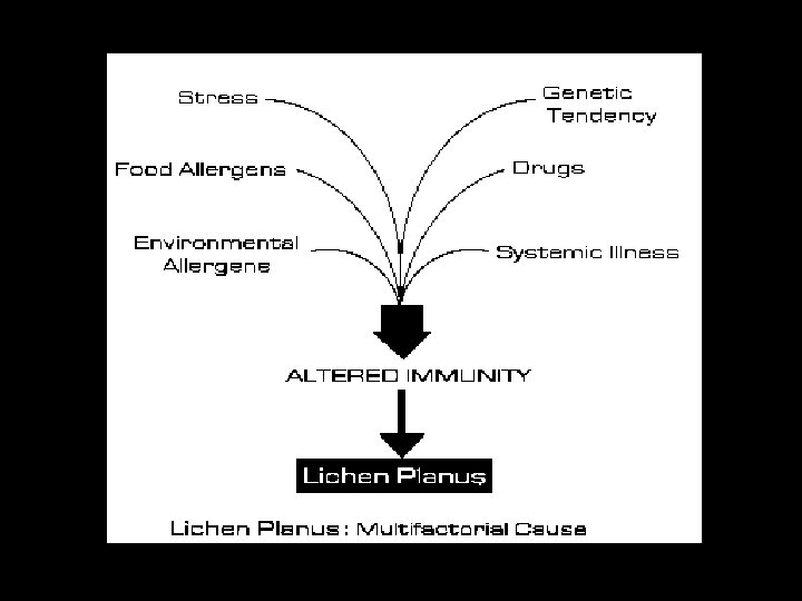
Pathophysiology : The cause of LP is unknown -LP may be a cell-mediated immune response of unknown origin -LP may be found with other diseases of altered immunity like ulcerative colitis, alopecia areata, vitiligo, dermatomyositis -An association is noted between LP and hepatitis C virus infection , chronic active hepatitis, and primary biliary cirrhosis -Familial cases -Drug may induce lichenoid reaction like thiazide, antimalarials, propranolol
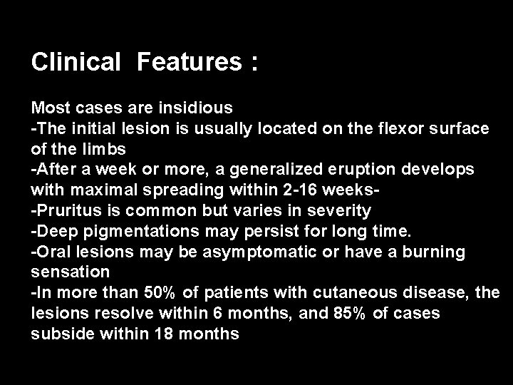
Clinical Features : Most cases are insidious -The initial lesion is usually located on the flexor surface of the limbs -After a week or more, a generalized eruption develops with maximal spreading within 2 -16 weeks-Pruritus is common but varies in severity -Deep pigmentations may persist for long time. -Oral lesions may be asymptomatic or have a burning sensation -In more than 50% of patients with cutaneous disease, the lesions resolve within 6 months, and 85% of cases subside within 18 months
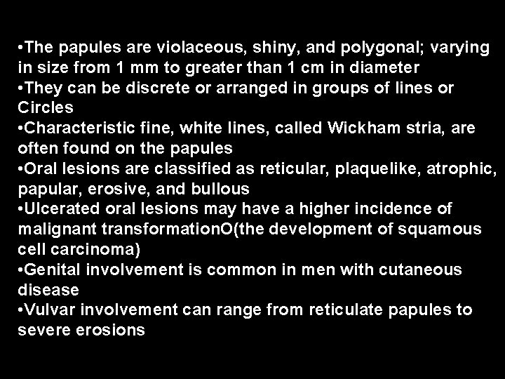
• The papules are violaceous, shiny, and polygonal; varying in size from 1 mm to greater than 1 cm in diameter • They can be discrete or arranged in groups of lines or Circles • Characteristic fine, white lines, called Wickham stria, are often found on the papules • Oral lesions are classified as reticular, plaquelike, atrophic, papular, erosive, and bullous • Ulcerated oral lesions may have a higher incidence of malignant transformation. O(the development of squamous cell carcinoma)k for • Genital involvement is common in men with cutaneous disease • Vulvar involvement can range from reticulate papules to severe erosions
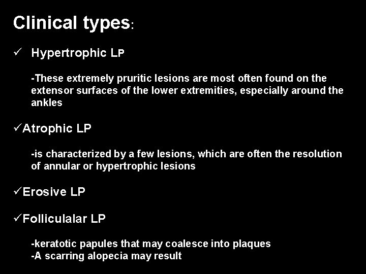
Clinical types: ü Hypertrophic LP -These extremely pruritic lesions are most often found on the extensor surfaces of the lower extremities, especially around the ankles üAtrophic LP -is characterized by a few lesions, which are often the resolution of annular or hypertrophic lesions üErosive LP üFolliculalar LP -keratotic papules that may coalesce into plaques -A scarring alopecia may result
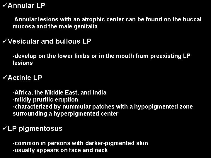
üAnnular LP -Annular lesions with an atrophic center can be found on the buccal mucosa and the male genitalia üVesicular and bullous LP -develop on the lower limbs or in the mouth from preexisting LP lesions üActinic LP -Africa, the Middle East, and India -mildly pruritic eruption -characterized by nummular patches with a hypopigmented zone surrounding a hyperpigmented center üLP pigmentosus -common in persons with darker-pigmented skin -usually appears on face and neck
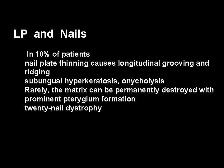
LP and Nails IIn 10% of patients nail plate thinning causes longitudinal grooving and ridging subungual hyperkeratosis, onycholysis Rarely, the matrix can be permanently destroyed with prominent pterygium formation twenty-nail dystrophy
Management : self-limited disease that usually resolves within 8 -12 months -Anti-histamine -topical steroids, particularly class I or II ointments -systemic steroids for symptom control and possibly more rapid resolution -Oral acitretin -Photo-therapy -Others