Orientation History taking and eye examination Essam Osman




- Slides: 107
Orientation, History taking, and eye examination Essam Osman PROFESSOR College Of Medicine King Saud University
Orientation Course OPH 432
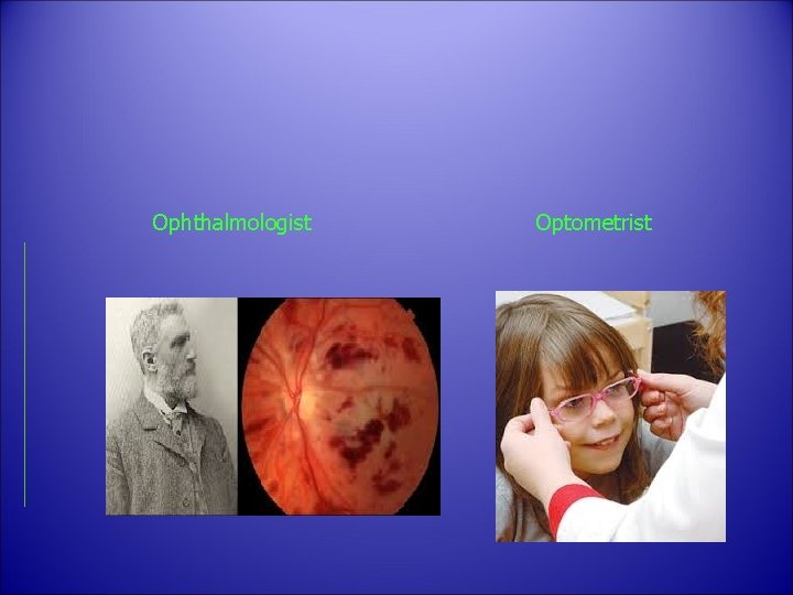
Ophthalmologist S Optometrist
Why should you be interes ted in the eye? Internet is a window to the world
Objectives a. To know the basic ophthalmic anatomy and physiology. b. To recognize assessment and management of common ophthalmic diseases.
Objectives c. To know how to handle common ophthalmic emergencies. d. To handle simple ophthalmic diagnostic instruments. e. To be aware of common ophthalmic operations.
Components of the course • Lectures • Clinical sessions • ER
Lectures 1. History taking and physical examination in ophthalmology 2. Basic Anatomy and Physiology of the Eye 3. Lid, Lacrimal, and Orbit Disorders 4. Ocular emergencies and red eye 5. Strabismus, Amblyopia and Leukocoria 6. Acute Visual Loss
Lectures 7. Chronic Visual Loss 8. Refractive Errors 9. Ocular manifestations of systemic diseases 10. Neuro-ophthalmology 11. Ocular Pharmacology and Toxicology
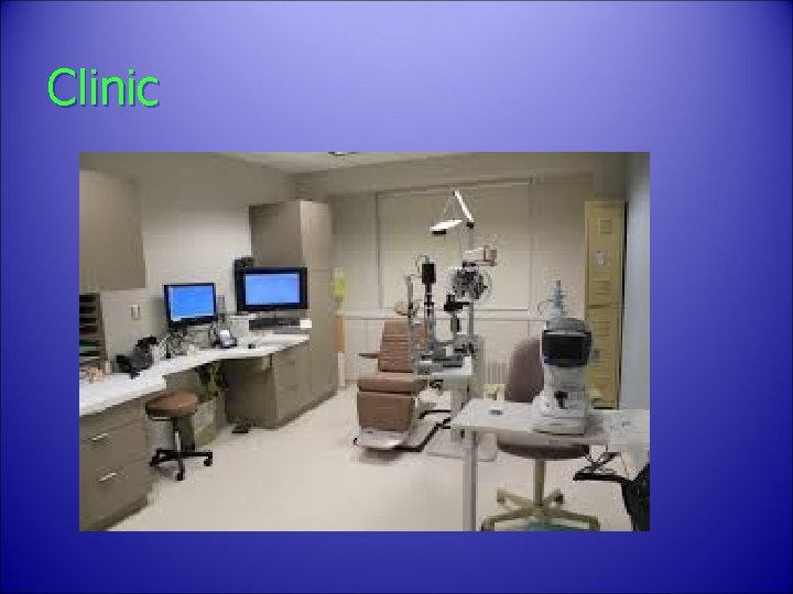
Clinic
Clinical skill sessions v External Ocular Examination, Ocular motality and Alignment v Visual acuity and Ophthalmoscopy v Visual field, Tonometry, Pupil Examination
Marks distribution • 30 marks for the MCQs • 40 short answer question One day exam • 20 marks for the OSCE (2 -3 cases) One day exam • 10 evaluation in clinical sessions
Recommended textbooks 1. Required Text(s) a. Lecture notes Ophthalmology (latest edition) By: Bruce James (published by Blackwell Science) b. Basic Ophthalmology (latest edition) By: Cynthia A. Bradford (latest edition) (published by American Academy of Ophthalmology) c. Practical Ophthalmology: A manual for Beginning Residents (latest edition) By: Fred M. Wilson (published by American Academy of Ophthalmology
2. References • Vaughan and Asbury’s general Ophthalmology • By: Paul Riordan-Eva (published by LANGE) Clinical Ophthalmology: A Systematic Approach By : Jack T. Kanski (published by Butterworth Heinemann) Ophtho. Book www. Ophtho. Book. com Written and Illustrated by Timothy Root, M. D.
c. Electronic Materials, Web Sites 1. Department internet website. 2. Department’s teaching staff personal websites on University site. 3. University and KKUH/KAUH Library. 4. Audiovisual Unit of the Ophthalmology Department. 5. Pub. Med 5. Medscape 6. The digital journal of ophthalmology (www. djo. harvard. edu)
EYE
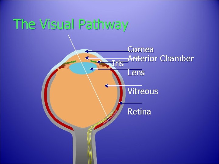
The Visual Pathway Iris Cornea Anterior Chamber Lens Vitreous Retina
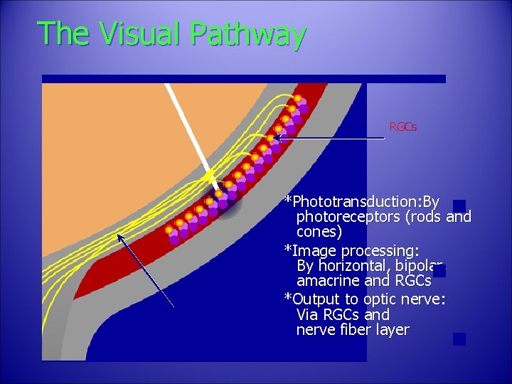
The Visual Pathway RGCs Nerve Fibers *Phototransduction: By photoreceptors (rods and cones) *Image processing: By horizontal, bipolar, amacrine and RGCs *Output to optic nerve: Via RGCs and nerve fiber layer
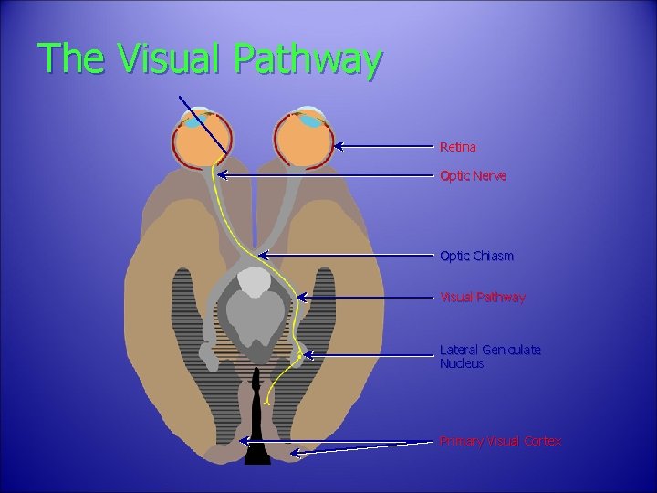
The Visual Pathway Retina Optic Nerve Optic Chiasm Visual Pathway Lateral Geniculate Nucleus Primary Visual Cortex
Diagnostically and functionally, it is the most important square inch of the body surface.
• The eye is so intimately connected with the rest of the body that it reveals enormous amount of general information. • Eye is the only part of the body where blood vessels and central nervous system tissues can be viewed directly.
Examples
Neurological connections
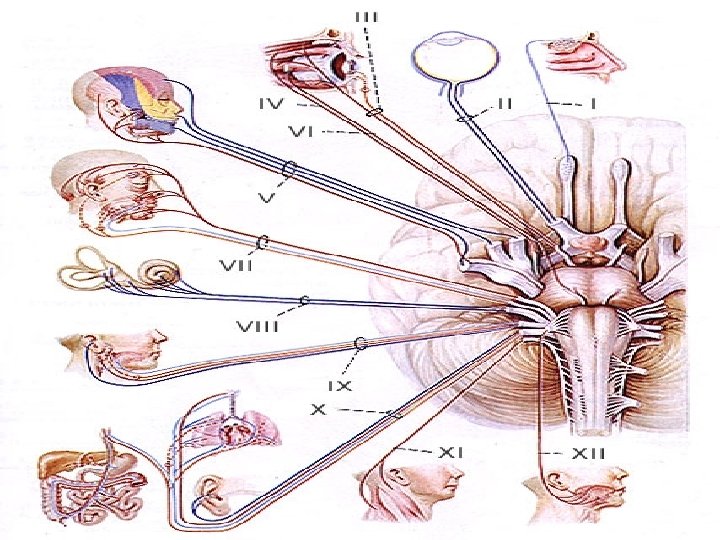
• The 12 cranial nerves provide us with a large part of our information about the brain. Of these , the eye examination evaluates CN II, IV, V, VII, VIII. • In addition, it provides information about the autonomic pathways. (sympathetic /parasympathetic)
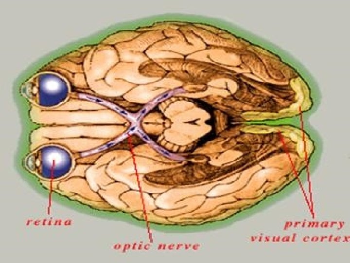
The best known connection between the brain and the eye is the ON. The visual pathways, which extends from front to back across the brain can be studied easily and safely using perimetry. It can differentiates accurately between lesions of the temporal, parietal, and occipital lobes.
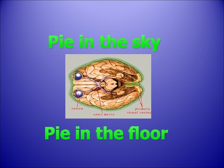
Pie in the sky Pie in the floor
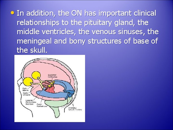
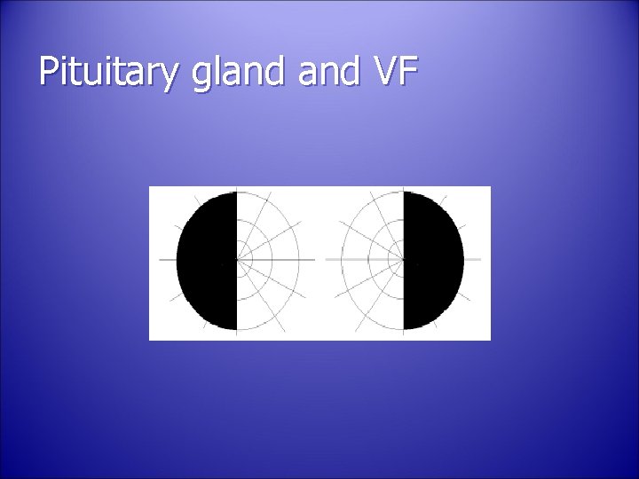
• In addition, the ON has important clinical relationships to the pituitary gland, the middle ventricles, the venous sinuses, the meningeal and bony structures of base of the skull.
Pituitary gland VF
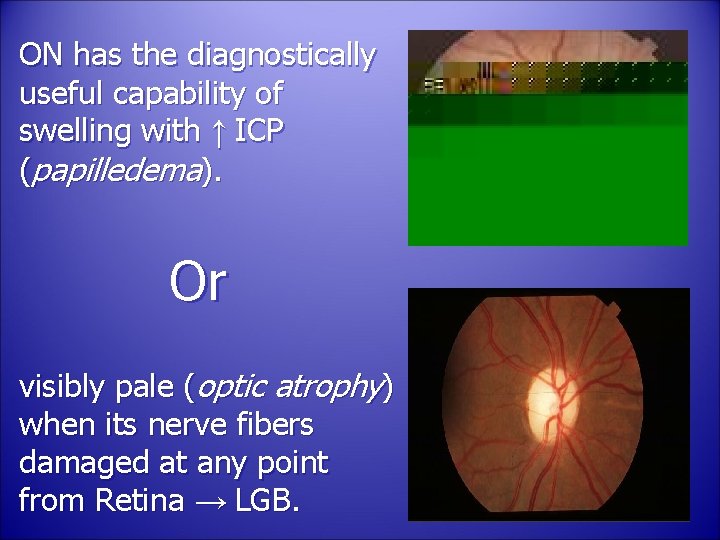
ON has the diagnostically useful capability of swelling with ↑ ICP (papilledema). Or visibly pale (optic atrophy) when its nerve fibers damaged at any point from Retina → LGB.
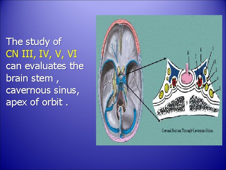
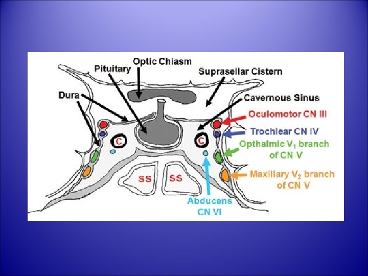
The study of CN III, IV, V, VI can evaluates the brain stem , cavernous sinus, apex of orbit.
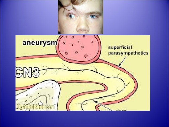
Unilateral dilated pupil after head injury can occur due to pressure on pupil constrictor fibers of CN III. CN VI involved in mastoid infection (petrous ridge) Parotid gland, Inner ear disease → CN VII. Nystagmus → CN VIII
Focal brain lesions like: Vascular occlusions Hemorrhage Neoplasm Diffuse brain lesions like: Infections Demyelinating disorders → nerve damage.
Vascular connections
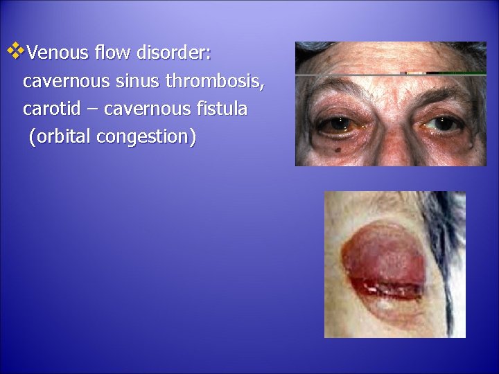
v. Venous flow disorder: cavernous sinus thrombosis, carotid – cavernous fistula (orbital congestion)
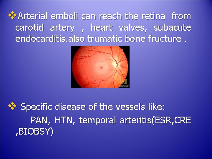
v. Arterial emboli can reach the retina from carotid artery , heart valves, subacute endocarditis. also trumatic bone fructure. v Specific disease of the vessels like: PAN, HTN, temporal arteritis(ESR, CRE , BIOBSY)
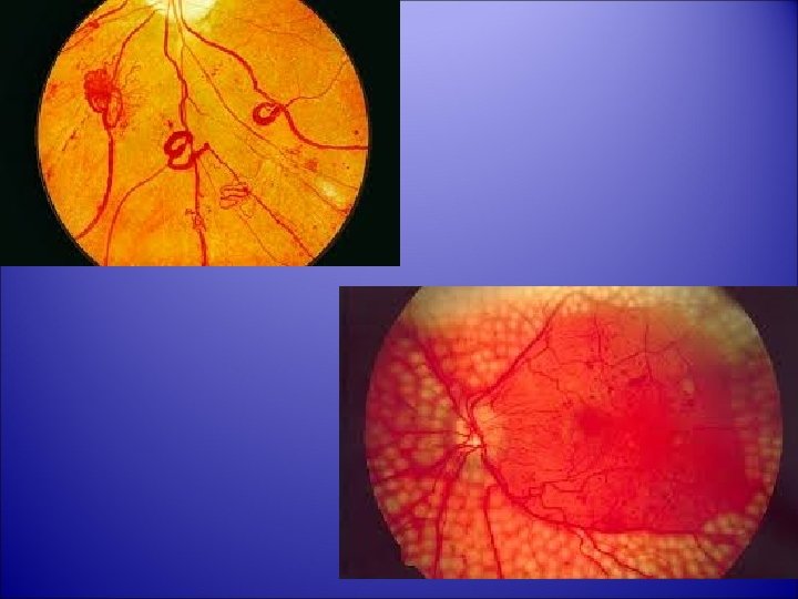
v. Hematological disorders of all types can manifest in the fundus. v. Almost all metabolic disorders can affects the eye: DM : DR, cataract, RE, ophthalmoplegia. Hypoparathyroidism : cataract Wilson’s disease.
v. Thyroid eye disease: Exophthalmos, Lid retraction. v. Infections: (Syphilis, Toxoplasmosis, Rubella)
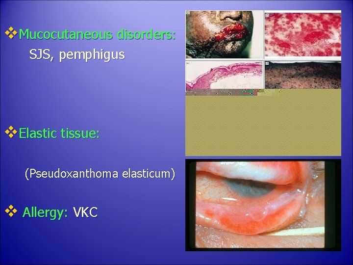
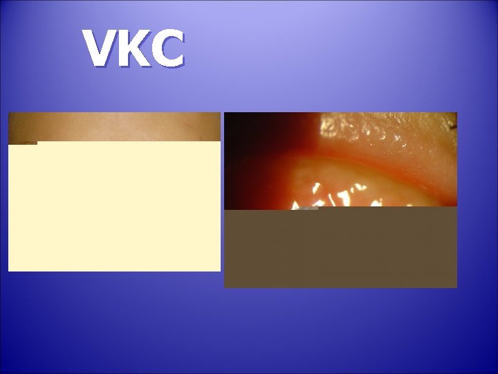
v. Mucocutaneous disorders: SJS, pemphigus v. Elastic tissue: (Pseudoxanthoma elasticum) v Allergy: VKC
VKC
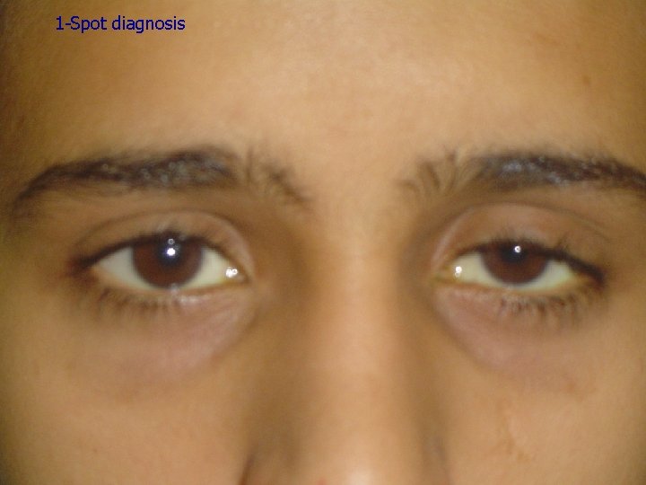
1 -Spot diagnosis
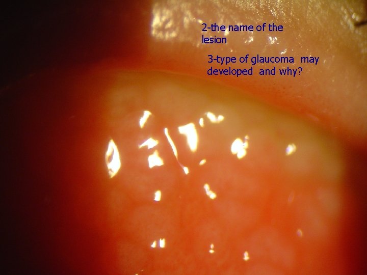
2 -the name of the lesion 3 -type of glaucoma may developed and why?
v. Chromosomal abnormalities: Trisomy: 13, 15, 21. v The eye is a delicate indicator of poisoning: -Morphine addict → meiotic pupil -Lead poisoning, vitamin A intoxication → papilledema
• I could go on and on about the discoveries possible in our most important square inch. ‘’EYE’’ 90% of our information reaches our brain via sight. Unfortunately, of all the parts of the body, the eye is the most vulnerable to minor injury.
What are the objectives of the comprehensive ophthalmic evaluation?
ØObtain an ocular and systemic history. ØDetermine the optical and health status of the eye and visual system. ØIdentify risk factors for ocular and systemic disease. ØDetect and diagnose ocular diseases.
ØEstablish and document the presence or absence of ocular symptoms and signs of systemic disease. ØDiscuss the nature of the findings and the implications with the patient. ØInitiate an appropriate response. e. g. further diagnostic tests, treatment, or referral.
HISTORY
• It is a gathering information process from the patient guided by an educated and active mind. • It is a selective guided and progressive elicitation and recognition of significant information • History by skilled person can arrive at the proper diagnosis in 90% of patients.
• It gives vital guidance for: (a) physical examination (b) laboratory work (c) Therapy v. Failure to take history can lead to missing vision or life threatening conditions.
Chief complaint: ’’The patient’s own words’’ ‘’she cannot see with the RE’’ You should not come to conclusion that her problem is nearsightedness and write down “Myopia of RE”. • The patient needs will not be satisfied until he/she has received an acceptable explanation of the meaning of the chief complaint and its proper management.
History of the Present Illness: • Detailed description of the chief complaint to understand the symptoms and course of the disorder. • Listen and question and then write down in orderly sequence that make sense to you.
* The time sequence when, How fast, what order did events occur? * Frequency, intermittency * location, Laterality * Severity * Associated symptoms * Documentation (old records, photo) e. g ptosis, proptosis, VII N palsy. Ø gradual painless decrease vision both eyes for 1 y. Ø Sudden painless decrease vision RE for 10 min.
“cannot see with RE”!! • ? Only distance vision blurred. • ? Blind spot is present in the center of VF • ? Right side of VF of the RE lost • ? Right VF of both eyes lost • ? A diffuse haze obscures the entire field of RE v. Each of these has different diagnostic implication v. Most pt. has difficulty providing precise and concise description
Disturbances of vision: • Blurred or decreased central vision • Decreased peripheral vision. • Altered image size. (micropsia, macropsia, metamorphopsia). • Diplopia (monocular, binocular) • Floaters • Photopsia (flash of light)
• Color vision abnormalities. • Dark adaptation problems. • Blindness (ocular, cortical). • Oscillopsia (shaking of images).
Ocular pain or discomfort: • Foreign body sensation • Ciliary pain (aching, severe pain in or around the eye, often radiating to the ipsilateral forehead, molar area) • Photophobia • Headache • Burning • Dryness • Itching: patient rub the eye vigorously (allergy) • Asthenopia (eye strain)
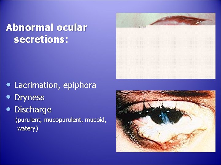
Abnormal ocular secretions: • Lacrimation, epiphora • Dryness • Discharge (purulent, mucoid, watery)
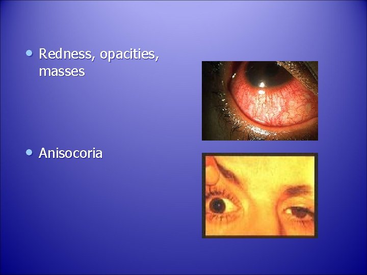
• Redness, opacities, masses • Anisocoria
Family history: • Many eye conditions are inherited RE, glaucoma, strabismus, retinoblastoma, neoplastic, vascular disorders • Familial systemic disease can be helpful in ophthalmic evaluation and diagnosis Atopy, thyroid diseases, DM, certain malignancies.
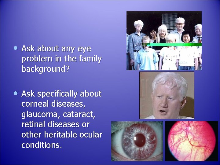
• Ask about any eye problem in the family background? • Ask specifically about corneal diseases, glaucoma, cataract, retinal diseases or other heritable ocular conditions.
• Ask questions designed to confirm or exclude your tentative diagnosis - significant positive - significant negative ‘’Significant is equal to expected’’ • predict the physical and lab. finding likely to be present. • any discrepancy between the history and physical examination requires explanation
Ophthalmic examination
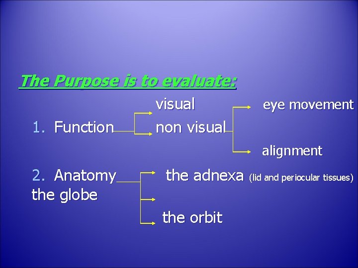
The Purpose is to evaluate: 1. Function visual non visual eye movement alignment 2. Anatomy the globe the adnexa (lid and periocular tissues) the orbit
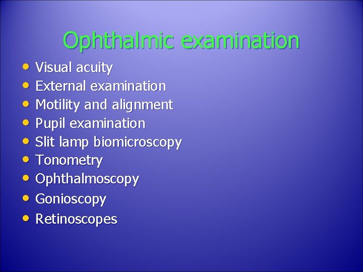
Ophthalmic examination • Visual acuity • External examination • Motility and alignment • Pupil examination • Slit lamp biomicroscopy • Tonometry • Ophthalmoscopy • Gonioscopy • Retinoscopes
Vision: • Vital sign (MUST) • Good vision intact neurological visual pathology structurally healthy eye Proper focus • Subjective
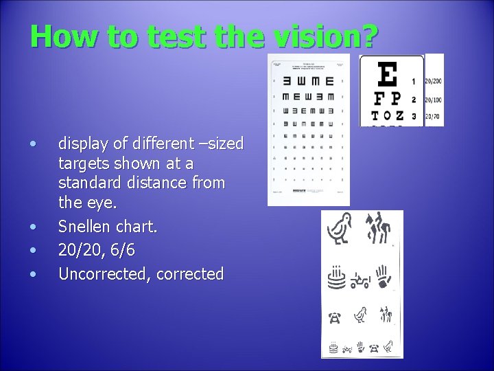
How to test the vision? • • display of different –sized targets shown at a standard distance from the eye. Snellen chart. 20/20, 6/6 Uncorrected, corrected
Testing poor vision: • If the patient is unable to read the largest letter <(20/200) • Move the patient closer e. g. 5/200 • If patient cannot read: count fingers (CF) hand motion (HM) Light perception (LP) No light perception (NLP)
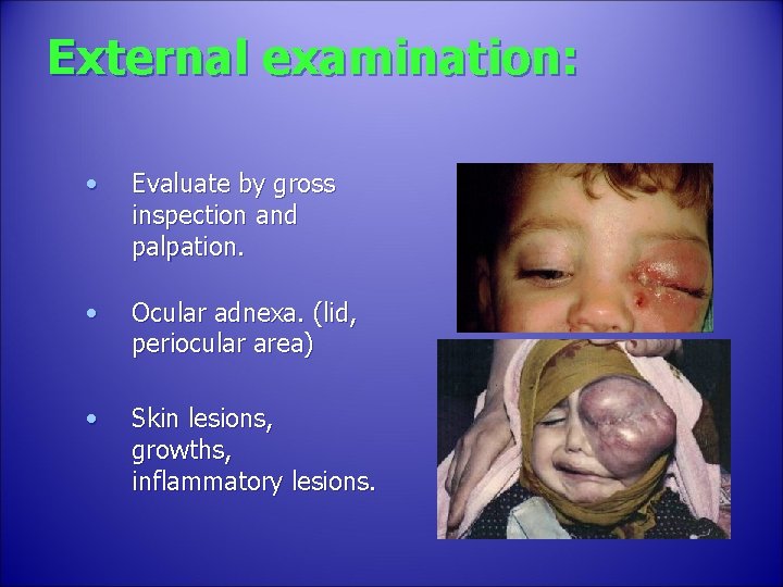
External examination: • Evaluate by gross inspection and palpation. • Ocular adnexa. (lid, periocular area) • Skin lesions, growths, inflammatory lesions.
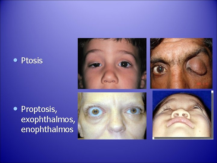
• Ptosis • Proptosis, exophthalmos, enophthalmos
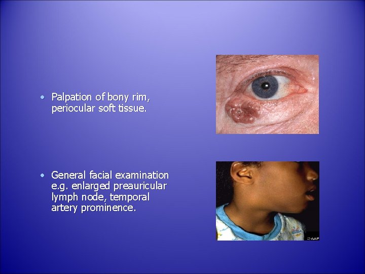
• Palpation of bony rim, periocular soft tissue. • General facial examination e. g. enlarged preauricular lymph node, temporal artery prominence.
Ocular motility: Evaluate - Alignment Movements
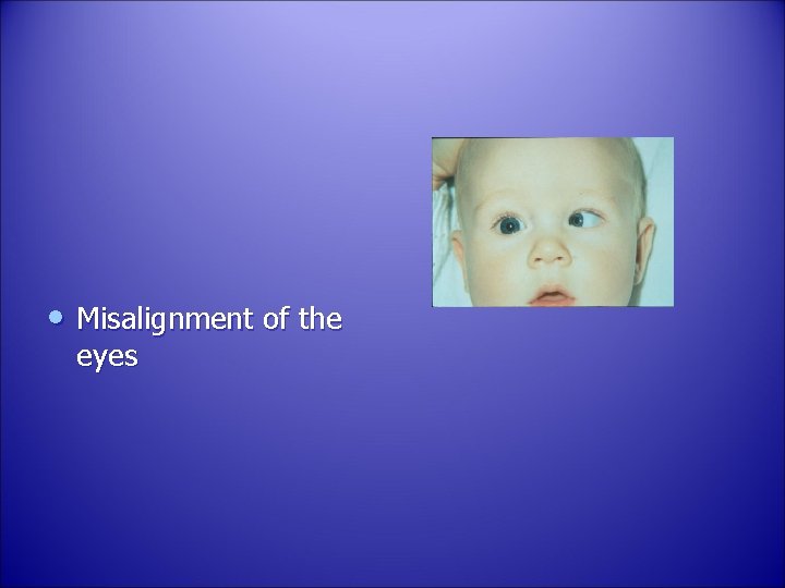
• Misalignment of the eyes
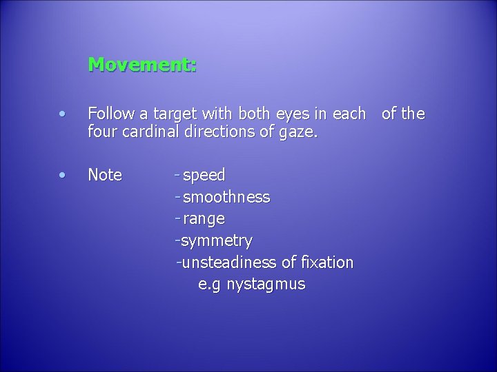
Movement: • Follow a target with both eyes in each of the four cardinal directions of gaze. • Note - speed - smoothness - range -symmetry -unsteadiness of fixation e. g nystagmus
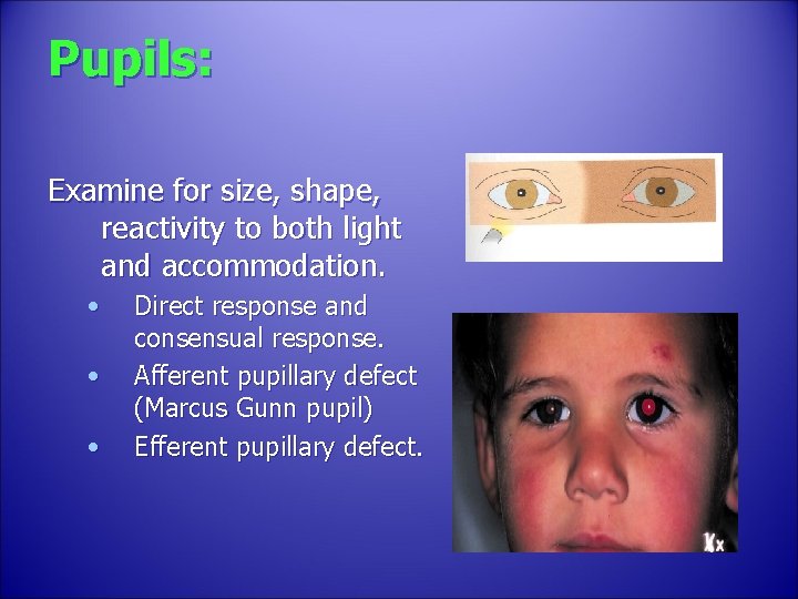
Pupils: Examine for size, shape, reactivity to both light and accommodation. • • • Direct response and consensual response. Afferent pupillary defect (Marcus Gunn pupil) Efferent pupillary defect.
• Pupillary abnormalities: neurologic disease previous inflammation – adhesion acute intraocular inflammation - spasm - atony prior surgical trauma effect of systemic or eye medication benign variation of normal
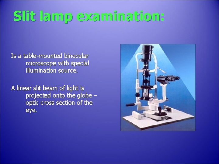
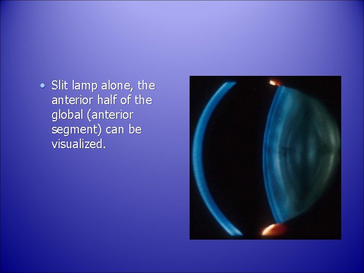
Slit lamp examination: Is a table-mounted binocular microscope with special illumination source. A linear slit beam of light is projected onto the globe – optic cross section of the eye.
• Slit lamp alone, the anterior half of the global (anterior segment) can be visualized.
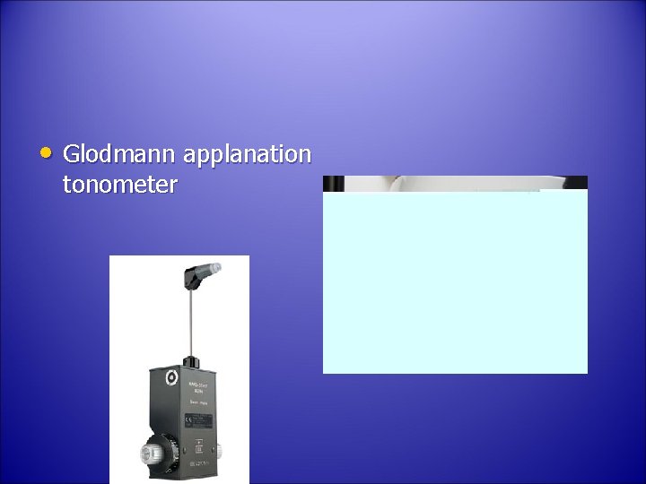
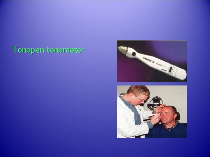
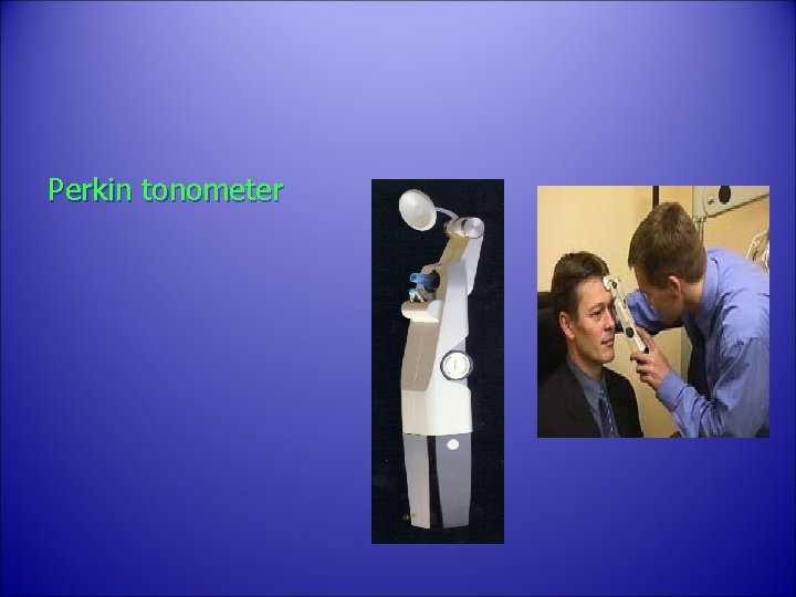
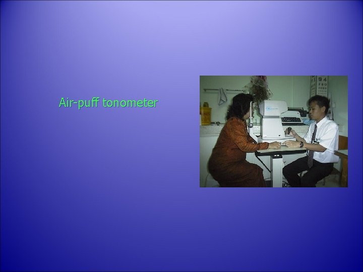
Tonometry: – The globe is a closed compartment with constant circulation of aqueous humor. – This maintains the shape, and relatively uniform pressure within the globe. – Normal pressure 10 – 21 mm. Hg.
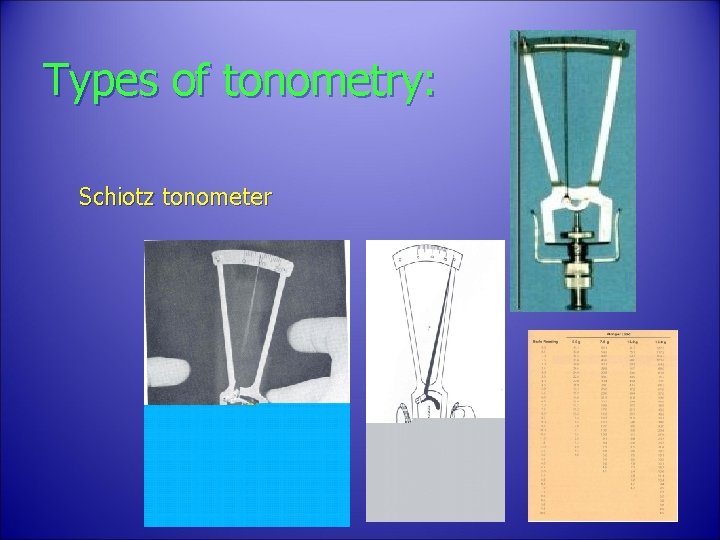
Types of tonometry: Schiotz tonometer
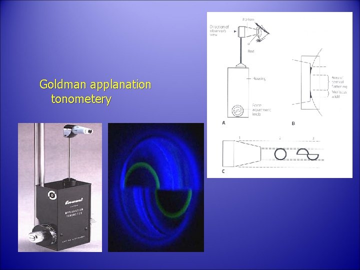
Goldman applanation tonometery
• Glodmann applanation tonometer
Tonopen tonometer
Perkin tonometer
Air-puff tonometer
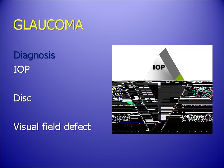
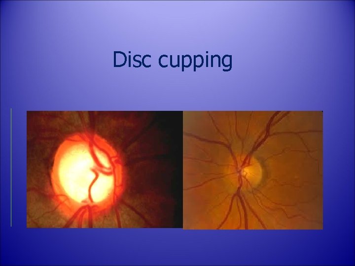
GLAUCOMA Diagnosis IOP Disc Visual field defect
Disc cupping
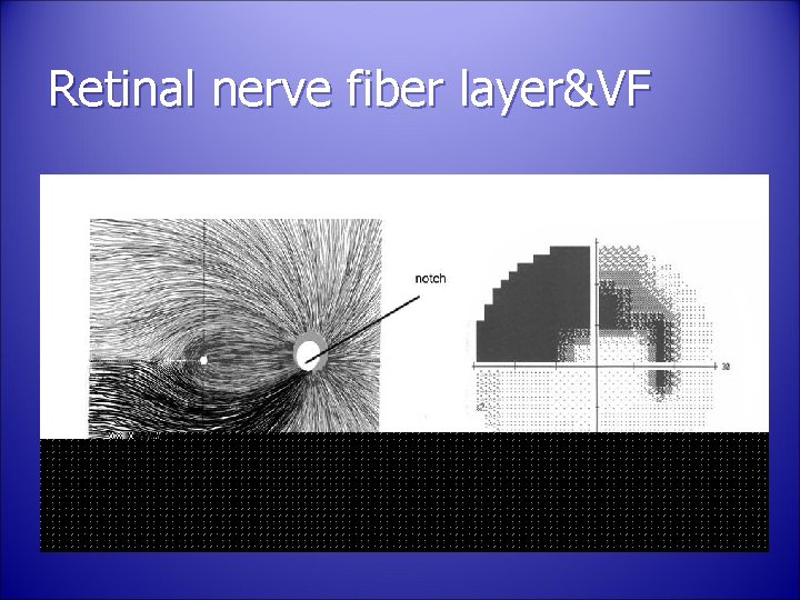
Retinal nerve fiber layer&VF
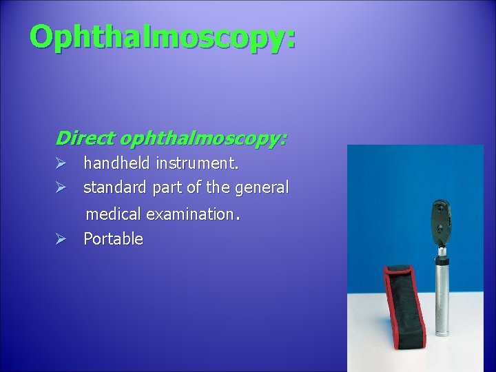
Ophthalmoscopy: Direct ophthalmoscopy: Ø handheld instrument. Ø standard part of the general medical examination. Ø Portable
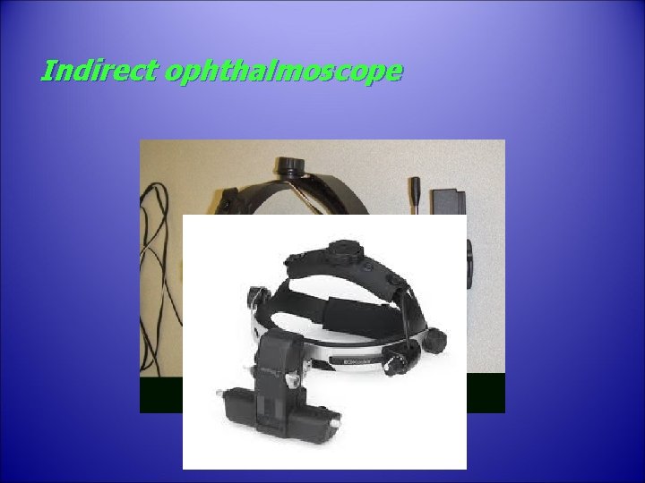
Indirect ophthalmoscope
Indirect Ophthalmoscoy: 1. 2. 3. 4. 5. provide much wider field of view less magnification (3. 5 X with 20 D lens) brighter light source – better view. Binocular – steroscopic view. Allow entire retina examination till the periphery.
Disadvantage: 1. Inverted retinal image. 2. Brighter light is uncomfortable to the patient.
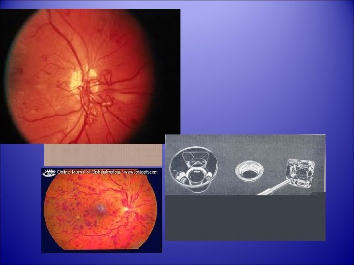
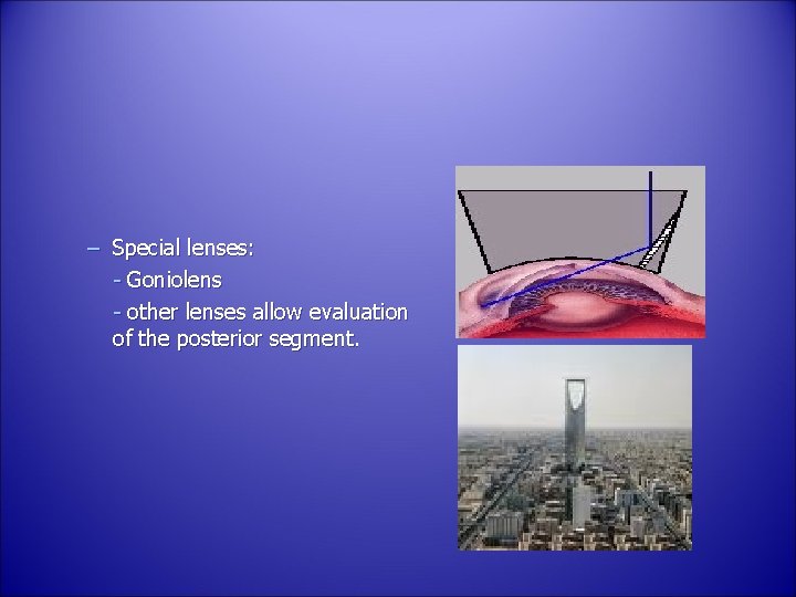
– Special lenses: - Goniolens - other lenses allow evaluation of the posterior segment.
IMPORTANCE OF GONIOSCOPY
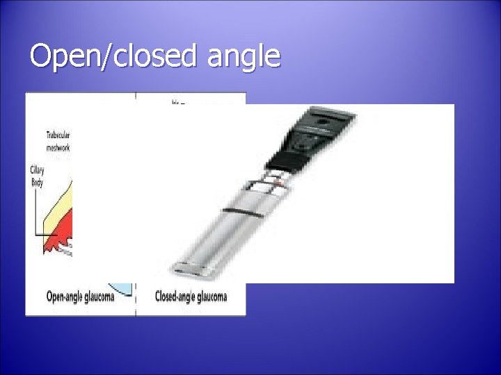
Open/closed angle
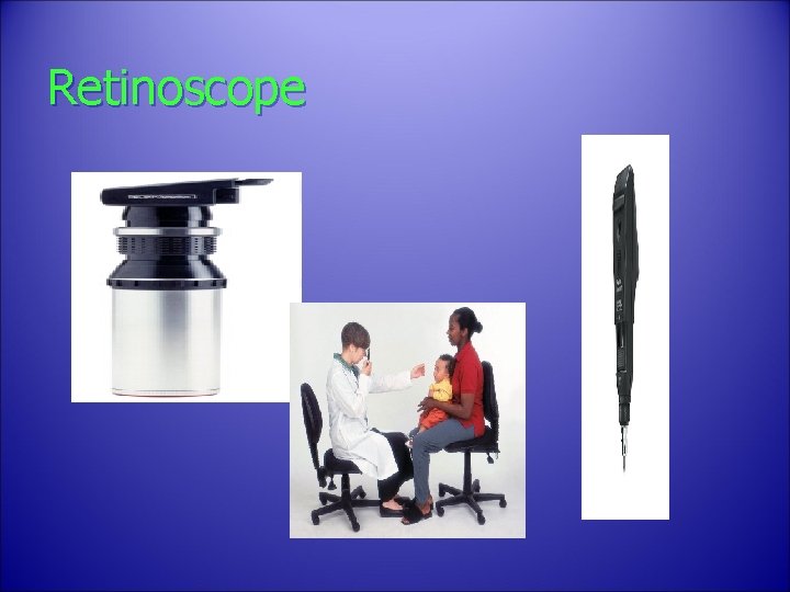
Retinoscope
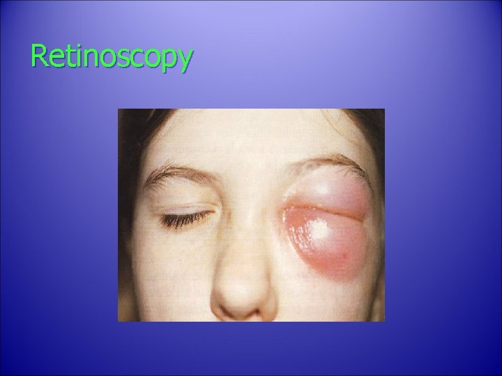
Retinoscopy
Exam • MCQ A 10 -year-old boy came for his annual eye check-up. Upon examination, the doctor noticed cherry red spot in the macular area. What could this sign represent? • Hydroxychloroquine toxicity • Niemann pick disease • Recurrent Optic neuritis • Severe Central Retinal Vein Occlusion
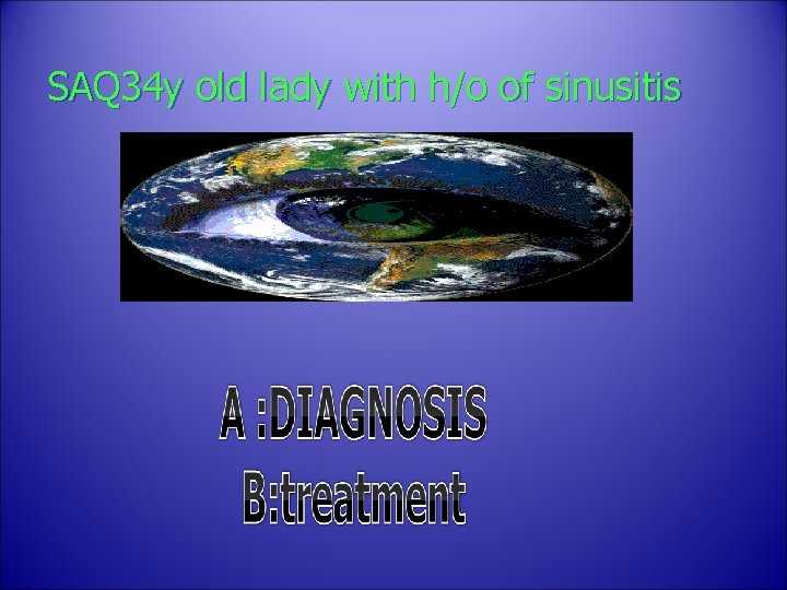
SAQ 34 y old lady with h/o of sinusitis