An Approach to Abdominal Pain in the ED

An Approach to Abdominal Pain in the ED Nisarg Shah MD, FACEP

Introduction Ø Complaints related to abdominal pain comprise between 5 -7% of all visits to the ED. Ø Of those, the most common discharge diagnosis is Abdominal Pain NOS. Ø Although most abdominal pain is non-emergent and selflimited in nature, attention must be paid to not miss medical and/or surgical emergencies.

Important Factors Ø Patients rarely present with the classical signs/symptoms of acute abdominal pain. Ø Three important factors to consider are age, gender, and comorbidities.

Age differences ØGreater than 50 Ø Biliary disease Ø NOS Ø Appendicitis Ø Bowel obstruction Ø Pancreatitis Ø Diverticular disease Ø Cancer ØLess than 50 Ø NOS Ø Appendicitis Ø Biliary tract disease Ø Gynecologic Ø Pancreatitis Ø Bowel obstruction

Gender differences ØMales Ø Perforated ulcer Ø Gastritis Ø Appendicitis ØFemales Ø Nonspecific Ø Diverticulitis Ø Acute cholecystitis Ø Gynecologic

Comorbidities ØImmunocompromised i. e. age, steroids, HIV, sickle cell disease, malignancy ØCAD, Atrial fibrillation, Peripheral vascular disease ØDiabetes ØDementia ØMultiple surgeries

Types of Pain Ø Visceral Pain: caused by stretching of fibers innervating the walls of hollow organs or capsules of solid organs, described as crampy or dull pain Ø Parietal Pain: caused by irritation of fibers that innervate the parietal peritoneum, pain is more sharp and localized Ø Referred Pain: pain at a location distant to the diseased organ based on embryonological origin

History ØMost diagnoses can be made by history alone. ØCareful attention must be paid to: ØTime/mode of onset ØDuration ØLocation ØCharacter/progression ØMedical history ØContributing symptoms

Caveat Although we can agree that history is usually the most important part of the encounter, urgent determination of potential surgical emergencies is essential.

History – time/mode of onset ØWoken up from sleep ØAbrupt/severe versus gradual ØLess severe but increasing ØGradual onset with slow progression ØIntermittent pain ØAssociated with certain activities – eating or exercise

History-Duration ØAcute onset of pain vs. chronic pain ØAcute, severe, and worsening pain suggests a surgical disorder ØVery long duration often, but not always, suggests a less acute cause

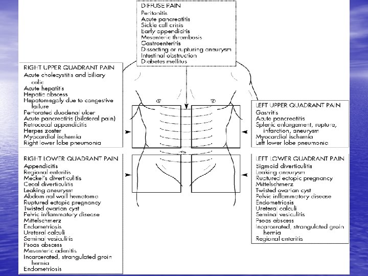
History-Location ØFour basic quadrants Ø Right upper quadrant Ø Right lower quadrant ØThree central areas Ø Epigastric Ø Periumbilical Ø Left upper quadrant Ø Suprapubic Ø Left lower quadrant

History-Character/Progression ØSeverity/magnitude of stimulus ØIntermittent crampy ØSevere and colicky ØSudden increase ØSudden change in sensation or location

Medical History ØPrevious surgery ØSexual activity ØMenstrual history ØTravel ØExposure risk/occupation ØPsychiatric ØMedications ØComorbidities
/Nausea ØDiarrhea ØBleeding ØConstipation ØObstipation ØDysuria ØSOB ØChest")
History-Contributing Symptoms ØAnorexia ØVomiting (bilious? blood? )/Nausea ØDiarrhea ØBleeding ØConstipation ØObstipation ØDysuria ØSOB ØChest pain

Physical Examination ØThe exam serves several important purposes ØTo confirm suspicions from the history ØTo localize the area of disease ØTo avoid missing extra-abdominal causes of pain

Physical Examination ØGeneral appearance including facial expression, diaphoresis, pallor, and degree of agitation to distinguish the intensity of the pain ØVital signs

Physical Examination ØInspection: look for distention, ecchymosis, scars, hernias ØAuscultation: listen for bowel sounds, pitch, bruits ØPalpation: feel for guarding, masses, tenderness, rebound ØPercussion: liver size, tympany

Differential Diagnosis ØThe next important step in the evaluation of abdominal pain is to formulate a differential diagnosis ØIt is helpful to construct a list based upon location of abdominal pain

Laboratory Evaluation ØDependent upon initial history and physical examination ØMost frequently ordered study is the CBC ØAdditional studies may include electrolytes, amylase, lipase, LFTs, BUN, creatinine, urinalysis, Beta Hcg, lactic acid ØEKG

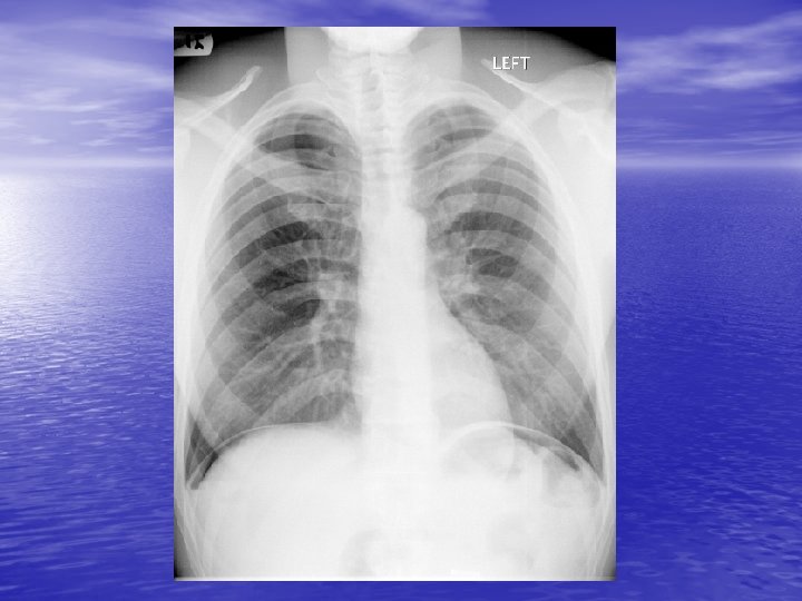
Imaging Studies ØPlain films: ØCXR or Upright p. CXR ØAbdominal series

Imaging studies cont’d ØCXR help determine the following: ØAbdominal pain of pulmonary origin - pneumonia with diaphragmatic irritation ØFree air under diaphragm - perforated viscous ØAir filled viscera in chest – diaphragmatic or hiatal hernia ØMediastinal air - Boerhave’s tear

Imaging studies cont’d ØAbdominal films can help with: ØFluid filled loops/air fluid levels – obstruction ØRenal calculi ØGallstones or air in the biliary tree ØMassive dilation of colon ØLots of stool
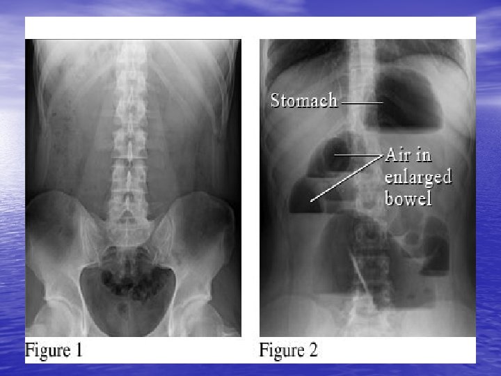

Imaging Ø This person most likely has Ø Large bowel obstr. Ø Small bowel obstr. Ø Generalized ileus Ø Localized ileus Ø Normal bowel gas pattern

Imaging Ø This person most likely has Ø Large bowel obstr. Ø Small bowel obstr. Ø Generalized ileus Ø Localized ileus Ø Normal bowel gas pattern

Imaging Ø This person most likely has Ø Large bowel obstr. Ø Small bowel obstr. Ø Generalized ileus Ø Localized ileus Ø Normal bowel gas pattern

Imaging Ø This person most likely has Ø Large bowel obstr. Ø Small bowel obstr. Ø Generalized ileus Ø Localized ileus Ø Normal bowel gas pattern

Imaging Ø This person most likely has Ø Large bowel obstr. Ø Small bowel obstr. Ø Generalized ileus Ø Localized ileus Ø Free intraperitoneal air

Imaging ØPneumobilia after passage of a gallstone. Take a good look at the liver where the biliary tract is outlined by air.

Imaging

Imaging

Imaging studies cont’d ØLikelihood ratio of finding abnormality on xray is increased by ØIncreased/high pitched bowel signs ØDistention ØHistory of abdominal surgery ØBlood in urine/history of kidney stones ØSevere abdominal pain and tenderness ØAbdominal pain for less than one day

Imaging studies cont’d ØSonography is the study of choice for: ØBiliary/hepatobiliary disease ØPregnant women ØEvaluation of gynecologic structures – ovarian as well as testicular ØRapid evaluation of hemoperitonium ØAAAs

Imaging studies cont’d ØCT scanning is now the test of choice for: Ø Intraabdominal infections such as diverticulitis, appendicitis, and post operative infections Ø Vasculature of the abdomen Ø Kidney stones Ø Abdominal hernias Ø Defining obstructions, neoplasms

Special Considerations ØPatients bearing special consideration ØWomen of childbearing age ØElderly patients ØChildren ØPatients on immunosuppressives

Women of childbearing age ØChildbearing women – atypical presentations – pregnant women with appendicitis may present with RUQ pain when uterus displaced other organs in 2 nd/3 rd trimesters

Elderly patients ØA low threshold should be used for admitting elderly patients. ØTheir presentation is rarely typical. ØTheir history is rarely clear. ØTheir comorbidities are many.

Children ØYoung children often have difficulty localizing their pain. ØHistory is limited. ØObtaining imaging is sometimes difficult but imaging has cut down on improper diagnoses.

Immunosuppressives ØAnyone on prednisone or other immunosuppressive medications be more careful with as they often present atypically. ØCorticosteroids may mask pain.

Aside About Radiation ØWe now image a lot in abdominal pain or chest pain. Try to keep in mind the large amount of radiation that we are exposing people to when we are making our diagnostic plan.

Time course of ailment ØEven if you image someone and the results are normal remember to tell people to still watch for warnings.

Abdominal Catastrophes ØThings not to miss ØMI ØAAA ØMesenteric ischemia ØEctopic pregnancy ØRuptured viscous

MI ØConsider Øin patients with risk factors ØIn patients with epigastric pain ØIn patients who are vomiting, particularly inferior wall MIs ØDiaphoresis is often common Ødiabetics

AAA ØUsed to be misdiagnosed commonly as nephrolithiasis. ØConsider in any patient with CAD, hypertension, testicular pain, flank pain. ØCheck for pulsatile mass, abdominal bruits.

Mesenteric Ischemia ØPain out of proportion to exam is the classical description. ØHigh morbidity/mortality ØConsider in older patients with comorbidities such as A. Fib. , severe CAD, CHF ØAngiography is test of choice but can be hard to set up in a timely manner so early consultation is essential.

Ectopic pregnancy ØPerform a pregnancy test in any woman of child bearing age. ØIf positive get a BQuant. ØDepending on the number and your clinical suspicion obtain a pelvic sonogram.

Extraabdominal causes ØCardiopulmonary – MI, angina, ptx, pna ØAbdominal wall – cellulitis, shingles ØHernias ØMetabolic – DKA, AKA or adrenal crisis, sickle cell crisis

Questions? ? ?
- Slides: 51