Nucleotides Nucleic Acid and Heredity What Are the


 in terms of an enzyme (protein) led")


 serves as a common currency into which the energy")


 is always")
 and Francis")


 Containing from 73 to 93 nucleotides per chain, ü")
 Ribosomes, which are small spherical bodies located in the")
 A recently discovered RNA molecule")
 A very recent discovery is another type of small")
 Short stretches of RNA (20 30 nucleotides long),")
. (RNA enzyme or catalytic RNA) RNA molecule possessing")


 has been hence")

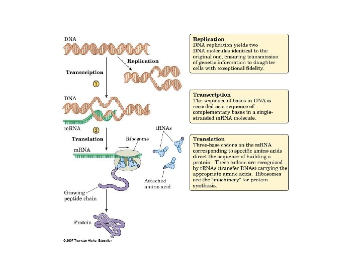
 In order to be decoded by the cell, DNA")


- Slides: 109
Nucleotides. Nucleic Acid, and Heredity
What Are the Molecules of Heredity? After scientists became aware of the differences in amino acid sequences, their next quest was to determine how cells know which proteins to synthesize out of the extremely large number of possible amino acid sequences. The answer is that an individual gets the information from its parents through heredity. Heredity is the transfer of characteristics, anatomical as well as biochemical, from generation to generation. We all know that a pig gives birth to a pig and a mouse gives birth to a mouse.
From about the end of the nineteenth century, biologists suspected that the transmission of hereditary information from one generation to another took place in the nucleus of the cell. More precisely, they believed that structures within the nucleus, called chromosomes, have something to do with heredity.
Thus the expression of the gene (DNA) in terms of an enzyme (protein) led to the study of protein synthesis and its control. The information that tells the cell which proteins to manufacture is carried in the molecules of DNA. We now know that not all genes lead to the production of protein, but all genes do lead to the production of another type of nucleic acid, called ribonucleic acid (RNA).
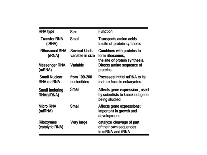
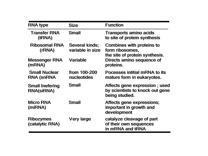
What Are Nucleic Acids ? Two kinds of nucleic acids are found in cells: Ø ribonucleic acid (RNA) is not found in the chromosomes, but rather is located elsewhere in the nucleus and even outside the nucleus, in the cytoplasm. And there are six types of RNA, all with specific structures and functions. Ø deoxyribonucleic acid (DNA), is present in the chromosomes of the nuclei of eukaryotic cells Each has its own role in the transmission of hereditary information.
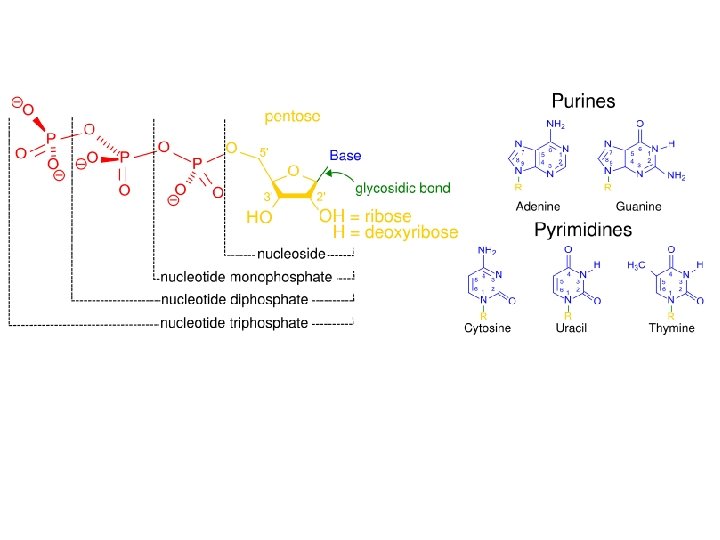
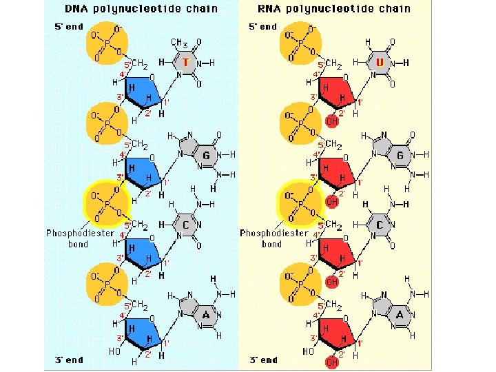
q. Both DNA and RNA are polymers. q nucleic acids are chains. q The building blocks (monomers) of nucleic acid chains are nucleotides. Ø Nucleotides themselves, however, are composed of three simpler units: a base, a monosaccharide, and a phosphate.
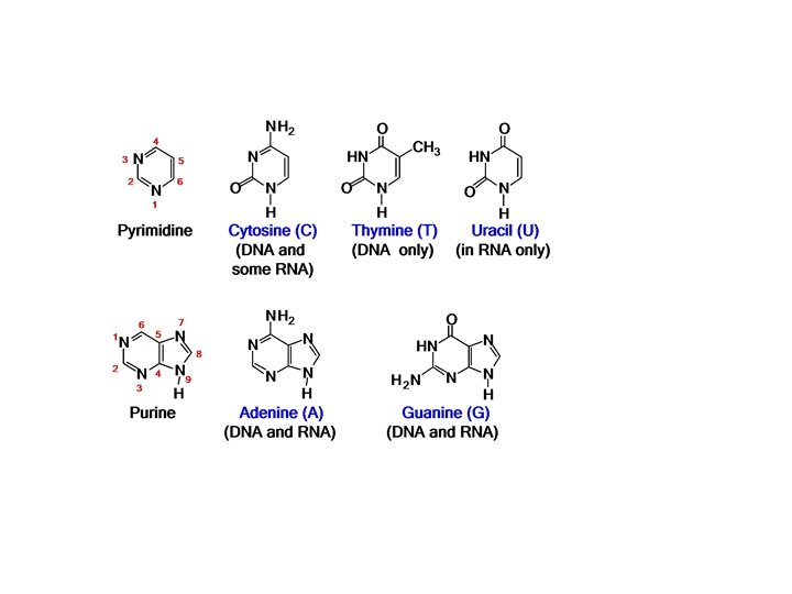
A. Bases The bases found in DNA and RNA are chiefly those. All of them are basic because they are heterocyclic aromatic amines. Ø Two of these bases-adenine (A) and guanine (G)-are purines; Ø the other three-cytosine (C), thymine (T), and uracil (U) -are pyrimidines. Note that thymine differs from uracil only in the methyl group in the 5 position. Thus both DNA and RNA contain four bases: two pyrimidines and two purines. For DNA, the bases are A, G, C, and T; for RNA, the bases are A, G, C, and U.
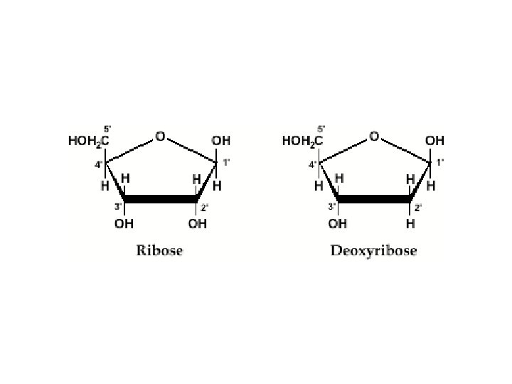
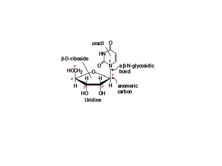
B. Sugars The sugar component of RNA is D ribose DNA, is 2 deoxy D ribose (hence the name deoxyribonucleic acid). The combination of sugar and base is known as a nucleoside. The purine bases are linked to C-l of the monosaccharide through N-9 (the nitrogen at position 9 of the five-membered ring) by a β-N-glycosidic bond:
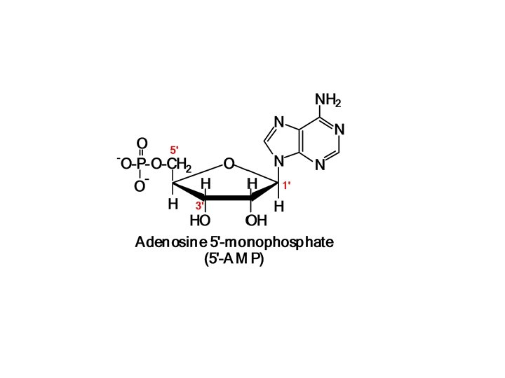
C. Phosphate The third component of nucleic acids is phosphoric acid. When this group forms a phosphate ester bond with a nucleoside, the result is a compound known as a nucleotide. q For example, adenosine combines with phosphate to form the nucleotide adenosine 5' monophosphate (AMP):
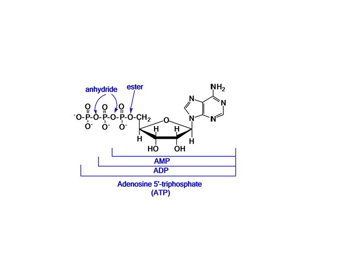
Most notably, adenosine 5'-triphosphate (ATP) serves as a common currency into which the energy gained from food is converted and stored. v In ATP, two more phosphate groups are joined to AMP in phosphate anhydride bonds. In adenosine 5' -diphosphate (ADP), only one phosphate group is bonded to the AMP.
A nucleoside = Base + Sugar A nucleotide = Base + Sugar + Phosphate A nucleic acid = A chain of nuclcotides
3 rd. Home Work GTP is an important store of energy. Draw the structure of guanosine triphosphate.
What Is the Structure of DNA and RNA? Nucleic acids, which arc chains of monomers, also have primary, secondary, and higher order structures.
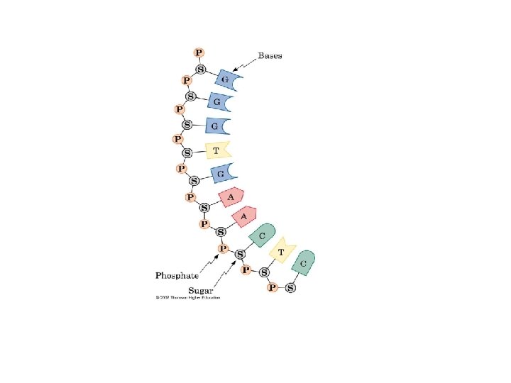
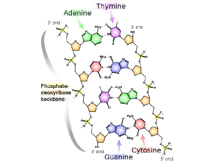
A. Primary Structure Nucleic acids are polymers of nucleotides. Their primary structure is the sequence of nucleotides. Note that it can be divided into two parts: (1) the backbone of the molecule (2) the bases that are the side chain groups.
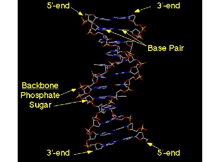
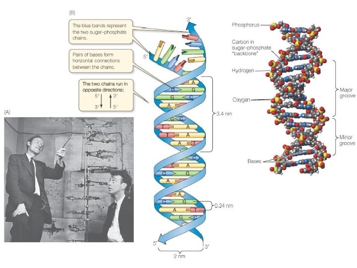
The backbone of the polynucleotide is made from alternating phosphate and sugar residues. Ø The sugars are joined together by phosphate groups that form phosphatediester bonds between the third and fifth carbon atoms of adjacent sugar rings. Ø The ends of polynucleotide strands are called the 5′ (five prime) and 3′ (three prime) ends, 5' end having a terminal phosphate group and the 3' end a terminal hydroxyl group. Ø One major difference between DNA and RNA is the sugar, with the 2 deoxyribose in DNA being replaced by the alternative pentose sugar ribose in RNA.
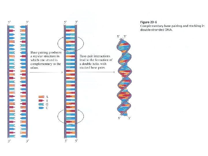
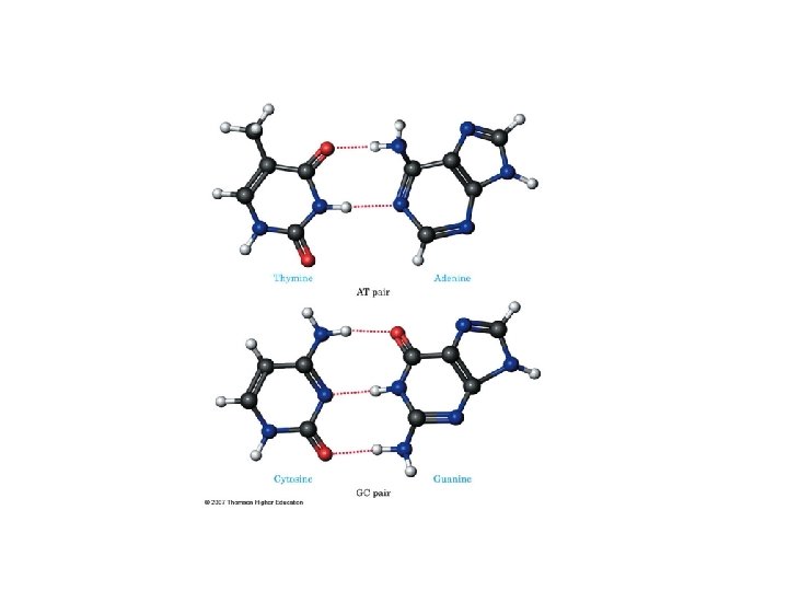
DNA taken from many different species, the quantity of adenine (in moles) is always approximately equal to the quantity of thymine, and the quantity of guanine is always approximately equal to the quantity of cytosine, although the adenine/guanine ratio varies widely from species to species
B. Secondary Structure of DNA • In 1953, James Watson (1928 ) and Francis Crick (1916 2004) established the three dimensional structure of DNA. The model of DNA developed by Watson and Crick was based on two important pieces of information obtained by other workers: (1) the Chargaffrule that (A and T) and (G and C) are present in equimolar quantities and (2) x ray diffraction photographs obtained by Rosalind Franklin and Maurice Wilkins
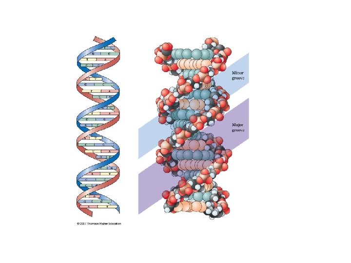
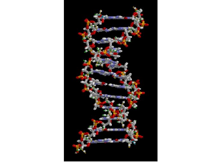
Watson and Crick concluded that DNA is composed of two strands entwined around each other in a double helix In the DNA double helix, the two polynucleotide chains run in opposite directions. Thus, at each end of the double helix, there is one 5' OH and one 3' OH terminus. The sugar phosphate backbone is on the outside, exposed to the aqueous environment, The bases point inward. The bases are hydrophobic, so they try to avoid contact with water. Through their hydrophobic interactions, they stabilize the double helix.
The bases so paired form hydrogen bonds with each other, two for A=T and three for GΞC, thereby stabilizing the double helix The entire action of DNA-and of the heredity mechanism-depends on the fact that, wherever there is an adenine on one strand of the helix, there must be a thymine on the other strand because that is the only base that fits and forms strong hydrogen bond, with adenine, and similarly for G and C.
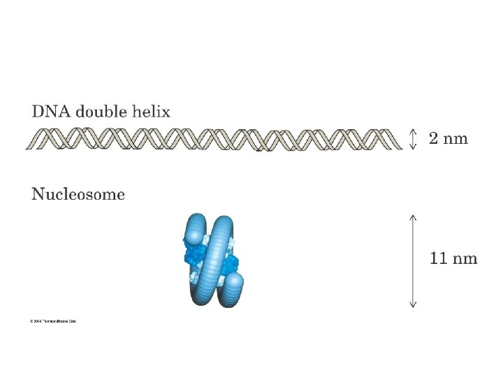
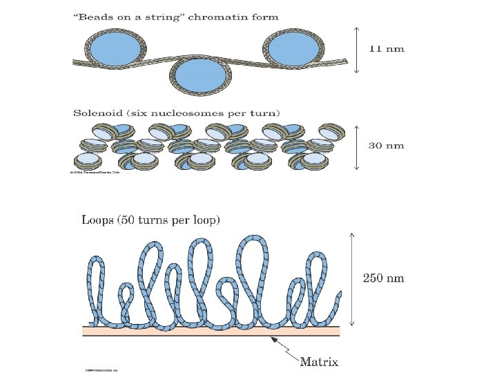
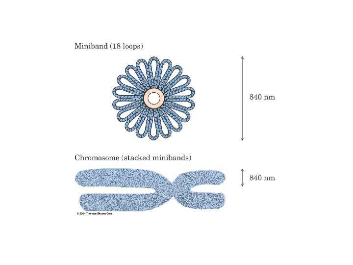
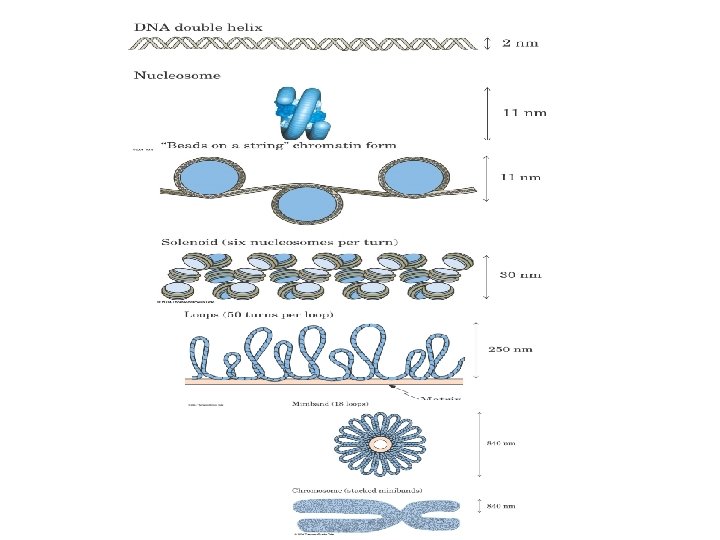
C. Higher-Order Structures of DNA If a human DNA molecule were fully stretched out, its length would be perhaps 1 m. However the DNA molecules in the nuclei are not stretched out, but rather coiled around basic protein molecules called histones. Ø The acidic DNA and the basic histones attract each other by electrostatic (ionic) forces, combining to form units called nucleosomes. In a nucleosome, eight histone molecules form a core, around which a 147 base pair DNA double helix is wound. Nucleosomes are further condensed into chromatin when a 30 nm widefiber forms in which nucleosomes are wound in a solenoid fashion, with six nuc 1 eosomes forming a repeating unit. Chromatin fibers are organized still further into loops, and loops are arranged into bands to provide the superstructure of chromosomes.
1 -DNA has four bases: A, G, C, and T. RNA has three of these bases-A, G, and C-but its fourth base is D, not T. 2 - In DNA, the sugar is 2 deox. Y-D-ribose. In RNA, it is D-ribose. 3 - DNA is almost always double-stranded, with the helical structure
What Are the Different Classes of RNA? We previously noted that' there are six types of RNA. 1 Messenger RNA (m. RNA) m. RNA molecules are produced in the process called transcription, and they carry the genetic information from the DNA in the nucleus directly to the cytoplasm, where most of the protein is synthesized. ü Messenger RNA consists or a chain of nucleotides whose sequence is exactly complementary to that of one of the strands of the DNA. ü This type of RNA is not long lived, however. It is synthesized as needed and then degraded. so its concentntration at any given time is rather low. ü The size of m. RNA varies widely, with the average unit containing perhaps 750 nucleotides.
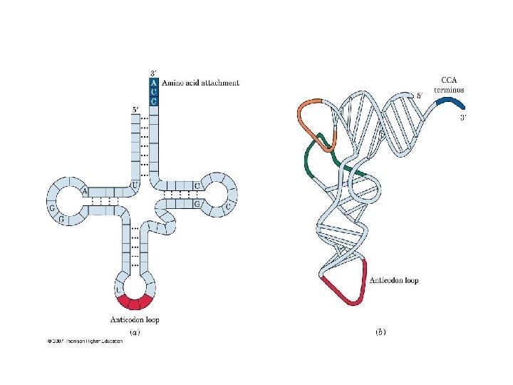
2 Transfer RNA (t. RNA) Containing from 73 to 93 nucleotides per chain, ü t. RNAs are relatively small molecules. ü There is at least one different t. RNA for each of the 20 amino acids from which the body makes its proteins. ü The three dimensional t. RNA molecules are L shaped, but they are conventionally represented as a clover leaf in two dimensions. ü Transfer RNA molecules contain not only cytosine, guanine, ; adeninc, and uracil, but also several other modified nucleotides, such as 1 methylguanosine.
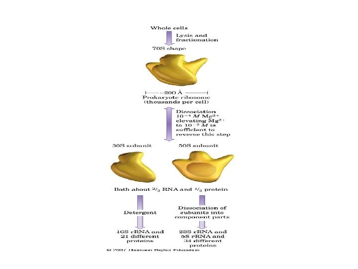
3 Ribosomal RNA (r. RNA) Ribosomes, which are small spherical bodies located in the cells but outside the nuclei, contain r. RNA. They consist of about 35% protein and 65% ribosomal RNA (r. RNA). These large molecules have molecular weights up to 1 million. v protein synthesis takes place on the ribosomes. v In both prokaryotes and eukaryotes, a ribosome consists of two subunits, one larger than the other. Ø In turn, the smaller subunit consists of one large RNA molecule and about 20 different proteins; Ø the larger subnit consists of two RNA molecules in prokaryotes (three in eukaryotes) and about 35 different proteins in prokaryotes (about 50 in eukaryotes). Ø The subunits are easily dissociated from one another in the laboratory by lowering the Mg 2+ concentration of the medium. Raising the Mg 2+ concentration to its original level reverses the process, and active ribosomes can be reconstituted by this method.
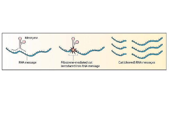
ü ü ü 4 Small Nuclear RNA (sn. RNA) A recently discovered RNA molecule is sn. RNA, which is found, as the name implies, in the nucleus of eukaryotic cells. This type of RNA is small, about 100 to 200 nucleotides long, but it is neither a t. RNA molecule nor a small subunit of r. RNA. In the cell, it is complexed with proteins to form small nuclear ribonucleo protein particles, sn. RNPs, pronounced ·'snurps. " Their function is to help with the processing of the initial m. RNA transcribed from DNA into a mature form that is ready for export out of the nucleus. This process is often referred to as splicing, and it is an active area of research. While studying splicing, scientists realized that part of the splicing reaction involved catalysis by the RNA portion of a sn. RNP and not the protein portion. This recognition led to the discovery of ribozymes, RNA based enzymes, for which Thomas Cech received the Nobel Prize.
5 Micro RNA (mi. RNA) A very recent discovery is another type of small RNA, mi. RNA. These RNAs are only 22 nucleotides long but are important in the timing of an organism's development. They bind to sections of m. RNA and prevent their translation.
6 Small Interfering RNA (si. RNA) Short stretches of RNA (20 30 nucleotides long), called small interfering RNA, have been found to have an enormous control over gene expression. This process serves as a protective mechanism in many species, with the si. RNAs being used to eliminate expression of an undesirable gene, such as one that causes uncontrolled cell growth or one that came from a virus.
Ø Ribozyme (from ribonucleic acid enzyme). (RNA enzyme or catalytic RNA) RNA molecule possessing a well defined tertiary structure that enables it to catalyze a chemical reaction. Many natural ribozymes catalyze either the hydrolysis of one of their own phosphodieste bonds (self cleaving ribozymes), or the hydrolysis of bonds in other RNAs, but they have also been found to catalyze the aminotransferase activity of the ribosome.
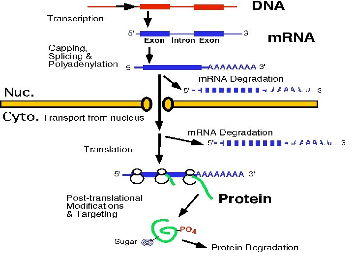
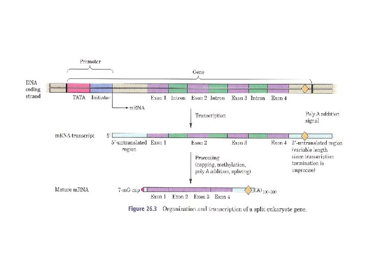
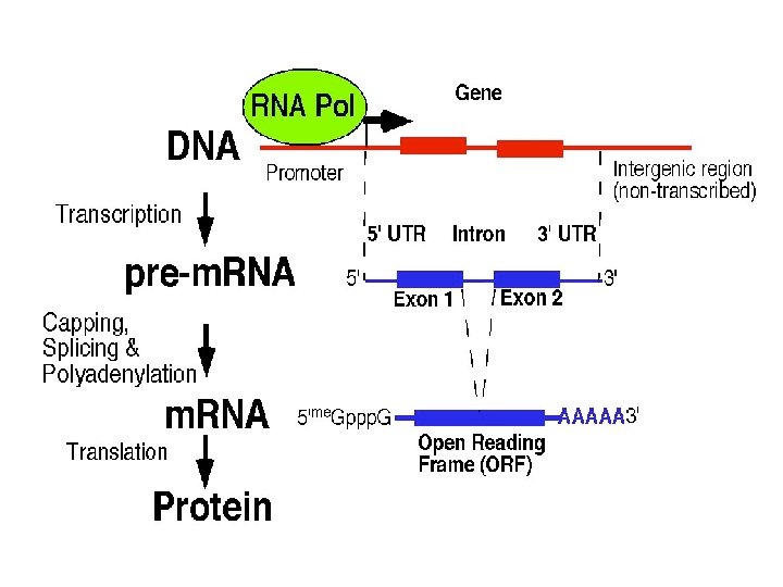
What Are Genes? A gene is a segment of DNA molecule that carries the sequence of bases that directs the synthesis of one particular protein or RNA molecule. A gene is a stretch of DNA, containing a few hundred nucleotides, that carries one particular message for example, "make a globin molecule" or "make a t. RNA molecule. " One DNA molecule may have between 1 million and 100 million bases. Therefore, many genes are present in one DNA molecule. Stretches of DNA that spell out (encode) the amino acid sequence to be assembled are interrupted by Ø long stretches that seemingly do not code for anything. the coding sequences are called exons, Ø short for "expressed sequences, ” the noncoding sequences are called introns, short for "intervening sequences.
Because DNA contains both exons and introns, the m. RNA transcribed from it also contains both exons and introns. The introns are spliced out by ribozymes, ; and as enzymes, catalyzing the splicing of exons into mature m. RNA
5 Home Work What is DNA fingerprinting
Gene Expression and Protein Synthesis
Genomes, Chromosomes and Genes How Does DNA Lead to RNA and Protein? The DNA present in each cell of an organism, the total information content, is called the genome. Genomes are made up of very long DNA molecules housed on chromosomes. Chromosomes include many genes. A gene is that section of the chromosomal DNA that contains the information to make a specific polypeptide through the production of a specific RNA.
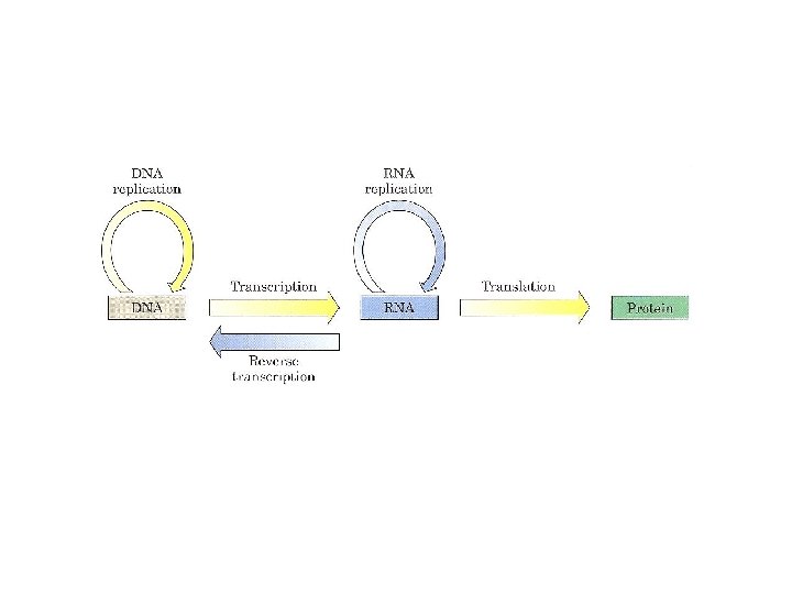
A. Information Transfer: DNA —> RNA —> Protein The genome (DNA) has been hence likened to an architect’s set of master drawings. These drawings are copied into blueprints (RNA synthesis=transcription) that are bought out to the construction site where they are acted upon (translation) to make the rooms, doors, windows, etc. of a house (cellular proteins).
Gene expression The activation of a gene to produce a specific protein; it involves both transcription and translation. Ø Transcription The process in which information encoded in a DNA molecule is copied into an m. RNA molecule. Ø Translation The process in which information encoded in an m. RNA molecule is used to assemble a specific protein.
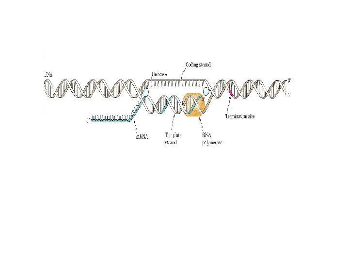
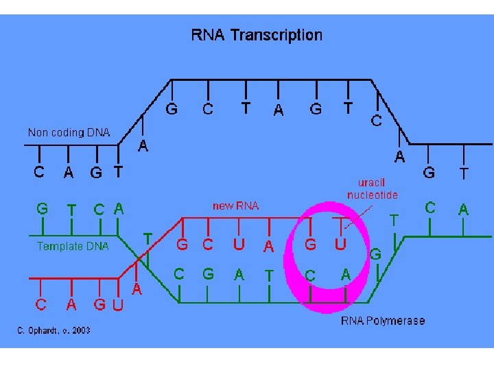
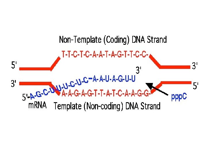
Transcription DNA —> RNA a) In order to be decoded by the cell, DNA is first copied into messenger RNA (m. RNA). The process of synthesizing a specific m. RNA is called transcription. The m. RNA copy is called the transcript. Remember that RNA, ribonucleic acid, differs from DNA in several significant respects: 1. The chemical makeup of RNA includes a different base from DNA— the pyrimidine uracil is utilized instead of thymine. Uracil is very similar to thymine and base pairs with adenine. Additionally, the ribose sugar includes a hydroxyl group at the 2’ position. 2. RNA is usually single stranded.
The process of transcription is divided into several steps: 1 A transcription factor recognizes the start site (promoter) of a gene that is to be transcribed. 2 The enzyme that makes the RNA (RNA polymerase) binds to the transcription factor and recognizes the start region. 3 The enzyme proceeds down the DNA making a copy until the end of the gene is reached. 4 The enzyme falls off and the RNA is released. This copying process may be repeated numerous times. 5 If the RNA is one that codes for a protein, it will leave the nucleus and enter the cytosol.
Polymerization of the chain occurs via the same basic mechanism as DNA polymerization—the chain is extended from its 3' end as the 3' OH of the last ribose on the chain attacks the phosphate attached to the 5' OH of the ribose from a free nucleotide triphosphate. The RNA chain is thus synthesized in a 5' to 3' direction.
Added nucleotides are selected because they base pair to the template. Thus, the m. RNA is complementary to its DNA template. Ø The strand that serves as a template for RNA synthesis is called the template strand. Ø The DNA strand complementary to the template, the nontemplate strand, or coding strand, is identical in base sequence to the RNA transcribed from the gene, with U in place of T.
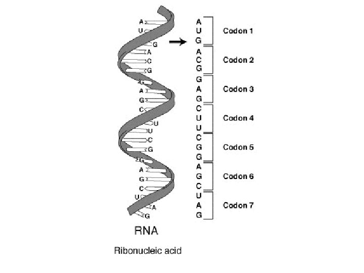
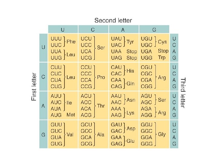
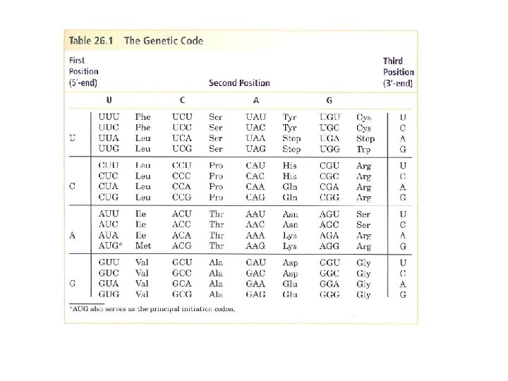
Protein Synthesis-Translation refers to the synthesis of a protein directed by instructions encoded within an m. RNA molecule. Ø The triplet code: A sequence of three consecutive nucleotides on the m. RNA specifies the addition of an amino acid to a growing polypeptide chain. The three ordered nucleotides comprise what is called a codon. Ø Clever experiments performed in the mid 1960 s resulted in the elucidation of the genetic code—the amino acid specified by each codon was determined. v This table can be used to predict the amino acid sequence of a protein if the DNA sequence is known.
The code defines a mapping between tri nucleotide sequences, called codons, and amino acids.
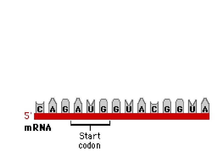
1. The code is degenerate—in other words, often more than one codon calls for the same amino acid to be added to the growing peptide chain. For example there are 6 codons that code for Leu and 6 that code for Arg. Only one codon codes for Met or Trp. 2. The same codon, AUG, is (ALMOST) always used to begin translation of an m. RNA. o AUG that is used to start protein synthesis is called an initiation codon. o AUG calls for the addition of the amino acid methionine both at the start and within the protein.
3. Certain codons result in the termination of a growing chain. These stop or termination codons are UAA, UAG and UGA. 4. It is essential to know where to start and stop reading the m. RNA. The protein translated from the sequence that follows is obviously quite different, depending upon where you start reading.
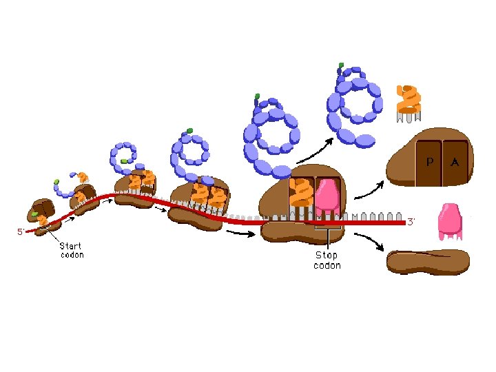
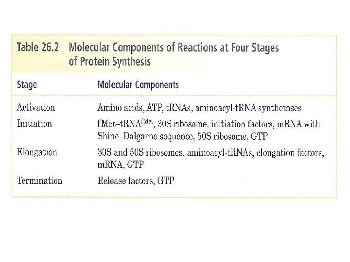
The Steps of Translation
I Initiation
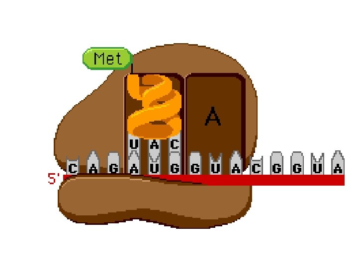
Initiation Step 1 The m. RNA joins to the small ribosomal unit at the 5' untranslated region. This binds to a special binding site on the small ribosomal subunit. The large ribosomal subunit has 3 binding sites, E, P, and A
Initiation Step 2. The large ribosomal subunit attaches to the small subunit such that the first codon is aligned at the P binding site
Initiation step 3 A t. RNA carrrying the amino acid methionine attaches to the start codon (AUG) on the messaenger RNA. This inititates elongation.
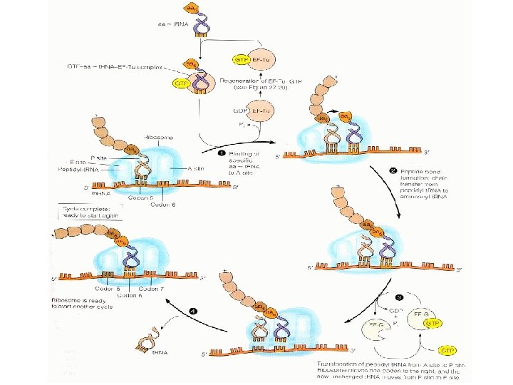
II Elongation
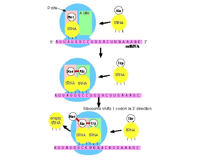
Elongation step 1. Attachment of first amino acid carrying t. RNA to A binding site. A t. RNA and its amino acid attaches to the A binding site
Elongation Step 2. Peptide bond formation between the met and the amino acid carried at the A bindiung site. Our polypeptide chain is now: Met Thr
Elongation Step 3. Ribosome moves in the 3' direction down the messenger RNA by three bases or one codon shifting the t. RNA and polypeptide chain to the P Binding site. The A binding site is open and a vacant t. RNA is in the E binding site
Elongation Step 4. t. RNA ejected from the E binding site
Repeat Elongation Step 1 4 until stop codon encountered.
Elongation ends with: Old t. RNA ejected from the E Binding site
III Termination
Termination Step 1. The polypeptide chain is at the P site. The stop codon at the A site
Termination Step 2. A Release factor protein binds to the stop codon at the A binding site
Termination Step 3. Release factor protein initiates separation of polypeptide chain: Met Thr His Asp Gly
Termination Step 4. Separation of translation machinary. Polypeptide chain may go to cytoplasm for further processing
Polysomes Several ribosomes can translate an m. RNA at the same time, forming what is called a polysome
Home Work What are the codons for histidine? What are the anticodons?