Nasal cavity The nose consists of the external


 is related above to the orbit ;")


- Slides: 62
Nasal cavity
The nose consists of the external nose and the nasal cavity, both of which are divided by a septum into right and left halves.
They open on the face by anterior nasal apertures. Posteriorly, they open into the nasopharynx through the posterior nasal apertures
Boundaries of the nasal cavity
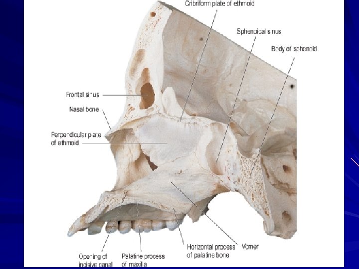
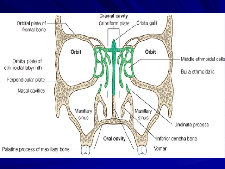
Medial wall The nasal septum consists of : The perpendicular plate of the ethmoid bone above; The vomer below & behind. The septal nasal cartilage anteriorly ;
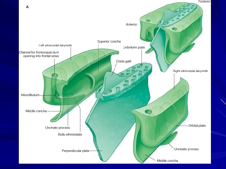
Roof Sloping anterior part frontal and nasal bones. Horizontal middle part: the cribriform plate of the ethmoid bone.
Sloping posterior part is the body of sphenoid bone and the ala of the vomer
The floor consists of palatine process of the maxilla, and the horizontal plate of the palatine bone, which together form the hard palate.
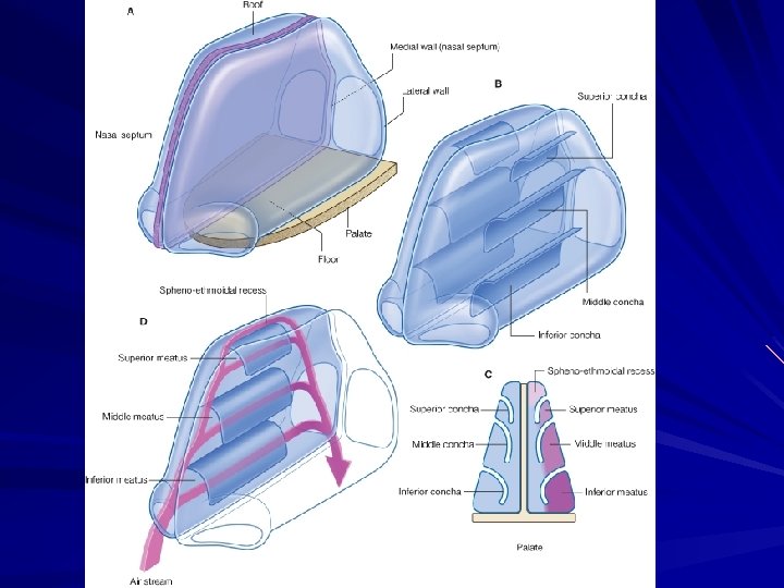
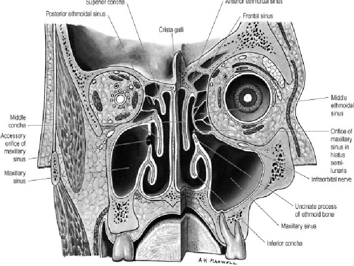
Lateral wall: Inside the nasal cavity the surface of the lateral wall is irregular in contour and is interrupted by the three bony projections, nasal conchae.
The inferior, middle, and superior conchae extend medially across the nasal cavity, separating it into four air channels, an inferior, middle, and superior meatus, and a spheno-ethmoidal recess.
The sphenoidal air sinus drains into the spheno -ethmoidal recess
The superior meatus lies below the superior concha The posterior ethmoidal cells usually open onto the lateral wall of the superior nasal meatus
The middle meatus lies below the middle concha. It has rounded prominence (the bulla ethmoidalis) This is formed by the underlying middle ethmoidal cells.
The middle ethmoidal cells open onto or just above the ethmoidal bulla; Inferior to the ethmoidal bulla is a curved gutter (the hiatus semilunaris),
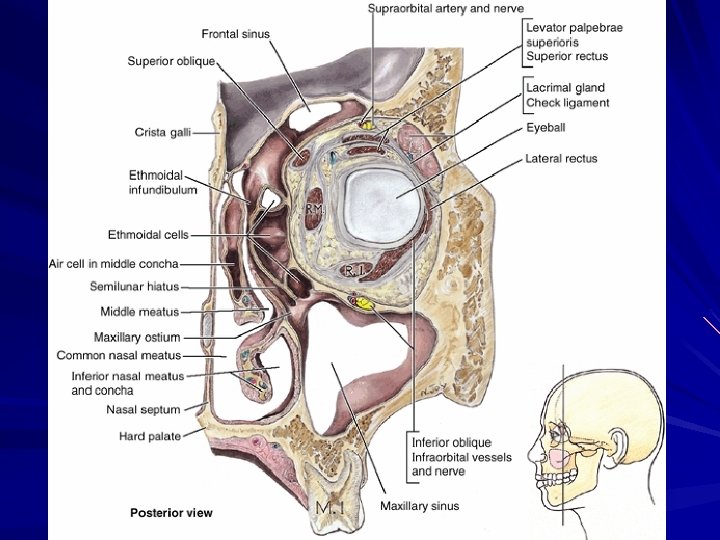
The large maxillary sinus opens into the hiatus semilunaris The anterior end of the hiatus semilunaris forms a channel (the ethmoidal infundibulum), which curves upwards
The frontal sinus and the anterior ethmoidal cells drain via the frontonasal duct and ethmoidal infundibulum into the anterior end of the hiatus semilunaris.
The nasolacrimal duct opens onto the lateral wall of the inferior nasal meatus under the anterior lip of the inferior concha-it drains tears from the conjunctival sac of the eye into the nasal cavity and originates at the inferior end of the lacrimal sac on the anteromedial wall of the orbit.
Mucosa of the nasal cavity Olfactory mucosa. It lines the upper surface of superior concha and the sphenoethmoidal recess. It also lines the corresponding area of the nasal septum and lines the roof. Its function is the reception of olfactory stimuli. It possesses specialized olfactory nerve cell. Respiratory mucosa. It lines the lower part of the nasal cavities. It is formed of columnar ciliated epithelium with goblet calls. Its function is to warm, moisten and clean the inspired air. Vestibular mucosa. The vestibule is lined by skin that bears short thick hairs (vibrissae).
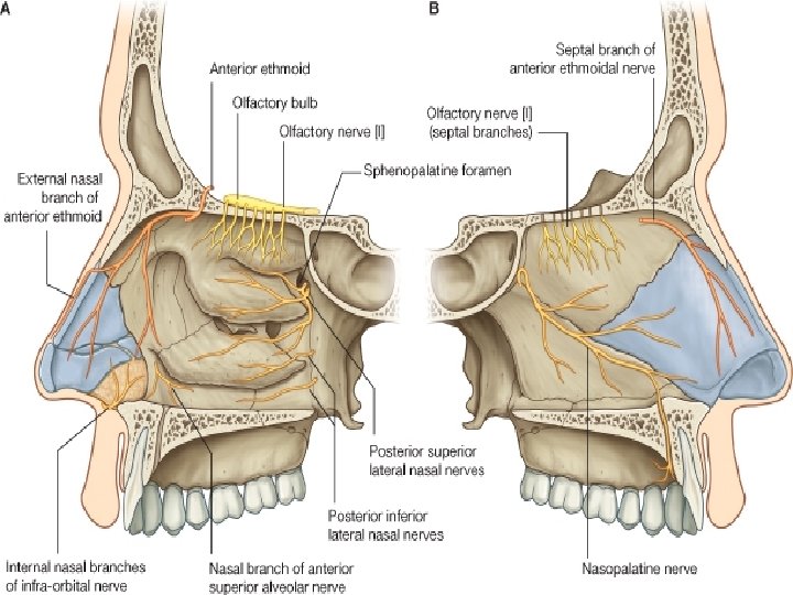
Nerve supply
The olfactory nerves arise from the special olfactory cells in the olfactory mucous membrane. They ascend through the cribriform, plate of ethmoid to reach the olfactory bulbs.
The nerves of ordinary sensation are derived from the ophthalmic and maxillary divisions of the trigeminal nerve The nerve supply to the anterior part of the nasal cavity comes from the anterior ethmoidal nerve.
The nerve supply to the posterior part of the nasal cavity comes from the nasal branches of the sphenopalatine ganglion, the nasoplatine nerve and the nasal branches of the greater palatine nerve.
Blood supply
It is derived from the anterior and posterior ethmoidal branches (branches of the ophthalmic artery) and the sphenopalatine artery which is the main source of the arterial blood to the nose (branch of the maxillary artery).
It is also supplied by branches of the superior labial artery (branch of the facial artery).
Clinical note: The sphenopalatine artery anastomoses with the septal branch of the superior labial artery in the region of the vestibule which is a very common site of bleeding from the nose (epistaxis).
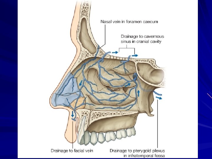
Venous drainge: The veins form a rich plexus in the submucosa. The veins are drained by the pterygoid Plexus, maxillary vein and by the facial veins.
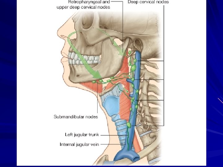
Lymphatic drainge: The lymphatic vessels draining the vestibule end in the submandibular nodes. The remainder of the nasal cavity is drained by vessels that pass to the upper deep cervical lymph nodes.
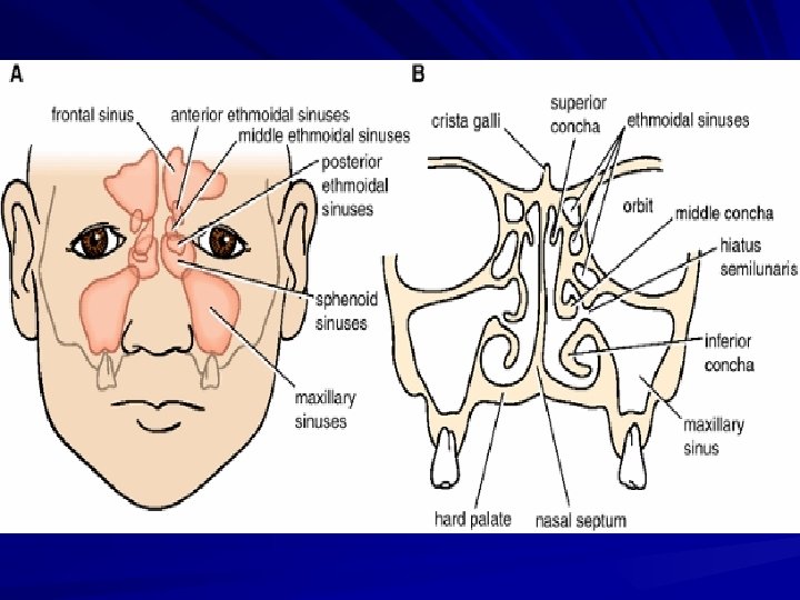
Paranasal sinuses
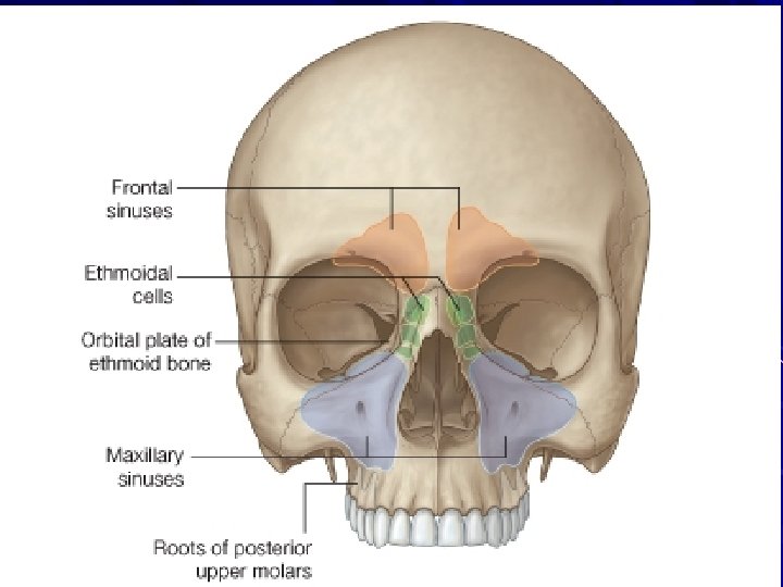
There are four paranasal air sinuses-the ethmoidal cells, and the sphenoidal, maxillary, and frontal sinuses Each is named according to the bone in which it is found.
Frontal sinuses
The frontal sinuses, one on each side, separated by bony septum are variable in size and are the most superior of the sinuses Each is triangular in shape and is in the part of the frontal bone under the forehead.
Each frontal sinus drains onto the lateral wall of the middle meatus via the frontonasal duct, which penetrates the ethmoidal labyrinth and continues as the ethmoidal infundibulum at the front end of the hiatus semilunaris
The frontal sinuses are innervated by branches of the supra-orbital nerve from the ophthalmic nerve. Their blood supply is from branches of the anterior ethmoidal arteries.
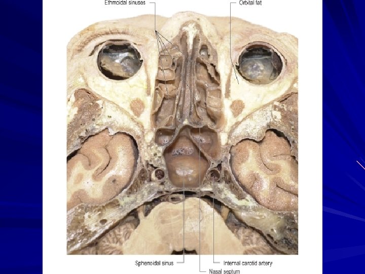
Ethmoidal cells
The ethmoidal cells on each side fill the ethmoidal labyrinth. Each cluster of cells is separated from the orbit by the thin orbital plate of the ethmoidal labyrinth, and from the nasal cavity by the medial wall of the ethmoidal labyrinth.
The ethmoidal cells are formed by a variable number of individual air chambers, which are divided into anterior, middle, and posterior ethmoidal cells based on the location of their apertures on the lateral wall of the nasal cavity :
The anterior ethmoidal cells open into the ethmoidal infundibulum or the frontonasal duct ;
The middle ethmoidal cells open onto the ethmoidal bulla, or onto the lateral wall just above this structure ; The posterior ethmoidal cells open onto the lateral wall of the superior nasal meatus.
The ethmoidal cells are innervated by : The anterior and posterior ethmoidal branches of the nasociliary nerve from the ophthalmic nerve [V 1] The maxillary nerve [V 2] via orbital branches from the pterygopalatine ganglion. The ethmoidal cells receive their blood supply through branches of the anterior and posterior ethmoidal arteries.
Maxillary sinuses The maxillary sinuses, one on each side, are the largest of the paranasal sinuses and completely fill the bodies of the maxillae.
Each is pyramidal in shape with the apex directed laterally and the base deep to the lateral wall of the adjacentnasalcavity. The medial wall or base of the maxillary sinus is formed by the maxilla, and by parts of the inferior concha and palatine bone that overlie the maxillary hiatus
The opening of the maxillary sinus is near the top of the base, in the center of the hiatus semilunaris, which grooves the lateral wall of the middle nasal meatus
Relationships of the maxillary sinus are as follows :
The superolateral surface (roof) is related above to the orbit ;
The anterolateral surface is related below to the roots of the upper molar and premolar teeth and in front to the face;
The posterior wall is related behind to the infratemporal fossa.
The maxillary sinuses are innervated by infra-orbital and alveolar branches of the maxillary nerve {V 2} and receive their blood through branches from the infraorbital and superior alveolar branches of the maxillary arteries.
Sphenoidal sinuses The sphenoidal sinuses, one on either side within the body of the sphenoid, open into the roof of the nasal cavity via apertures on the posterior wall of the spheno -ethmoidal recess. Innervation of the sphenoidal sinuses is provided by : the posterior ethmoidal branch of the ophthalmic nerve [V 1]