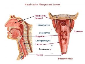
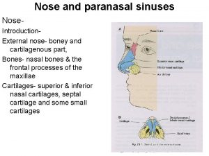
STRUCTURE Nose Nasal septum divides nose into R


- Slides: 12

* STRUCTURE


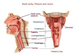
* *Nose *Nasal septum divides nose into R and L sides * Both sides are lined with ______ membranes *Cilia – hairs in nose that trap dirt and particles

* *Cavities in skull filled with air * Frontal, maxillary, sphenoid, and ethmoid *Connected to nasal cavity by ducts *Lined with mucous membrane * Helps to warm and moisten air passing through them *Give resonance to the voice *SEE FIGURE 17 -3 pg 355

* *Throat *Serves as a common passageway for air and food *5” long *Can be subdivided into: * Nasopharynx * Oropharynx * laryngopharynx

* *Voice box *Triangular chamber below pharynx *Composed of nine fibrocartilaginous plates *Adams apple – largest of the plates *Contains vocal cords * Become larger in males during puberty which makes Adams apple more prominent *Lined with mucous membrane *Epiglottis – flap of cartilage that covers larynx during swallowing *SEE FIGURE 17 -4 pg 356

* *Windpipe *Tubelike passageway that extends from the larynx, passes in front of esophagus, and continues to form the 2 bronchi *4 ½” long *Walls have bands of C-shaped cartilage *Lined with ciliated mucous membrane *FIGURE 17 -4

* *Lower end of trachea divides into R and L bronchus * Right bronchus is shorter, wider, and more vertical *Become bronchial tubes and bronchioles as branches enter lungs *FIGURE 17 -5 pg 358

* *Clusters of thin-walled sacs made of single layer epithelial tissue *Inner surfaces covered with surfactant * What does surfactant do? *Each alveolus surrounded by capillaries * Why? *FIGURE 17 -5

* *Fill thoracic cavity *Upper part = apex *Lower part = base *Lung tissue porous and spongy, it floats * Due to alveoli and air it contains *R lung larger and broader but shorter, 3 lobes (superior, middle, and inferior) *L lung smaller, narrower, and longer, has 2 lobes (superior and inferior) *FIGURE 17 -6 pg 359

* *Membrane that covers lungs (thin, moist, and slippery tough endothelial cells) *Double-walled sac *Space is pleural cavity *Pleural cavity filled with pleural fluid to prevent friction

* *Interpleural space *Between lungs along the medial plane of the thorax and extends from the sternum to vertebrae *Contains the thoracic viscera
 What divides the nasal cavity
 Medical terminology
 Nose obstruction causes
 Membranous septum of nose
 Venous drainage of nose
 Ethmoid
 Nasoseptoplasty
 The coronal plane divides the body into
 Latitudinal geographic zones
 A grid divides an area into small parts called
 Midsagittal plane
 Epigastric vs hypogastric
 Gummy bear dissection
