The Respiratory System Respiratory System Functions Gas exchange


 – Where air enters the nose")


 • Formed by division of trachea •")


 – Air move in and out")
 • Expiration – Air")


")


- Slides: 86
The Respiratory System
Respiratory System Functions • Gas exchange: Oxygen enters blood and carbon dioxide leaves • Regulation of blood p. H: Altered by changing blood carbon dioxide levels • Voice production: Movement of air past vocal folds makes sound and speech
Respiratory System Functions • Olfaction: Smell occurs when airborne molecules drawn into nasal cavity • Protection: Against microorganisms by preventing entry and removing them • Temperature regulation: Panting in some animals
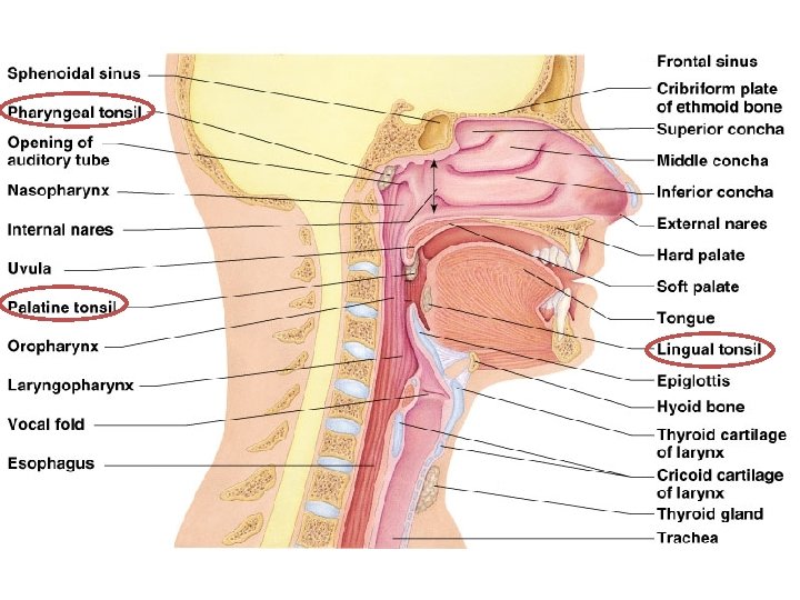
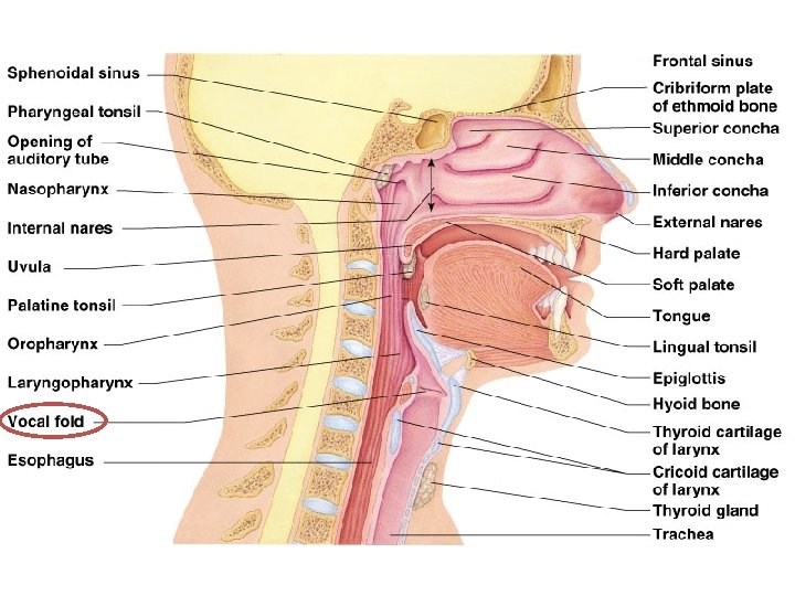
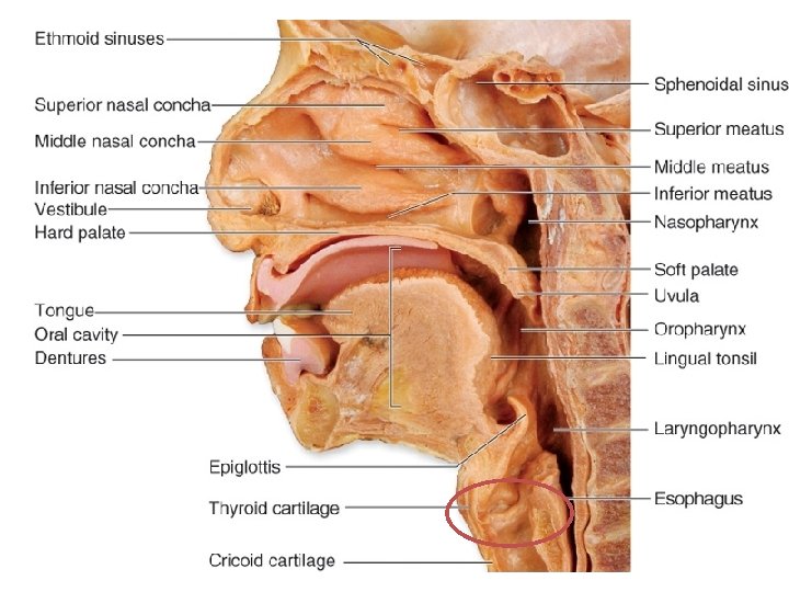
Parts of the Nose • Nose – Only externally visible part of the respiratory system – Job is to begin warming, purifying, and humidifying air
Parts of the Nose • External Nares (nostrils) – Where air enters the nose – The openings • Nasal Cavity – Interior of the nose – Has thin-walled blood vessels to begin warming air
Parts of the Nose • Nasal Septum – Midline dividing nasal cavity into two
Parts of the Nose • Respiratory Mucosa – Sticky mucous – Moistens air and traps bacteria & debris – Ciliated cells move mucous back toward throat
Parts of the Nose • Conchae – Mucosa-covered projections – Increase surface area & air turbulence – Provide more opportunity for warming & filtration
Parts of the Nose • Hard Palate – Anterior separation of nasal & oral cavity – Supported by bone • Soft Palate – Posterior separation of nasal & oral cavity
Cleft Palate • Palate does not fuse together • Can also affect the lip
Parts of the Nose • Paranasal Sinuses – Openings in skull bones – Lighten the skull – Resonance chambers for speech – Produce mucus
Parts of the Pharynx • Pharynx – Muscular passageway for food & air – “Throat” – About 5 inches long – Broken into 3 parts
Parts of the Pharynx • Internal Nares – Opening between nasal cavity and pharynx
Parts of the Pharynx • Nasopharynx • Oropharynx • Laryngopharynx – Three divisions of Pharynx – Listed from superior to inferior – After passing through, air enters larynx, food enters esophagus
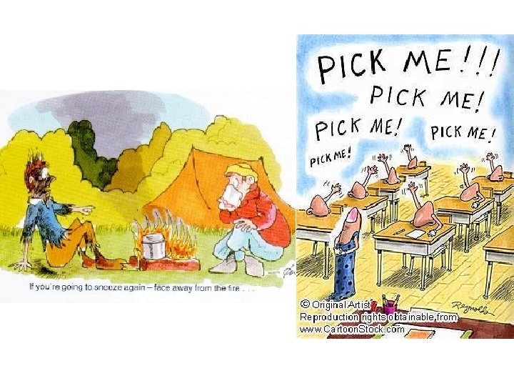
Parts of the Pharynx • Pharyngeal Tonsils: – AKA adenoids – High in the nasopharynx – Trap bacteria/pathogens • Palatine Tonsils – In oropharynx, end of soft palate – When you get your tonsils out, this is what is removed – Trap bacteria/pathogens • Lingual Tonsils – Base of the tongue – Trap bacteria/pathogens
FYI: Tonsilitis • Catch too much bacteria; palatine tonsils can’t keep up! EEW.
• http: //video. about. com/coldflu/Tonsillitis. htm
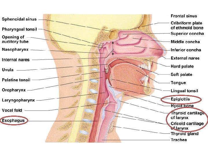
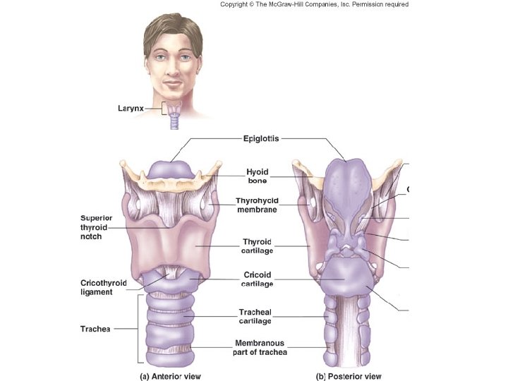
Larynx • • AKA Voice Box Routes air and food into proper channels Inferior to pharynx Formed by 8 rigid cartilages and a spoonshaped flap of elastic cartilage (epiglottis) – Thyroid cartilage = Adam’s apple
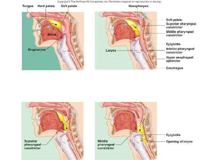
Epiglottis • Cartilage flap of larynx; protector! • When not swallowing: – Epiglottis flapped up – Does not block larynx • When you are swallowing: – Larynx rises – Epiglottis falls – Larynx closed off – This means that food is directed into esophagus
• FYI: If anything other than air tries to enter the larynx, a cough reflex is triggered to get it out and prevent it from going into the lungs!
Vocal Folds • Vocal Folds – Formed from folds in larynx membrane – Vibrate with expelled air • Glottis – Slit-like passageway between vocal folds
• http: //video. about. com/coldflu/Laryngitis. htm • You. Tube - Video Stroboscopy of the Vocal Cords • You. Tube - Mythbusters - Helium and Sulfur Hexafluoride
Trachea • AKA windpipe • Has cartilage rings around it to keep it open during pressure changes • About 4 inches long • Lined with ciliated mucosa to propel mucus (with dust particles & debris) away from the lungs to the throat
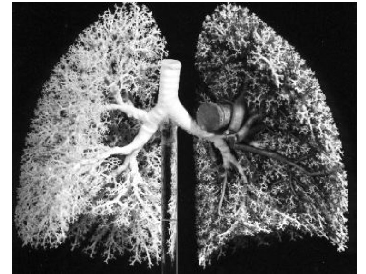
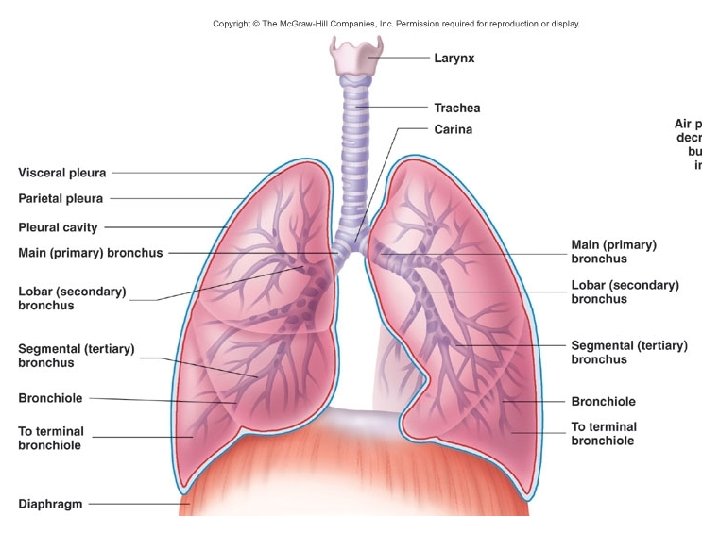
Primary Bronchi • Two (right & left) • Formed by division of trachea • Enters the lung, and then breaks off into secondary bronchi
Mediastinum • Most central area of the thoracic cavity • Includes heart, great blood vessels, bronchi, esophagus, etc. (everything except lungs)
Lungs • Site of gas exchange • Soft & Spongy, only weigh about 2 ½ pounds • Each lung divided into lobes – Left: 2 lobes – Right: 3 lobes
Parts of Lungs • Apex – Narrow superior portion – By clavicle • Base – Wide inferior portion – Rests on diaphragm • Visceral Pleura – Covers surface of lung – Along with parietal pleura, provides attachment and eliminates friction
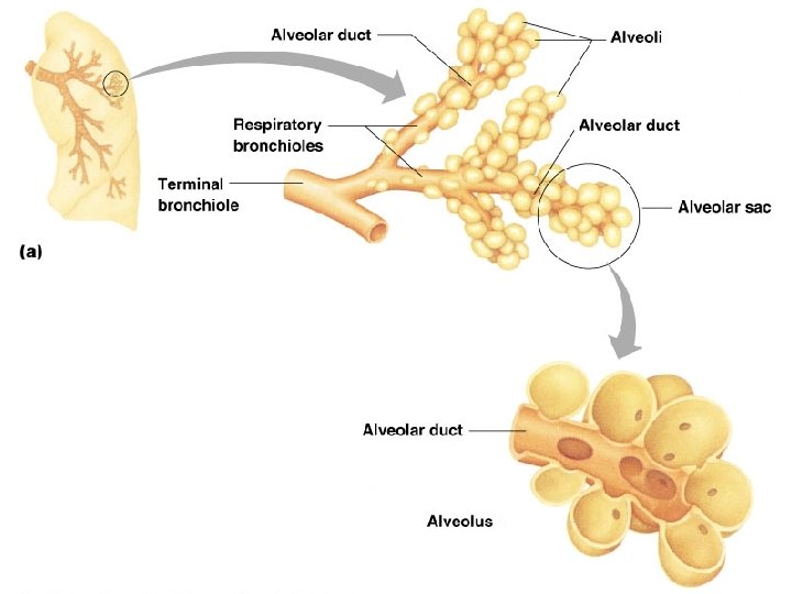
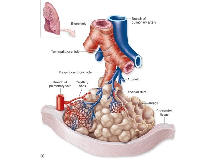
Bronchial Tree • Bronchioles – All of the branching of respiratory passageways in the lungs – Divisions include • • • Primary bronchi Secondary bronchi Tertiary bronchi Bronchioli Terminal bronchioli (end in alveoli)
This image is showing the carina – the point where the primary bronchi break off from each other.
Alveoli • “Air Sacs” • Resemble bunches of grapes • Make up bulk of lungs
Respiratory Zone • Includes respiratory bronchioles, alveolar ducts, alveolar sacs, and alveoli • Only places where gas exchange occurs
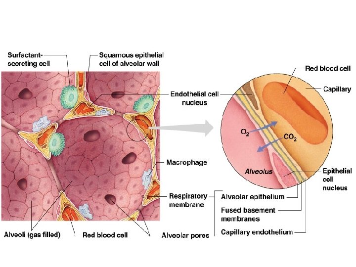
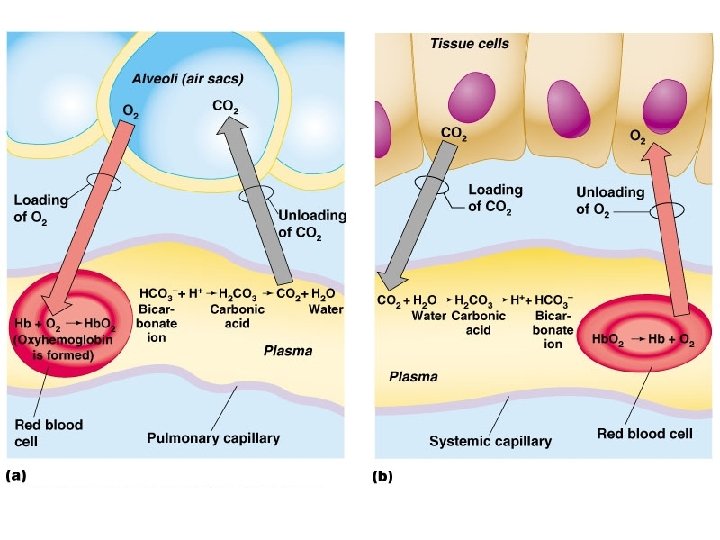
Walls of Alveoli • Made of single, thin layer of simple squamous epithelial cells • Covered with a cobweb of pulmonary capillaries
Respiratory Membrane • Made up of alveolar and capillary walls • Has air flowing on one side, blood flowing on the other • Gas exchange occurs through simple diffusion – Oxygen from alveolar air to capillary blood – Carbon dioxide leaving blood to air FUN FACT: total surface of alveoli walls is about 50 -70 square meters - about the surface of a tennis court!
• http: //highered. mcgrawhill. com/sites/0072495855/student_view 0/ch apter 25/animation__gas_exchange_during_re spiration. html • http: //video. about. com/asthma/How-Lungs. Function. htm
Steps to Respiration • 1. Pulmonary Ventilation (breathing) – Air move in and out of lungs so alveoli air is refreshed • 2. External Respiration – Gas exchange between blood and alveoli air • 3. Respiratory Gas Transport – Gas transport between lungs and body by bloodstream • 4. Internal Respiration (Cellular Respiration!) – In body, at capillaries, gas exchange between blood and tissue cells
Respiration • Inspiration – Air flowing into lungs (breathe in!) • Expiration – Air leaving the lungs (breathe out. ) • Volume changes lead to pressure changes, which lead to the flow of gases to equalize the pressure!
Inspiration • Diaphragm and external intercostals contract – Diaphragm actually moves DOWN when contracts • • Thoracic cavity size increases Rib cage lifts Intrapulmonary volume increases Gas molecules spread out due to increased volume! This causes a decrease in pressure, which pulls in more gas.
Inspiration
Expiration • Diaphragm and external intercostals relax – Internal intercostals and abdominal muscles contract if expiration is forced • • Thoracic cavity size decrease Rib cage descends Intrapulmonary volume decreases Gas molecules get closer together due to the decrease in volume! This causes an increase in pressure, which forces gas out.
Expiration
Non-Respiratory Air Movements • • • Cough Sneeze Crying – release of air in short breaths Laughing – release of air in short breaths Hiccups Yawn
Lung Development • Fetus – All respiratory exchange made by placenta – Lungs filled with fluid • Birth – Fluid drained, passageways fill with air – Surfacant: fatty molecule that lines each alveolar sac to prevent from collapsing (don’t have enough of this until fetus is 30 weeks) – Lungs do not fully inflate for 2 weeks after birth – 20 to 40 respirations/min
Lung Development • Teens – 12 to 18 respirations/min (rates differ depending on source) – Continue to develop alveoli (smoking stops this production!) • Elderly – Chest wall becomes rigid, lungs lose elasticity, cilia in trachea less effective – Vital capacity decreases – More at-risk for respiratory tract conditions – 18 to 20 respirations/min
Respiratory Conditions • Pulmonology – study of diseases of the lungs and respiratory tract • Hypoxia – inadequate oxygen delivery to body tissues, causes cyanosis (bluish-tone of skin)
Upper Respiratory Tract Infection • Includes things like the common cold, sinusitis, tonsillitis, and laryngitis • Symptoms include nasal congestion, cough, running nose, sore throat, fever, facial pressure, and sneezing
• http: //video. about. com/coldflu/Upper. Respiratory-Infection. htm • http: //video. about. com/coldflu/Sinusitis. htm
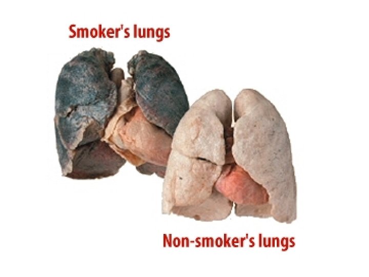
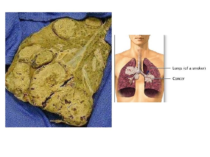
Lung Cancer • 1/3 of all cancer deaths in the US • Very low survival rate, because usually not caught until very advanced • Growth usually occurs in the bronchial tree • Problem with smoking… – Kills off cilia that cleans & moves mucus out of airway – Mucus collects – Traps carcinogens from cigarettes
COPD • Chronic Obstructive Pulmonary Disease – Characterized by limited airflow – Symptoms include coughing, wheezing, and shortness of breath (dyspnea) – Usually have a history of smoking, but can also be caused by other inhalants (coal, asbestos, air pollution)
COPD – Patients retain carbon dioxide, meaning blood becomes slightly acidic – Usually results in respiratory failure – Two major types (there are other types, but these are the most common)…
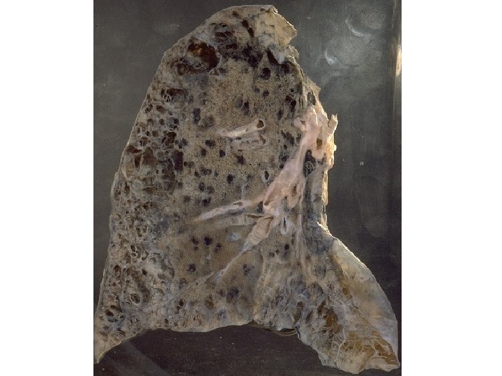
Emphysema • Characterized by breakdown of elastin in connective tissue of lungs • Leads to destruction of alveolar walls • Lungs become less elastic, which makes it difficult to exhale
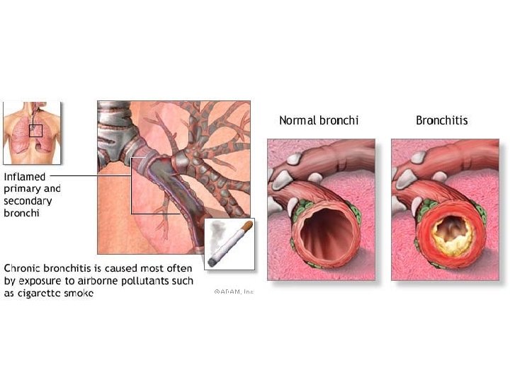
Chronic Bronchitis • Characterized by inflammation of the bronchi • Mucosa lining of lower respiratory passages become inflamed and produces excess mucus, which leads to blockage • Symptoms include persistent coughing with sputum, cyanosis very common
Acute Bronchitis • Caused by virus or bacteria • Only last days/weeks • Symptoms same as chronic bronchitis
• http: //video. about. com/asthma/Bronchitis. ht m
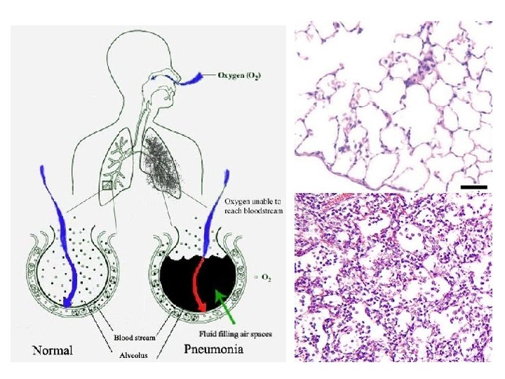
Pneumonia • Characterized by: Inflammatory illness of the lungs, alveoli become filled with fluid • Varied causes, including bacteria, virus, fungi, chemical, & physical • Often diagnosed with chest xray
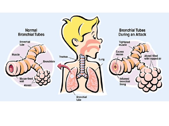
Asthma • Characterized by airways constricting due to trigger (exposure to allergens, pollutants, cold air, warm air, moist air, exercise, or emotional stress) • Symptoms include wheezing, shortness of breath, chest tightness, and coughing • Scientists believe cause is combination of genetics and environment • Inhalers act as bronchodilator
Sleep Apnea • Characterized by pauses of breathing during sleep • 5 or more events, lasting at least 10 seconds each • Caused by blockage of the airways due to causes such as decreased muscle tone, increased soft tissue around airway (obesity), and structural features (deviated septum, enlarged tonsils)
Carbon Monoxide Poisoning • Competes with oxygen for same binding sites on hemoglobin • Binds VERY easily, and so it pushes out oxygen • Leading cause of death from fire • Victim usually becomes confused, has headache, and blushing of skin
Cystic Fibrosis • Cystic Fibrosis is a recessive genetic disease that effects both the digestive and respiratory system. • Abnormally thick mucus gets stuck in many places. Most commonly in the lungs & pancreatic duct • Symptoms can include persistent coughing, shortness of breath and frequent upper respiratory infections. 1 in 20 people are unaffected carriers!
Cystic Fibrosis • Easily diagnosable with a sweat test • Treatment for the GI tract involves taking digestive enzymes before eating. • Treatment for the lungs is called CPT, Chest Physiotherapy. – A light clapping of the chest, back, and under the arms to loosen mucus in the lungs