Some conditions of external nose CONGENITAL MIDLINE NASAL


")


- Slides: 37
Some conditions of external nose
CONGENITAL MIDLINE NASAL MASSES
• Congenital midline nasal masses include: • Nasal dermoids. • Nasal gliomas. • Encephaloceles.
• These are rare congenital anomalies, estimated to occur in 1: 20, 000 to 40, 000 births. • Although rare, these disorders are clinically important because of their potential for connection to the central nervous system.
• Biopsy of a lesion with an intracranial connection can lead to meningitis or cerebrospinal fluid leak. • The treatment of these masses is surgical excision. • Preoperative knowledge of an intracranial connection allows for neurosurgical consultation and planning for craniotomy.
• Inflammatory lesions • Traumatic deformity • Benign neoplasms • Malignant neoplasms • Congenital masses The differential of a midline nasal mass
Nasal Dermoids
• Nasal dermoids are epithelial-lined cavities or sinus tracts with variable numbers of skin appendages: • Hair follicles • Sebaceous glands • Eccrine glands. • They constitute the most common congenital nasal anomaly
• Nasal dermoids may arise from clusters of epithelium trapped during ectodermal processes, or they may signify failure of ectodermal extensions into the fetal nasal septum to disappear as the septum fuses and ossifies. • Dural attachments also may exist, creating a tract between the nasal skin and the dura that passes through the prenasal space (toward the foramen cecum) or fonticulus.
• Present as a mass on the dorsum of the nose or intra-nasally, with a pit or sinus tract opening on the nasal dorsum, hair around the external opening, and discharge of pus or sebaceous material. • Typically manifest within the first month of life, and 73% are diagnosed in the first year of life.
• Intermittent inflammation • Abscess • Osteomyelitis • Broaden nasal root • Meningitis • Cerebral Abscess Complications
CT scan reveals the bony defect, and MRI differentiates the dermoid element from brain tissue
CT scan reveals the bony defect, and MRI differentiates the dermoid element from brain tissue
Firm masses which are non-pulsatile, present on the nasal dorsum and/or arise from the lateral nasal wall, have telangiectasias of the overlying skin, and do not enlarge with bilateral compression of the internal jugular veins (Furstenberg test). Nasal Glioma
Nasal Encephaloceles
• Encephaloceles may present as: • Nasal broadening • A blue, pulsatile, compressible mass near the nasal bridge • Transilluminates • Enlarges with crying or with bilateral compression of the internal jugular veins, • An intranasal mass arising from the cribriform plate.
• Complete cyst and sinus tract excision. • If an intracranial cyst exists, a combined craniofacial approach with neurosurgical consultation is required. • Recurrence is attributed to incomplete excision. Treatment
RHINOPHYMA
It is a descriptive term derived from the Greek "rhis" meaning nose and "phyma" meaning growth.
• Typically afflicts white males between 40 and 60 years of age. • It is far more common in men than women, with the ration of 12: 1 generally reported. • This is very interesting when you consider that the disease is the end-stage of acne rosacea, which is three times more common in females.
• It begins as an accentuation of the normal flush reaction in and can involve the nose, central forehead, malar areas, and chin. • With time, the vessels of the nose become progressively dilated and the skin thickens and may become involved with cysts and pustules, and the skin can be quite oily. • The nose thickens at the tip and sebaceous glands hypertrophy. • As the deformity worsens, pits, nodules, fissures, lobulations, and pedunculation contort the nose into monstrous cosmetic problems. • There is no uniformity in the end stage or final appearance.
• Partial thickness excision as the treatment of choice, and this has been termed "decortication. " • Decortication has been performed using cryosurgical techniques, chemical peels, dermabrasion, the cold scalpel, the Shaw knife (a thermally heated scalpel), the Bovie, hot wire loops, and the Argon and CO 2 lasers. The scalpel and dermabrader often lead to significant bleeding that can compromise accuracy. • Electrosurgical techniques are better, but most authors now recommend use of the CO 2 laser which can allow operation in a near bloodless field Treatment
LUPUS VULGARIS
• Lupus vulgaris is the most common morphological variant of cutaneous tuberculosis, accounting for approximately 59% of cases of cutaneous tuberculosis in India. • It is more common in females than males
Apple jelly nodules
• More often encountered than frank tuberculosis of nasal mucosa. • It is acquired: • Either exogenously by direct inoculation of the bacilli • Or endogenously by haematogenous • Or lymphatic, spread from an underlying distant focus. • Seen in patients with moderate or high degree of immunity.
• The classical lesions consist of reddishbrown plaque with “apple-jelly” colour on diascopy. • The lesions progress by peripheral extension and central healing, atrophy and scarring. and affects the nasal vestibule, the septm and the ala. • If untreated it is slowly destructive and can lead to gross and embarrssing deformity. • The symptoms include ulceration with sanguineous mucopurulent discharge.
• The diagnosis is based on histopathology of a deep biopsy from the ulcerated site. • A deep biopsy of the lesion supported with tests like PCR for TB is the correct approach if there is any doubt about the diagnosis. • The differential diagnosis of this clinical picture includes lupus erythematosus, rosacea.
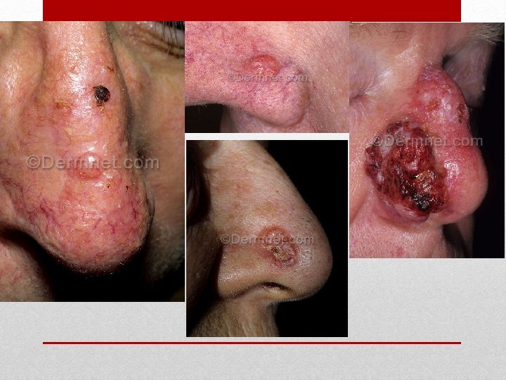
BASAL CELL CARCINOMA (BCC)
• BCC is the most common skin cancer in humans. • BCC tumors typically appear on sunexposed skin, are slow growing, and rarely metastasize. • Neglected tumors can lead to significant local destruction and even disfigurement. • Tumors usually arise from the epidermis or the outer root sheath of a hair follicle
• Historically, men are affected twice as often as women. • The higher incidence in men is probably due to increased recreational and occupational exposure to the sun, although these differences are becoming less significant with changes in lifestyle. • The likelihood of developing basal cell carcinoma increases with age. • BCC is rarely found in patients younger than 40 years.
• • • Waxy papules with central depression Pearly appearance Erosion or ulceration, often central Bleeding, especially when traumatized Crusting Rolled (raised) border Translucency Telangiectases over the surface Slow growing (0. 5 cm in 1 -2 y) Chracteristic features
• Nodular - Cystic, pigmented, keratotic • Infiltrative • Micronodular • Morpheaform • Superficial Clinicopathological types
• The goal of therapy for patients with BCC is removal of the tumor with the best possible cosmetic result. • By far, surgical modalities are the most studied, most effective, and most used treatments for basal cell carcinoma. • Modalities used include: • Electrodesiccation and curettage • Excisional surgery • Mohs micrographically controlled surgery • Cryosurgery. Surgical modalities