ANATOMY OF NOSE External Nose Pyramidal shaped osteocartilagenous


 • U shaped cartilages • Medial crus form")
 Cartilages – Two or More in number Lie above")


")


 It is the Largest of Para Nasal Sinuses, pyramidal")


 Thin walled air cavities in lateral masses of")
 divides eth. Air")


- Slides: 57
ANATOMY OF NOSE
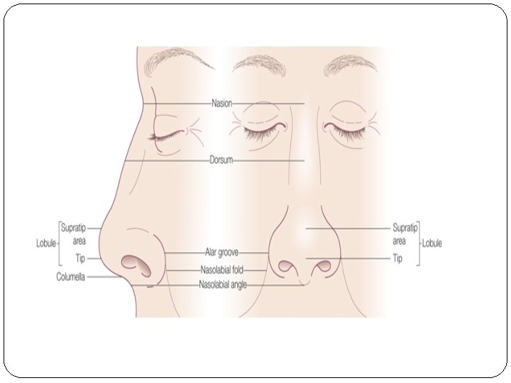
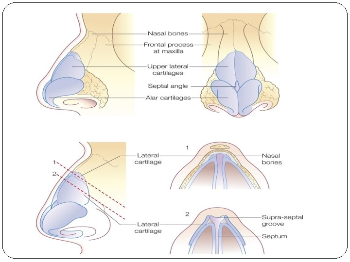
External Nose Pyramidal shaped osteocartilagenous framework Root- point where nose continues with forehead Base- directed downwards opens into ant. nares Bridge- where lateral surfaces meet in midline .
Columella- separates the two nares Nasal ala- forms lateral boundary of nose Nasal Tip Dorsum- connects tip to root of nose .
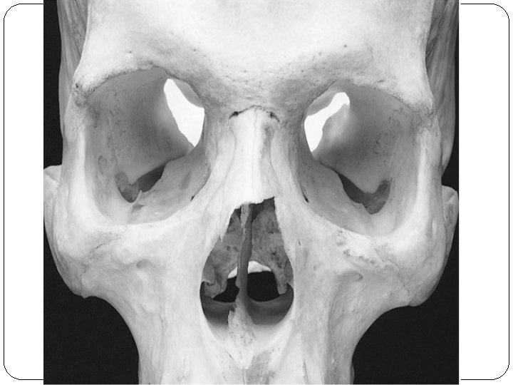
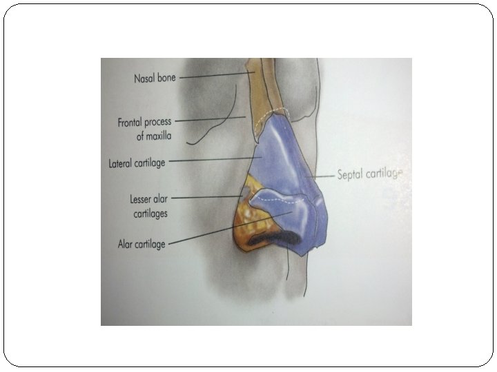
Bony Part q. Paired nasal bones – Upper 1/3 rd of ext. nose – bony - Lower 2/3 rd of ext. nose - cartilaginous - Nasal bones meet in midline forming bridge - Articulate - Superiorly with nasal process of frontal bone - Laterally with frontal Process of maxilla - Inferiorly with upper lateral cartilages.
Cartilaginous Part q. Upper lateral cartilages • Extend from under surface of nasal bones to alar • • cartilage. Articulate with nasal bone & frontal process of maxilla above & lower lat. cartilages below Fuse with each other & septal cartilage in midline Limen nasi-groove between upper & lower lat. cartilages, ( site of intercartilagenous incision) Limen vestibuli (Nasal Valve)– lower free edge of upper lateral cartilage.
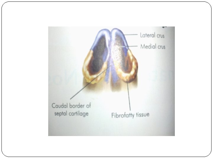
q. Lower lateral cartilages- (Alar cartilages) • U shaped cartilages • Medial crus form collumela • Lateral crus (wider) form ala of nose .
q Lesser Alar (Sesamoid ) Cartilages – Two or More in number Lie above and lateral to alar cartilage. Various cartilages are connected with adjoining bones and with one another by perichondrium and periosteum. q Septal cartilage • Supports dorsum of cartilaginous part of nose • Anterosuperior border runs from under nasal bones to nasal tip.
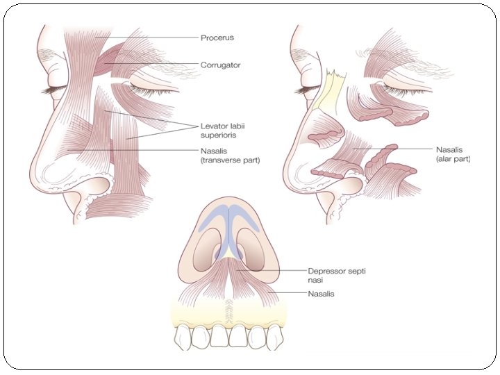
q. Skin over nasal bones & upper lat. Cartilages is thin &mobile while over lower cartilages is thick & adherent & Contains many sebaceous glands. q. Muscles- Procerus Nasalis – Transverse & Alar Ant. & Post. dilator nares Leveator labii superioris alaque nasi Ant. & Post. Dilator nares Ant. & Post. Depressor septi.
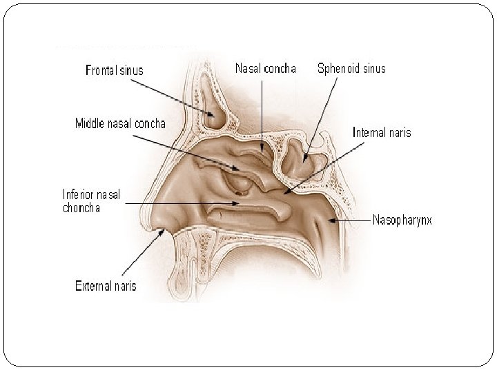
q Nasal cavity- Medial Wall Lateral Wall Roof Floor Vestibule- ( Lined by skin) Ant. inf. part of nasal cavity &contains sebaceous gland & vibrissae.
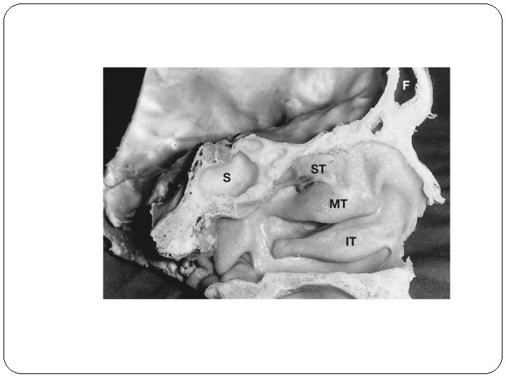
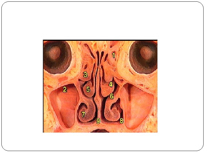
Lateral Nasal Wall- Marked by three turbinates & coresponding meatus Some times a fourth turbinate – Concha suprema may be present q. Formed by. Maxilla – Medial surface, Frontal process, Ethmoidal & lacrimal bones Perpendicular plate of palatine bone Medial pterygoid plate Inferior turbinate Sphenoid.
q. Turbinates • Inferior turbinate : - Separate bone which covers inf. meatus with opening of nasolacrimal duct in ant. Part of meatus - 1 cm behind the posterior end of inf. turbinate, pharyngeal opening of eust. tube is present • Inferior meatus - Present along length of nasal wall - Nasolacrimal duct opens in Ant. part - Nasolacrimal duct is guarded by Hasner’s mucosal valve.
q. Middle Turbinate : Part of ethmoid bone - Concha Bullosa – pneumatised middle turbinate - Paradoxical middle turbinate- Turbinate concave towards septum.
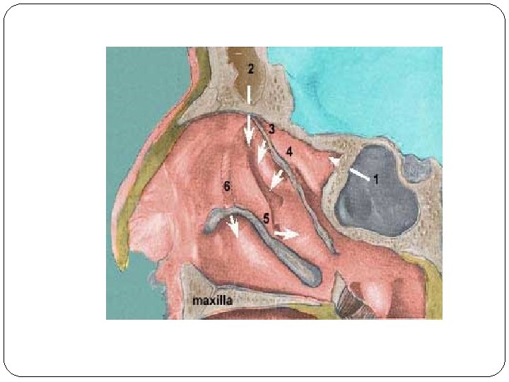
q Middle Meatus : Runs in post. Half of lateral wall. • • • Bulla Ethmoidalis- Is a round bulge present in middle meatus due to middle ethmoidal air cells, which open on or above it. Hiatus semilunaris is a gap below & infront of bulla which leades to ethmoidal infundibulum Uncinate Process -sickle shaped thin bonyplate, attached superiorly to middle tur. , /base of skull/lamina papyracea, & Inf. Attachment to inf. turbinate. Floor & medial wall of infundibulum are formed by uncinate process of the ethmoid. Atrium of the middle meatus – depression above the vestibule Agger nasi is a ridge above the atrium & may contain air cells.
Osteomeatal complex : Frontal Sinus, Ant. Ethmoidal, Maxillary Sinus opens in infundibulum Area of middle meatus into which maxillary, ant. Ethmoidal & frontal sinuses open & here malfunction of drainage, will have the maximum impact on these dependent sinuses. .
q Superior meatus – limited to Post. Third of lat. Wall Post. Ethmoidal sinus opens in it. Sphenoethmoidal recess – Lies above sup. Turbinate Sphenoid sinus opens in it. .
Medial wall – Is formed by nasal septum
Parts of Nasal Septum : - Columaellar Septum – Covered with skin Contains medial crura of lower lat. Cartilage - Membranous Septum – It consists of double layer of skin - Septum proper – Osteocartilaginous frame work of : Perpendicular plate of ethmoid Vomer Quadrilateral cartilage Crest of nasal bones, Platine & Maxilla Nasal spine of frontal bone Antrior nasal spine of Maxilla.
Roof - Ant. – Nasal Bone Post. – Body of sphenoid Middle – Cribriform plate of ethmoid. .
Floor – Palatine process of maxilla in ant. Three fourths Horizontal part of palatine bone in post. One fourth.
Blood supply of nose
Nasal Septum
Lateral wall
Venous drainage Sphenopalatine &ant. Facial vein Ophthalmic vein Sup. Saggital sinus.
Nerve Supply Olfaction – Olfactory nerves – • 12 -20 nerves pass through cribriform plate. • These nerves can carry sheaths of dura, pia & arachnoid in to the nose. .
Nerves of common sensation – • Ant. Ethomidal nerve – supplies ant. &sup. Part of nasal cavity • Branches of Infraorbital nerve – supply vestibule • Branches of sphenopalatine ganglion – post. 2/3 rd of nasal cavity. .
Autonomic nerves • Parsympathetic Nerves- supply nasal glands & control secretions. • Sympathetic – caroticotympanic plexus, deep petrosal nerve, vasoconstriction.
Anatomy of PNS
Clinically there are 2 groups of Para Nasal Sinuses Ant. Group- Maxillary, Frontal & ant. Ethmoidal Post. Group – Post. Ethmoidal, Sphenoid sinus. .
Maxillary Sinus (Antrum of Highmore) It is the Largest of Para Nasal Sinuses, pyramidal structure with a volume of 15 -30 ml in adults. Base– is towards lat. Nasal wall Maxillary ostia opens in the medial wall. Apex– Directed towards the zygomatic process. .
Walls of the pyramid: Anterolateral wall § Formed by ant. Surface of body of maxilla § Covered by periostium, soft tissue, skin of cheek. § Thinner at canine fossa. § Infraorbital foramen is present in relation to infraorbital margin. .
ROOF § Roof of antrum is formed by orbital plate of maxilla. § It has groove for infra Orbital nerve & vessels. Posterior wall § Formed by thin bone plate of post. Surface of maxilla & is related to pterygopalatine fossa & infratemporal fossa. § Fossa contains 3 rd part of internal maxillary artery, vidian nerve & sphenopalatine ganglion. .
Floor § Formed by alveolar & palatine process of maxilla. § It lies at level of floor of nasal cavity in children & about 1 cm. below the level of floor of nose in adults. § The roots of all molars, sometime premolars & canine are in relation to floor. - Oro- antral fistula can form on dental extraction - Dental infection can go to maxillary sinus. .
Drainage: Drains in middle meatus through natural ostium in medial wall. Accessary ostia are found in 4 -5% of adults. .
Frontal sinus It is situated between inner & outer tables of frontal bone Pyramidal in shape with apex pointing upwards & base at the floor. Volume is 6 -7 ml in adults Two frontal sinuses are often asymmetrical & loculated Frontal sinus may be absent on one or both sides.
Inf. Wall / Floor- formed by orbital roof. Anterior wall- outer table of frontal bone & skin. Posterior wall- inner table of frontal bone, separates sinus from cranial cavity. Medial wall- formed by septa between two sinuses. Drainage: Opening of sinus is present in its floor & leads into a space called frontal recess leading to middle meatus. .
Ethmoidal Sinuses – (Ethmoid Air cells) Thin walled air cavities in lateral masses of ethmoid bone 3 -18 in number Present between upper third of lat. Nasal wall & medial wall of orbit. .
Attachment of middle turbinate to lamina papyracea (ground lamella ) divides eth. Air cells in two groups- ant. Ethmoid cells- open in middle meatus - The largest & constant ant. Ethmoidal cell is BULLA ETHMOIDALIS - Post. Ethmoid cells- open in post meatus & sphenoethmoial recess. .
- Some times the most posterior ethmoidal cells may extend lat. To sphenoid behind its ant. Wall. Onodi cell. - These cells are closely related to optic nerve. .
- Ethmoidal cells may pneumatise in surrounding area & give rise to- Agger nasi cells - Ant. To attachment of middle turbinate to ant. Nasal wall - Ant. Most eth. Air cell. - Haller cell – Pneumatisation of orbital floor - Concha Bullosa- Pneumatised middle meatus.
Roof is called fovea ethmoidalis & formed by ant. Cranial fossa. Lat. Wall is Lamina Papyracea. It is thin paper like bone which separates it from orbit. .
Sphenoid Sinus Occupies the body of sphenoid, vol. is about 7. 5 ml. Both sided sinuses are asymmetrical- separated by septum, which may be deficient. Ostium is situated in upper part of ant. Wall & drains in sphenoethmoidal recess.
Roof- ant. Part is related to olfactory tract, optic chiasma & frontal lobe of brain. - Post part is related to pituitary gland in the sella turcica.
Lateral wall- ant. Part is related to optic nerve, Int. carotid artery, maxillary nerve. - Post. Part is related to cavernous sinus, Int. carotid A. & III, IV, V & VI cranial nerves. .
Functions of Sinuses 1. Air conditioning of inspired air 2. Voice resonance 3. Thermal insulation for protection of delicate structures of Orbit/ brain from variations in intranasal temp. 4. Lighten the skull bones.