Journey to the bottom of the face Pterygopalatine

Journey to the bottom of the face: Pterygopalatine Fossa Infratemporal fossa Pterygopalatine fossa Pterygomaxillary fissure

Pterygopalatine Fossa Boundaries: Anteriorly – posterior surface of maxilla Posteriorly – pterygoid process Medially – perpendicular plate of palatine Contents: • V 2 and its branches • Pterygopalatine ganglion • Terminal portion of maxillary artery

inf t an d me t la sup post Head and neck arts and crafts !!!
 is a deep space within the face that")
PTERYGOPALATINE FOSSA The pterygopalatine fossa (PPF) is a deep space within the face that is shaped like an inverted cone. The PPF communicates with infratemporal fossa, orbit, and nasal, oral, and cranial cavities. Pterygomaxillary fissure-Lateral opening (infratemporal fossa) Foramen Rotundum & Pterygoid Canal- Posterior openings (cranial cavity) Sphenopalatine Foramen-Medial opening (nasal cavity) Infraorbital fissure (maxillary sinus and onto face through infraorbital foramen)-Anterior Opening (orbit) Palatine Canal-Inferior opening (oral cavity)

Distribution of External Carotid A. Anterior: superior thyroid, lingual, and facial aa. (some little fat) Medial: ascending pharyngeal a. (apple) Posterior: occipital and posterior auricular a. (papa’s only) Terminal: maxillary and superficial temporal aa. (man stole) Some Little Fat Man Stole Papa’s Only Apple

Maxillary Artery One of two terminal branches of the external carotid artery. It takes a serpentine course through the infratemporal fossa. It can be divided into 3 portions during its course: 1. Mandibular portion • Runs between the medial surface of the ramus of the mandible and the sphenomandibular ligament. • deep auricular a. , anterior tympanic a. , middle meningeal a. and the inferior alveolar a. (which supplies • 2. blood to the mandibular teeth). mylohyoid a. branches from the inferior alveolar a. and supplies blood to the mylohyoid muscles. Pterygoid portion • passes either superficial or deep to the lateral pterygoid m. • Branches to muscles of mastication, buccal a. (a branch and travels with the buccal n. The buccal a. and buccal n. pierce the buccinator m. The buccal n. does not innervate the buccinator m. but the buccal a. does supply this muscle with blood. 3. Pterygopalatine portion: • These arterial branches run with the branches of the maxillary nerve V 2. This part of the maxillary a. • passes through the pterygomaxillary fissure to enter the pterygopalatine fossa. Most branches are given off in the pterygopalatine fossa however one branch, the posterior superior alveolar a. (PSA), is given off just before the maxillary artery enters the pterygomaxillary fissure. The PSA a. supplies blood to the maxillary molars and premolars, the buccal gingiva of the maxillary molars and premolars and mucosa of the maxillary sinus.

posterior superior alveolar a. infraorbital a. Anterior superior alveolar a. 3 rd: Pterygopalatine portion Posterior superior alveolar a. -branches JUST BEFORE the maxillary artery enters the pterygomaxillary fissure. It enters the maxilla to supply more posterior teeth of the maxilla. Infraorbital a. – maxillary a. becomes the infraorbital a. when it enters the infraorbital canal. Within the infraorbital canal the infraorbital a. gives off the anterior superior alveolar a. which supplies the pulps of the maxillary anterior teeth. The infraorbital a. exits the skull onto the face at the infraoribital foramen and then sends branches to the lower eyelid, lateral nose and upper lip.

Lateral wall of right nasal cavity sphenopalatine a. greater palatine a. descending palatine a. lesser palatine a. 3 rd: Pterygopalatine portion Descending palatine artery (greater & lesser palatine)- divides into the greater and lesser palatine arteries. Greater palatine a. exits via greater palatine foramen and supplies hard palate. Lesser palatine a. exits via lesser palatine foramen and supplies the soft palate Sphenopalatine artery- supplies the nasal cavity. One of the branches, the posterior septal a. descends with the nasopalatine nerve to exit the skull through the incisive foramen and joins with the greater palatine a.

Hard Palate 1. 2.

Blood Supply to Palate Sphenopalatine

Nosebleeds occur commonly in Kiesselbach’s area where there is an anastomosis between arteries arising from the internal and external carotid arteries. Two types: anterior (~90%) and posterior (~10%). Causes: nose picking, trauma, idiopathic, neoplasm in nasal cavity or paranasal sinuses Treatment: stop picking your nose!!! Pressure, chemical or electrical cautery, vessel ligation

A. foramen roundum B. foramen ovale C. foramen spinosum D. foramen lacerum E. carotid canal A. B. C. D. E. A. B. D. C.

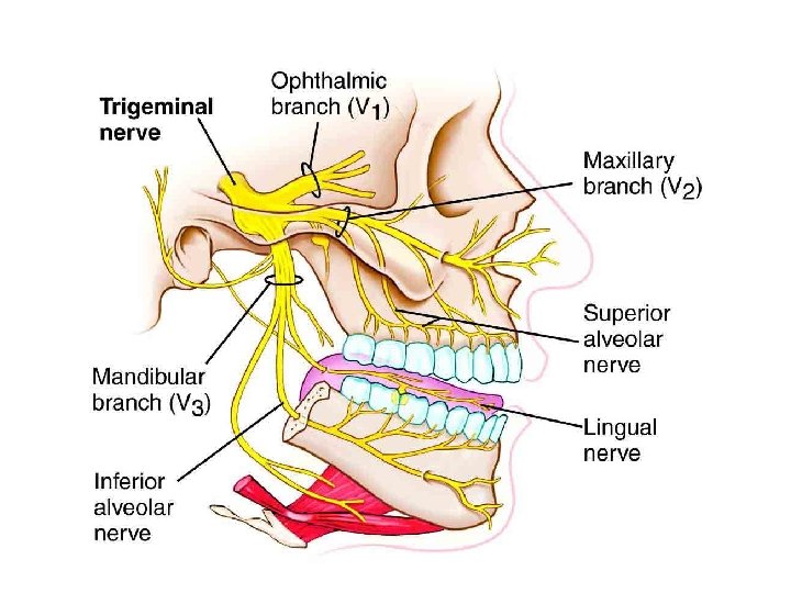
General Sensory Distribution of Maxillary Nerve • Mucous membrane of most of nasal cavity • Mucous membrane of roof of oral cavity = hard and soft palate • Skin of lower eyelid, lateral nose, prominence of cheek, skin and mucous membrane of upper lip, maxillary teeth

PTERYGOPALATINE FOSSA zygomatic infraorbital maxillary n. posterior superior alveolar Left lateral view, through the orbit and maxillary sinus Maxillary Nerve in Pterygopalatine Fossa Maxillary Nerve (division)-sensory to middle third of face. It enters the pterygopalatine fossa through foramen rotundum. It’s branches include: Infraorbital-sensory to face; has branches (anterior superior alveolar) to anterior maxillary teeth; leaves through inferior orbital fissure to enter maxillary sinus. Zygomatic-sensory to lateral side of face. Posterior superior alveolar-sensory to posterior maxillary teeth.

PTERYGOPALATINE FOSSA Deep view of nasal cavity Posterior nasal branches nasopalatine Sphenopalatine nerves (posterior nasal branches and nasopalatine) travel through sphenopalatine foramen to enter nasal cavity; sensory to nasal cavity

PTERYGOPALATINE FOSSA Nasopalatine n. Deep view of nasal cavity descending palatine nn. greater palatine lesser palatine Descending palatine nerves (greater and lesser palatine nerves) leave through the palatine canal for sensory innervation of hard & soft palates

Clinical correlate: Many of the arterial and venous branches of the ITF follow branches of V 2 and V 3. When dentists anesthetize branches of these nerves, arteries and veins may be punctured resulting in a facial hematoma. Also because the needle may inadvertently enter a vessel, dentists must always aspirate before injecting to prevent administering the anesthetic solution intravascularly. Sphenopalatine

Sympathetic fibers innervating the head and neck originate in upper thoracic levels (T 1 -T 4) of the intermediolateral cell column of the spinal cord. They ascend through the superior thoracic aperture to the base of the skull. The preganglionic sympathetic fibers terminate in the superior cervical ganglion, the most superior ganglion of the sympathetic chain at the base of the skull. It is posterior to the internal carotid artery. This is an external view.

Postganglionic sympathetic fibers from the superior cervical ganglion reach their target organs by: 1. Travel on the external and internal carotid arteries as the carotid plexus to follow these arteries to reach their target organs. 2. Follow internal carotid artery to the cranium and then "hitch a ride" with branches of CN V. Sympathetic fibers are not part of the trigeminal fibers. They are only following the trigeminal fibers to reach their target organs. 3) Travel on their own This is an internal view of the head.

Postganglionic sympathetic fibers from the superior cervical ganglion reach their target organs by: 1. Travel on the external and internal carotid arteries as the carotid plexus to follow these arteries to reach their target organs. 2. Follow internal carotid artery to the cranium and then "hitch a ride" with branches of CN V. Sympathetic fibers are not part of the trigeminal fibers. They are only following the trigeminal fibers to reach their target organs. 3) Travel on their own This is an internal view of the head.
 The superior salivary nucleus, located in the brainstem, is the")
FACIAL NERVE (CN VII) The superior salivary nucleus, located in the brainstem, is the parasympathetic origin for the first neuron in the two neuron chain that innervates the lacrimal gland, nasal and oral mucosa glands, submandibular salivary gland sublingual salivary gland. It travels with the remainder of the facial nerve and leaves the cranial cavity through the internal auditory meatus of the temporal bone to enter the facial canal.

In the facial canal the facial nerve has an initial horizontal course until it reaches its sensory ganglion, the geniculate ganglion. At the geniculate ganglion the majority of the facial nerve bends and descends through the temporal bone in the facial canal. At the geniculate ganglion a portion of the parasympathetic fibers, greater petrosal nerve, leave the remainder of the facial nerve and pass horizontally through the roof of the middle ear cavity.

The grater petrosal nerve reenters the cranial cavity through hiatus of the facial canal (hiatus for greater petrosal nerve). It is now on the floor of the cranial cavity. It leaves the cranial cavity a second time by entering the carotid canal and passing through a defect in its floor, the foramen lacerum.
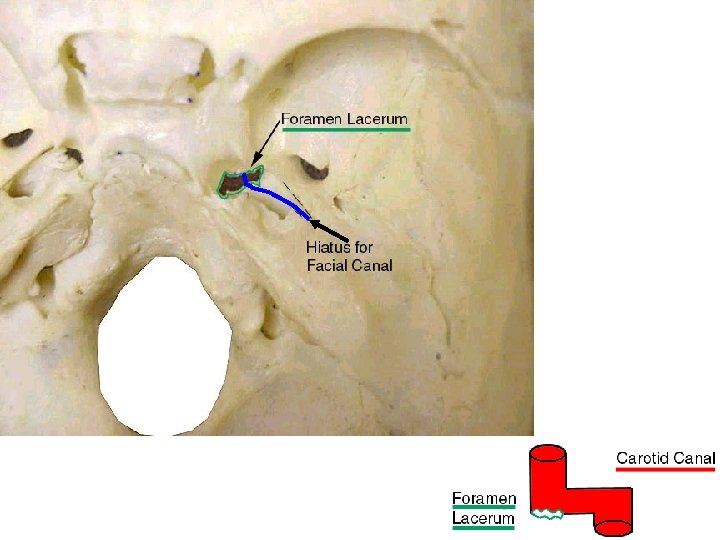

The greater petrosal nerve is now on the under side of the skull. It immediately enters the caudal opening of the pterygoid canal which is located immediately anterior to the foramen lacerum. As it enters the pterygoid canal the greater petrosal joins with deep petrosal nerve (postganglionic sympathetic fibers from the superior cervical ganglion that traveled on the internal carotid artery to the pterygoid canal’s posterior end). These two nerves form the nerve of the pterygoid canal.

The greater petrosal nerve portion of the nerve of the pterygoid canal terminates in the pterygopalatine ganglion. The postganglionic parasympathetic fibers and postganglionic sympathetic fibers will “hitch a ride” with branches of the maxillary nerve which enter the pterygopalatine fossa by way of the foramen rotundum. Branches destined to innervate nasal or oral mucosa glands join with sphenopalatine nerves (nasal cavity) or the greater & lesser palatine nerves of V 2.

Branches destined to innervate the lacrimal gland take a more indirect course. First they join with the zygomatic branch of V 2 and follow it into the floor of the orbit. Once in the orbit the autonomic fibers form a communicating branch that travels to the lacrimal nerve of V 1. They follow the lacrimal nerve to the lacrimal gland where they leave it to innervate the lacrimal gland.

Nose and Nasal Cavity

+ Broken Nose

The nasal septum is composed of the perpendicular plate of the ethmoid, vomer and nasal (septal) cartilage. Perpendicular Plate of the Ethmoid Nasal (septal) Cartilage Vomer

Nasal Septum = Medial Wall of Nasal Cavity

The Ethmoid crista gali ethmoidal air cells medial wall of orbit middle nasal concha perpendicular plate

Lateral Nasal Wall Nasal Conchae - bony protuberances extending into the nasal cavity along its lateral wall 1. superior nasal concha: part of the ethmoid bone; located superior and posterior in the nasal cavity 2. middle nasal concha-part of the ethmoid bone; lies inferior to the superior nasal concha 3. inferior nasal concha-its own bone; most prominent of the nasal concha There is a meatus (depression) that is named accordingly inferior to each concha.

Piriform aperture

Lateral Nasal Wall Meatuses: depression inferior to each concha 1. inferior meatus - nasolacrimal duct empties into it 2. middle meatus - frontal sinus, anterior and middle ethmoid air cells & maxillary sinus empty into it hiatus semilunaris: a crescent shaped slit for openings of frontal, maxillary sinuses & anterior ethmoid air cells) bulla ethmoidalis: raised area superior to the hiatus semilunaris; formed by middle ethmoid air cells which open on it 3. superior meatus - posterior ethmoid air cells open into it 4. sphenoethmoidal recess - superior to superior concha; sphenoid sinus opens into it

Nasal Septum & Sinuses The paranasal sinuses develop as diverticula of the lateral nasal wall, they extend into bone: 1. maxilla - 3 rd fetal month; extension of nasal sac 2. ethmoid - 5 th fetal month; extension of the middle meatus 3. sphenoid - 5 th postnatal month; extensions of the ethmoid sinuses 4. frontal - 5 th postnatal year; extensions of the middle meatus and of the ethmoid sinuses

+ Paranasal sinuses and the orbit
- Slides: 38