Genetics Lec 2 Dr Mohammed Hussein 1 2


 a a A a a a 50 % Disease 50")


 Male Y X X x X Y XX Normal")


 • Because the Y chromosome carries only about 50 protein-coding genes and the")


 Mitochondrial Encephalomyopathy, Lactic Acidosis, and Stroke (MELAS)")
")
, MRCPCH (Associate)")


, MRCPCH (Associate)")
- Slides: 71
Genetics Lec. 2 Dr. Mohammed Hussein
1. 2. 3. 4. Autosomal Dominant Inheritance Autosomal Recessive Inheritance Sex - linked Dominant Inheritance Sex - linked Recessive Inheritance
Autosomal Dominant Inheritance
Aa A Diseased (Hetrozygous) a a A a a a 50 % Disease 50 % Normal aa Normal Punnett Square
Autosomal Recessive Inheritance
Aa A Heterozygous carrier a A A a 75 % No Disease a A a a a 25 % Disease Aa Heterozygous carrier
• In codominant inheritance, two different alleles of a gene are expressed, and each version makes a slightly different protein. • Both alleles influence the genetic trait or determine the characteristics of the genetic condition
ABO blood group • There are three alleles that determine the type of the blood group; • A, B & O alleles • Both A & B alleles are Dominant over the O allele (the Recessive allele)
A A Blood group A A O Blood group A B B Blood group B B O Blood group B O O Blood group O A B Blood group AB
AO A Blood group A O 25% Blood group A B B O BO Blood group B 25% Blood group AB O A O O O 25% Blood group O
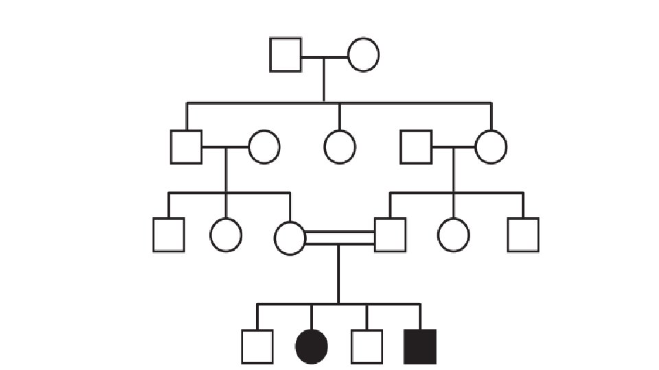
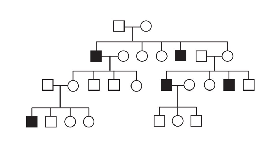
• X-linked = gene transmitted by the X chromosome • Recessive = required two copies of the gene to express the disease (Homozygous) • If the gene present with a normal gene then a carrier (Hetrozygous)
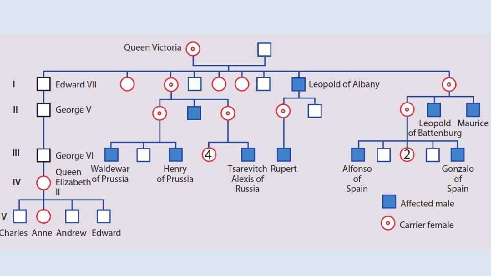
• Because males have only one copy of the X chromosome, they are said to be hemizygous for the X chromosome. • As recessive disease required two copies of gene to be expressed, X-linked recessive diseases are seen much more commonly in males than in females. • As male pass only the Y chromosome to his son, so male-to-male transmission is not seen in X-linked inheritance.
x. Y x Affected (Diseased) Male Y X X x X Y XX Normal Female 100 % of female are carriers 100 % of males are normal
XY X Normal Male Y X X Y x x X x Y Xx Female carrier 50 % of female are carriers 50 % of males are diseased
Hemophilia A and B Duchenne muscular dystrophy Lesch-Nyhan syndrome G 6 PD
Hemophilia A
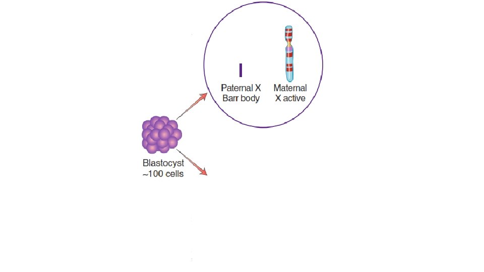
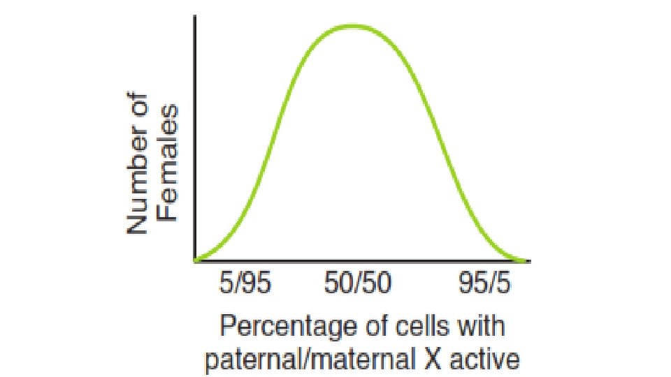
) • Because the Y chromosome carries only about 50 protein-coding genes and the X chromosome carries hundreds of protein-coding genes, a mechanism must exist to equalize the amount of protein encoded by X chromosomes in males and females. • This mechanism, termed X inactivation, occurs in the blastocyst (~100 cells) during the development of female embryos.
x X 95% 5%
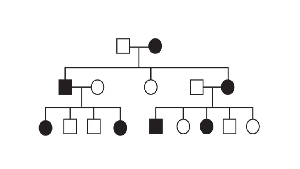
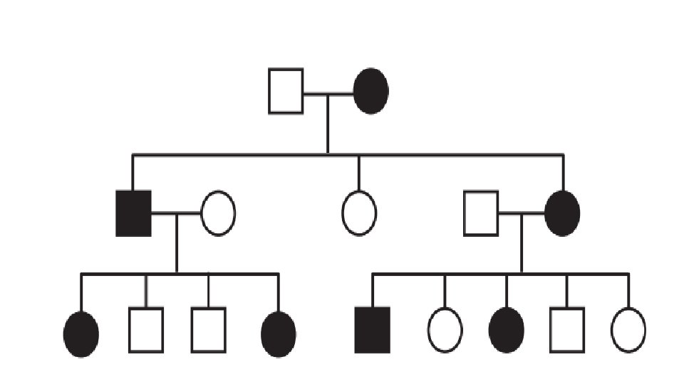
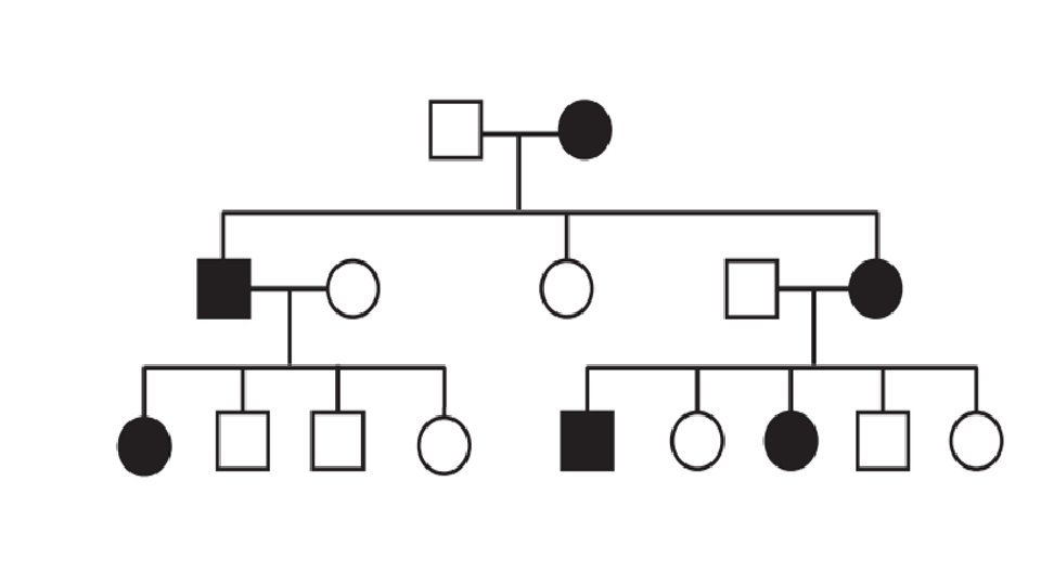
• Male–male transmission of the disease is not seen. • As the disease is dominant (one copy of the abnormal gene cause the disease), so Heterozygous females are affected. • Because females have two X chromosomes (and thus two chances to inherit an X-linked gene) and males have only one, X-linked dominant diseases are seen about twice as often in females as in males
XY X Affected Male Y X X X X Y XX Normal Female 100 % of female are diseased 100 % of males are normal
XY X X Normal Male Y X Y 50 % of offspring are diseased X Y 50 % of offspring are normal XX Affected Female X X X 50 % of females are diseased 50 % of males are disased
Hypophosphatemic rickets Fragile X syndrome
Fragile X syndrome
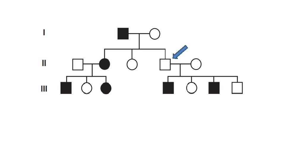
Summary • Ask yourself the following questions 1. Does an affected individual have an affected parent? Or (Multiple generations affected? ) 2. 3. 4. o Yes: Dominant disease o No: Recessive disease If it is recessive, then ask: Does all (or almost all) affected are males? o Yes: X-linked recessive disease o No: Autosomal recessive disease If it is dominant, then ask: Is there male-male transmission? o Yes: autosomal dominant disease o No: may be an X-linked dominant Are all daughters of an affected male also affected? o Yes: X-linked dominant o No: autosomal dominant disease
Does an affected individual have an affected parent? o Yes Dominant Is there male-male transmission? o Yes Autosomal Dominant 1. Does an affected individual have an affected parent? 2. If it is recessive, then ask: Does all affected are males? 3. If it is dominant, then ask: Is there male transmission? 4. Are all daughters of an affected male also affected?
Does an affected individual have an affected parent? o No Recessive Does all affected are males? o No Not X linked Autosomal Recessive 1. Does an affected individual have an affected parent? 2. If it is recessive, then ask: Does all affected are males? 3. If it is dominant, then ask: Is there male transmission? 4. Are all daughters of an affected male also affected?
Does an affected individual have an affected parent? o No Recessive Does all affected are males? o Yes X linked X-Linked Recessive 1. Does an affected individual have an affected parent? 2. If it is recessive, then ask: Does all affected are males? 3. If it is dominant, then ask: Is there male transmission? 4. Are all daughters of an affected male also affected?
Does an affected individual have an affected parent? o Yes Is there male-male transmission? o No Are all daughters of an affected male also affected? o Yes X-Linked Dominant 1. Does an affected individual have an affected parent? 2. If it is recessive, then ask: Does all affected are males? 3. If it is dominant, then ask: Is there male transmission? 4. Are all daughters of an affected male also affected? Dominant May be Autosomal May be X-linked
Does an affected individual have an affected parent? o Yes Dominant Is there male-male transmission? o No May be Autosomal May be X-linked Are all daughters of an affected male also affected? o No Not X-linked Autosomal Dominant 1. Does an affected individual have an affected parent? 2. If it is recessive, then ask: Does all affected are males? 3. If it is dominant, then ask: Is there male transmission? 4. Are all daughters of an affected male also affected?
Mitochondria
• Because a sperm cell contributes no mitochondria to the egg cell during fertilization, mitochondrial DNA is inherited exclusively through females.
• Pedigrees for mitochondrial diseases thus display a distinct mode of inheritance: 1. Transmission of the disease is only from a female. 2. All offspring of an affected female are affected. 3. None of the offspring of an affected male is affected.
Heteroplasmy
Heteroplasmy
Leber hereditary optic neuropathy (LHON) Mitochondrial Encephalomyopathy, Lactic Acidosis, and Stroke (MELAS)
Leber hereditary optic neuropathy (LHON)
Dr. Mohammed Hussein M. B. Ch. B, MSC, DCH (UK), MRCPCH (Associate)
Important principles that can characterize single-gene diseases 1. 2. 3. 4. 5. 6. 7. 8. 9. Variable expression Incomplete penetrance Pleiotropy Locus heterogeneity New mutations Delayed age of onset Anticipation Imprinting Uniparental disomy
Variable Expression • Most genetic diseases vary in the degree of phenotypic expression: Some individuals may be severely affected, whereas others are more mildly affected. • This can be the result of several factors: 1. Environmental Influences: exp xeroderma pigmentosum 2. Allelic Heterogeneity: exp, missense mutations in the factor VIII gene tend to produce less severe hemophilia than do nonsense mutations. 3. Heteroplasmy in mitochondrial pedigrees. 4. Modifier Loci. Disease expression may be affected by the action of other loci, termed modifier loci. Often these may not be identified.
Incomplete Penetrance • A disease-causing mutation is said to have incomplete penetrance when some individuals who have the disease genotype do not display the disease phenotype.
Pleiotropy • Pleiotropy exists when a single disease-causing mutation affects multiple organ systems. • Pleiotropy is a common feature of genetic diseases. • Marfan syndrome provides a good example of the principle of pleiotropy
Marfan syndrome Yao Ming
Locus Heterogeneity • Locus heterogeneity exists when the same disease phenotype can be caused by mutations in different loci. • For example, retinitis pigmentosa has autosomal dominant, autosomal recessive, and X-linked origins.
New Mutations • In many genetic diseases, a large proportion of cases are caused by a new mutation transmitted from an unaffected parent to an affected offspring. • There is thus no family history of the disease
Delayed Age of Onset • Many individuals who carry a disease-causing mutation do not manifest the phenotype until later in life. • This can complicate the interpretation of a pedigree because it may be difficult to distinguish genetically normal individuals from those who have inherited the mutation but have not yet displayed the phenotype. • Example: Familial breast cancer
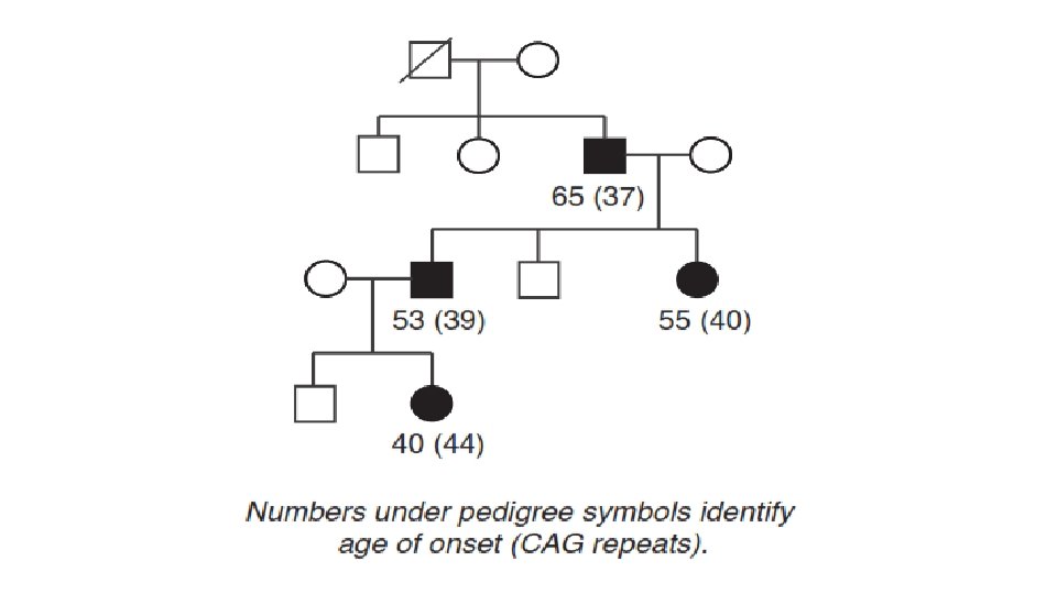
Genetic Anticipation • Anticipation refers to a pattern of inheritance in which individuals in the most recent generations of a pedigree develop a disease at an earlier age or with greater severity than do those in earlier generations. • For a number of genetic diseases, this phenomenon can be attributed to the gradual expansion of trinucleotide repeat polymorphisms within or near a coding gene.
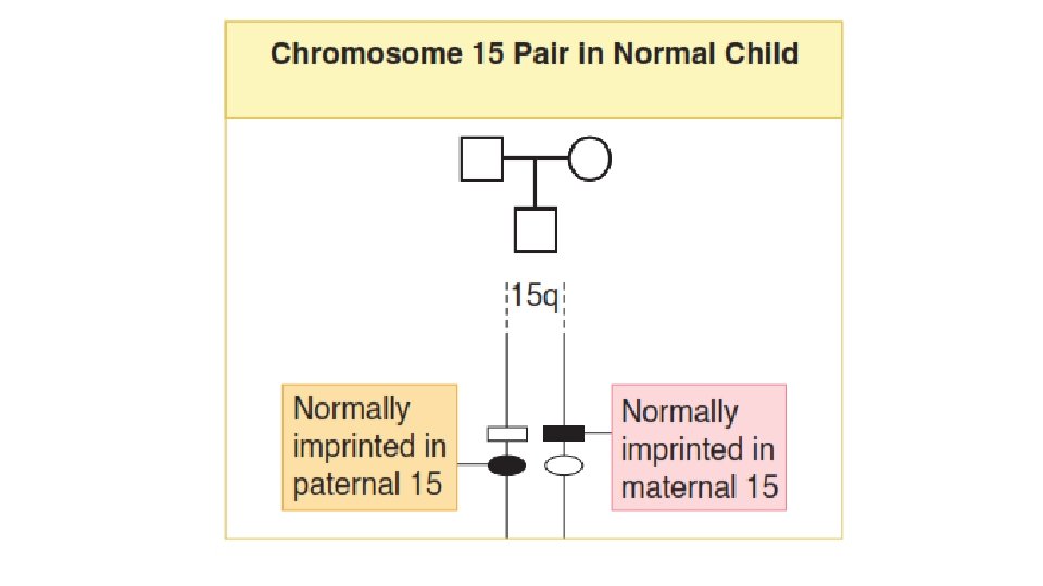
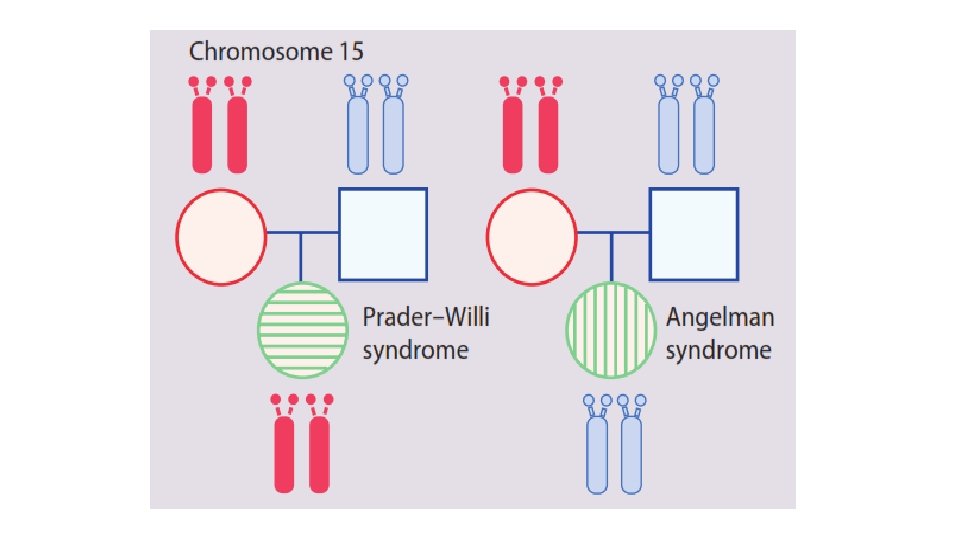
Imprinting • Imprinting refers to the fact that a small number of genes are transcriptionally active only when transmitted by one of the two sexes. • The homologous locus in the other parent is rendered transcriptionally inactive. • Thus, for imprinted loci, it is normal to have only the maternal (for some loci) active, or only the paternal (for other loci) active.
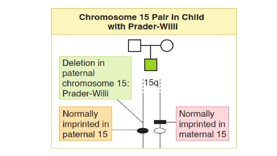
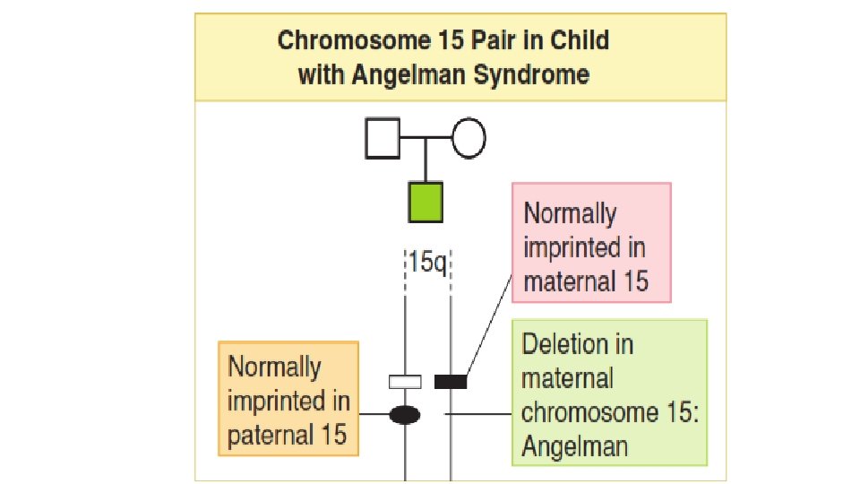
• On rare occasion, the transcriptionally active gene may be deleted from the chromosome during gametogenesis. • This leaves the offspring with no active gene at that locus. • The gene from one parent is inactivated due to normal imprinting, and the gene from the other parent deleted by a mutation. • This situation may result in a genetic disease.
Prader-Willi Syndrome and Angelman Syndrome
Prader-Willi Syndrome
Angelman Syndrome
Uniparental Disomy • Uniparental disomy is a rare condition in which both copies of a particular chromosome are contributed by one parent.
Dr. Mohammed Hussein M. B. Ch. B, MSC, DCH (UK), MRCPCH (Associate)