
Pneumonia By Dr Mona Mohammed Definition Pneumonia is

Pneumonia By Dr/ Mona Mohammed
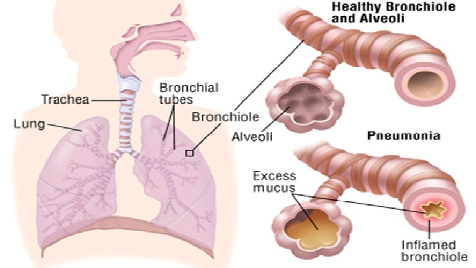
Definition: Pneumonia is an inflammation of the lung parenchyma may be caused by either infectious or noninfectious agents. Examples of infectious agents are bacteria, mycobacteria, fungi, and nonspecific viruses. Noninfectious agents may include irritating fumes, dust, or chemicals that are inhaled or foreign matter that is aspirated.
Classification: Pneumonias are classified as: - Community-acquired pneumonia (CAP): it is an acute infection of the lung occurring in patients who have not been hospitalized or resided in a long-term care facility within 14 days of the onset of symptoms. - Hospital-acquired (nosocomial) pneumonia (HAP): it is pneumonia that occurs 48 hours or longer after hospital admission and was not incubating at the time of hospitalization.
- Pneumonia in the immunocompromised host or opportunistic Pneumonia: Individuals at risk for opportunistic pneumonia include those with altered immune responses. - Aspiration pneumonia: it results from the abnormal entry of material from the mouth or stomach into the trachea and lungs.
Etiology: - When the defense mechanisms become incompetent or are overwhelmed by the virulence or quantity of infectious agents. - Decreased consciousness depresses the cough and epiglottal reflexes, which may allow aspiration of oropharyngeal contents into the lungs. - Tracheal intubation interferes with the normal cough reflex and the mucociliary escalator mechanism.
- Air pollution. - Cigarette smoking. - Viral URIs. - Normal changes that occur with aging can impair the mucociliary mechanism. - Chronic diseases can suppress the immune system’s ability to inhibit bacterial growth.
Methods By Which Organisms Can Reach The Lung: Organisms that cause pneumonia reach the lung by three methods: - Aspiration of normal flora from the nasopharynx or oropharynx. Many of the organisms that cause pneumonia are normal inhabitants of the pharynx in healthy adults. - Inhalation of microbes present in the air. Examples include Mycoplasma pneumoniae and fungal pneumonias. - Hematogenous spread from a primary infection elsewhere in the body. An example is Staphylococcus aureus.
Pathophysiology: Pneumonia is an acute infection of the lungs that occurs when an infectious agent enters and multiplies in the lungs of a susceptible person. When the microorganisms multiply, they release toxins that induce inflammation in the lung tissue, causing damage to mucous and alveolar membranes. This leads to the development of edema and exudate, which fills the alveoli and reduces the surface area available for exchange of carbon dioxide and oxygen. Some bacteria also cause necrosis of lung tissue.
Pneumonia may be confined to one lobe, or it may be scattered throughout the lungs. If it affects only one lobe, it is called lobar pneumonia. Generalized pneumonia is much more serious and is called bronchopneumonia. Bronchopneumonia occurs more often as a nosocomial (hospitalacquired) infection in hospitalized patients, the very young, or the very old.
Risk Factors: - Abdominal or thoracic surgery. - Age >65 yrs. - Air pollution. - Altered consciousness: alcoholism, head injury, seizures, anesthesia, drug overdose and stroke. - Bed rest and prolonged immobility. - Chronic diseases: chronic lung and liver disease, diabetes mellitus, heart disease, cancer and chronic kidney disease.
- Debilitating illness. - Exposure to bats, birds, rabbits, farm animals. - Immunosuppressive chemotherapy, disease human and/or therapy immunodeficiency (corticosteroids, virus [HIV] immunosuppressive therapy after organ transplant). - Inhalation or aspiration of noxious substances. - Intestinal and gastric feedings via nasogastric or nasointestinal tubes. cancer infection,
- IV drug use. - Malnutrition. - Recent antibiotic therapy. - Resident of a long-term care facility. - Smoking. - Tracheal intubation (endotracheal intubation, tracheostomy). - Upper respiratory tract infection.
Clinical Manifestations: - Shortness of breath due to inflammation within the lungs, impairing gas exchange. - Difficulty breathing (dyspnea) due to inflammation and mucus within the lungs. - Fever due to infectious process. - Chills due to increased temperature. - Cough due to mucous production and irritation of the airways. - Crackles due to fluid within the alveolar space and smaller airways. - Rhonchi due to mucus in airways; wheezing due to inflammation within the larger airways.
- Discolored, possibly blood-tinged, sputum due to irritation in the airways or microorganisms causing infection. - Tachycardia and tachypnea as the body attempts to meet the demand for oxygen. - Pain on respiration due to pleuritic inflammation, pleural effusion, or atelectasis development. - Headache, muscle aches (myalgia), joint pains, or nausea may be present depending on the infecting organism.
Complications: Potential complications include the following: - Pleurisy (inflammation of the pleura) is relatively common. - Pleural effusion (fluid in the pleural space) can occur. - Atelectasis (collapsed, airless alveoli) of one or part of one lobe may occur. - Bacteremia (bacterial infection in the blood) is more likely to occur in infections with Streptococcus pneumonia and Haemophilus influenzae.
- Lung abscess (not common). - Empyema (the accumulation of purulent exudate in the pleural cavity). - Pericarditis. - Meningitis. - Sepsis can occur when bacteria within alveoli enter the bloodstream. - Acute respiratory failure. - Pneumothorax.
Prognosis: Prognosis will vary depending on patient’s age, preexisting lung disease, infecting organism and response to antibiotics. Patients at greater risk for complications from pneumonia will be treated within the hospital, while those at lower risk may be treated at home.
Diagnostic Findings: - History: history (particularly of a recent respiratory tract infection). - Physical Examination: On physical examination, rhonchi and crackles may be auscultated over the affected region. If consolidation is present, bronchial breath sounds, and increased fremitus (vibration of the chest wall produced by vocalization) may be noted. Patients with pleural effusion may exhibit dullness to percussion over the affected area.
- Chest X-Ray: Shadows on chest x-ray, indicating infiltration, may be in a lobar or segmental pattern or more scattered. - Gram stain of sputum: to identify the organism is obtained before beginning antibiotic therapy. - Sputum culture and sensitivity test: (to identify the infective agent and the appropriate antibiotics). - Pulse oximetry (Low oxygen saturation) or ABGs (may show low oxygen and elevated carbon dioxide levels).
- Complete blood count, WBC differential {Elevated WBC (leukocytosis) showing sign of infection}, and routine blood chemistries (if indicated) - Blood cultures (bloodstream invasion occurs frequently). - Auscultation of the chest reveals wheezing, crackles, and decreased breath sounds. The nail beds, lips, and oral mucosa may be cyanotic. - Bronchoscopy is often used in patients with acute severe infection, in patients with chronic or refractory infection, in immunocompromised patients when a diagnosis cannot be made from an expectorated or induced specimen, and in mechanically ventilated patients.
Treatment: - Administer oxygen as needed. - Antibiotics are given for the most likely organism (empirically) until the sputum culture results are returned. - Administer antipyretics when fever >38 for patient comfort (as Acetaminophen, ibuprofen).
- Administer brochodilators to keep airways open, enhance airflow if needed (as Albuterol, metaproterenol, levalbuterol via nebulizer or metered dose inhaler). - Increase fluid intake to help loosen secretions and prevent dehydration. - Instruct the patient on how to use the incentive spirometer to encourage deep breathing; monitor progress.
Nutritional Therapy: - Hydration is important in the supportive treatment of pneumonia to prevent dehydration and loosen secretions. - Individualize and carefully monitor fluid intake if the patient has heart failure. - If the patient cannot maintain adequate oral intake, IV administration of fluids and electrolytes may be necessary.
- Weight loss often occurs in patients with pneumonia because of increased metabolic needs and difficulty eating due to shortness of breath and pleuritic pain. - Small, frequent meals are easier for dyspneic patients to tolerate. - Offer foods high in calories and nutrients.
Prevention: - Promote coughing and expectoration of secretions if client experiences increased mucus production. - Change position frequently if client is immobilized for any reason. - Encourage deep-breathing and coughing exercises at least every 2 hours. - Administer chest physical therapy as indicated. - Suction client if he or she cannot expectorate.
- Prevent aspiration in clients at risk. - Prevent infections. - Cleanse respiratory equipment on a routine basis. - Promote frequent oral hygiene. - Administer sedatives and opioids carefully to avoid respiratory depression. - Encourage client to stop smoking and reduce alcohol intake. - Vaccination against pneumococcal infection.
Nursing Management: Assessment: Nursing assessment is critical in detecting pneumonia. Respiratory assessment further identifies the clinical manifestations of pneumonia. The nurse monitors the patient for the following: changes in temperature and pulse; amount, odor, and color of secretions; frequency and severity of cough; degree of tachypnea or shortness of breath; changes in physical assessment findings and changes in the chest x-ray findings.
Nursing Diagnoses: Nursing diagnoses for the patient with pneumonia may include, but are not limited to, the following: - Impaired gas exchange related to fluid and exudate accumulation at the capillary-alveolar membrane. - Ineffective breathing pattern related to inflammation and pain. - Acute pain related to inflammation and ineffective pain management and/or comfort measures.
Planning: The overall goals are that the patient with pneumonia will have: 1. Clear breath sounds. 2. Normal breathing patterns. 3. No signs of hypoxia. 4. Normal chest x-ray. 5. No complications related to pneumonia.
Intervention: - Monitor respiration for rate, effort, use of accessory muscles, skin color, and breathing sounds. - Record fluid intake and output for differences, signs of dehydration. - Record sputum characteristics for changes in color, amount, and consistency. - Properly dispose of sputum. - Explain to the patient: Take adequate fluids— 3 liters per day—to prevent excess fluid loss through the respiratory system with exhalation. Use of incentive spirometer.
Evaluation: The expected outcomes are that the patient with pneumonia will: - Have effective respiratory rate, rhythm, and depth of respirations. - Lungs clear to auscultation.

- Slides: 33