Part 2 6 25 PM Dr Mohammed Hussein


 2. Deletions (del) 3. Inversions (inv)")


")

 • A male with")


 • Chronic Myelogenous Leukemia")


")
")


")

 Down syndrome (parent carries Robertsonian translocation) 47, XX, +21")


 • 46, XY, del(5 p) • Cri-du-chat syndrome")


(p 21; q 13)")

")


")

 • Spectral Karyotyping")
 • a chromosome-specific DNA segment is labelled with a")

")


- Slides: 55
Part 2 6: 25 PM Dr. Mohammed Hussein
• Structural alterations of chromosomes occur when chromosomes are broken by agents termed clastogens, example o Radiation o Some viruses o Some chemicals
Balanced & Unbalanced alterations • Balanced alterations: do not result in a gain or loss of genetic material and usually have fewer clinical consequences • Unbalanced alterations: alterations result in a loss or gain of genetic material
Germ cell & Somatic cell alterations • Germ line alterations can be transmitted to offspring. • Somatic cells alterations not transmitted to offspring, but can alter genetic material such that the cell can give rise to cancer.
Types of Structural Chromosome Abnormalities 1. Translocations (t) 2. Deletions (del) 3. Inversions (inv) 4. Ring Chromosome (r) 5. Isochromosome (i) Common Less Common
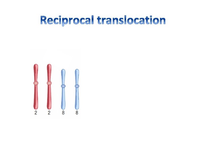
• Translocations occur when chromosomes are broken and the broken elements reattach to other chromosomes. • Translocations can be classified into two major types: 1. Reciprocal 2. Robertsonian
• Reciprocal translocations occur when genetic material is exchanged between nonhomologous chromosomes (other than acrocentric chromosomes) • Robertsonian translocations occur only in the acrocentric chromosomes (13, 14, 15, 21, and 22).
46, XY, t(2 p; 8 p)
Female has translocation between short arm of chromosome 1 and long arm of chromosome 12, write the karyotype? 46, XX , t( 1 p ; 12 q )
• 46, XY, t(5 p 13; 10 q 25) • A male with translocation between 5 p 13 (band 3 of region 1 of short arm of chromosome 5) and 10 q 25 (band 5 of region 2 of long arm of chromosome 10) • 46, XY, t(5; 10)(p 13; q 25)
Fertilization with normal egg
• Reciprocal translocations may occur by chance at the somatic cell level throughout life. • Because these translocations involve only a single cell and the genetic material is balanced, there is often no consequence. • Rarely, however, a reciprocal translocation may alter the expression or structure of an oncogene or a tumor suppressor gene, conferring an abnormal growth advantage to the cell.
Philadelphia chromosome • t(9; 22) • Chronic Myelogenous Leukemia
c myc oncogene • Reciprocal translocation involving 8 q 24 and 14 q 32 associated with Burkitt's lymphoma • 46, XX, t(8; 14)(q 24; q 32)
• • Much more common than reciprocal translocations occur in approximately 1 in 1, 000 live births. They occur only in the acrocentric chromosomes. Involve the loss of the short arms of two of the chromosomes and subsequent fusion of the long arms.
q. Acrocentric chromosomes: have the centromere far toward one end
45, XY, – 14, – 21, +t(14 q; 21 q)
• The extra chromosome 21 may result from 1. Meiotic non disjunction (aneuploidy) 94% 2. Robertsonian translocation 5% 3. Mosaicism 1%
karyotype 47, XX, +21 47, XY, +21
• About 5% of Down syndrome cases are the result of a Robertsonian translocation affecting chromosome 14 and chromosome 21.
1/3 Fertilization with normal egg X X X
46, XY, – 14, +t(14 q; 21 q)
• 1% of Down syndrome cases results from Mosaicism. • In which some of the cells are normal and some have trisomy 21. • It can arise by later mitotic nondisjunction. • The phenotype is sometimes milder in Down syndrome mosaicism.
Down syndrome (nondisjunction during meiosis) Down syndrome (parent carries Robertsonian translocation) 47, XX, +21 or 47, XY. +21 46, XX, – 14, +t(14 q; 21 q) 46, XY, – 14, +t(14 q; 21 q) No association with prior pregnancy loss May be associated with prior pregnancy loss Older mother May be a younger mother Very low recurrence rate Recurrence rate 10– 15% if mother is translocation carrier; 1– 2% if father is translocation carrier Parent chromosomal analysis is not required Parent chromosomal analysis is recommended
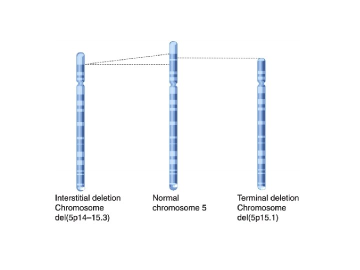
• A deletion occurs when a chromosome loses some of its genetic information. • Two types: 1. Terminal deletions (the end of the chromosome is lost) 2. Interstitial deletions (material within the chromosome is lost)
• 46, XX, del(5 p) • 46, XY, del(5 p) • Cri-du-chat syndrome
Cri-du-chat syndrome
• Deletions can be large and microscopically visible in a stained preparation • Some deletions may be so small that they are not readily apparent microscopically without special fluorescent probes (FISH) where they called microdeletions.
• Their frequency and clinical consequences tend to be less severe than those of translocations and deletions. 1. Inversions (inv) 2. Ring Chromosome (r) 3. Isochromosome (i)
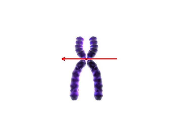
• Inversions occur when the chromosome segment between two breaks is reinserted in the same location but in reverse order. 1. Pericentric inversions include the centromere 2. Paracentric inversions do not include the centromere
46, XY, inv(3)(p 21; q 13)
• A ring chromosome can form when a deletion occurs on both tips of a chromosome and the remaining chromosome ends fuse together
46, X, r(X)
Consequences • Ring chromosomes are often lost, resulting in a monosomy, example: • loss of a ring X chromosome would produce Turner syndrome (45, XO)
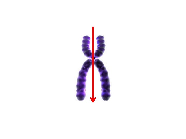
• When a chromosome divides along the axis perpendicular to its normal axis of division, an isochromosome is created (i. e. , two copies of one arm but no copy of the other).
46, X, i(Xq)
Consequences • Autosomal isochromosomes are lethal • X chromosome isochromosome results in Turner syndrome
Advances in Molecular Cytogenetics • Fluorescence in situ Hybridization (FISH) • Spectral Karyotyping
Fluorescence in situ Hybridization (FISH) • a chromosome-specific DNA segment is labelled with a fluorescent tag to create a probe. • This probe is then hybridized with the patient’s chromosomes, • which are visualized under a fluorescence microscope. • Because the probe will hybridize only with a complementary DNA sequence, • the probe will mark the presence of the chromosome segment being tested.
• For example, a probe that is specific for chromosome 21 will hybridize in three places in the cells of a trisomy 21 patient, providing a diagnosis of Down syndrome.
Fluorescence in situ Hybridization (FISH)
• FISH is also commonly used to detect deletions: An analysis using a probe that hybridizes to the region of 15 q corresponding to Prader-Willi syndrome will show only a single signal in a patient, confirming the diagnosis of this deletion syndrome.
• An advantage of FISH is that chromosomes do not have to be in the metaphase stage for an accurate diagnosis
Spectral Karyotyping • Spectral karyotyping involves the use of five different fluorescent probes that hybridize differentially to different sets of chromosomes. • In combination with special cameras and image-processing software, this technique produces a karyotype in which every chromosome is “painted” a different colour. • This allows the ready visualization of chromosome rearrangements, such as small translocations, e. g. , the Philadelphia chromosome rearrangement t(9; 22) involved in chronic myelogenous leukaemia.
Spectral Karyotyping
6: 28 PM