Medical Parasitology Assistant Prof Dr Ahmed A Mohammed


 is ingested")


- Slides: 21
Medical Parasitology Assistant Prof. Dr. Ahmed A. Mohammed Lecture 5 Helminths
• Special adaptations for parasitic mode of life and for species survival are more apparent in the helminths than in the protozoa. • The complete or partial loss of the digestive tract in certain parasitic helminths is presumed to be because of their location in the host’s intestine or tissue, where predigested nutrients are abundant. • A related adaptation in the trematodes and cestodes is evident in the tegument, which on its outer surface has a coat of microvilli morphologically similar to that of the intestinal mucosa of vertebrates. • While most of the vital systems of the parasitic helminths have been modified toward simplification, the reproductive system has been modified toward increased capacity.
• In some helminths, the life cycle is direct and relatively simple, involving only one host species and a brief period of development of an infective transfer stage. Groups of Helminths I- Phylum: Platyhelminths This phylum includes the following classes: 1 Class: Turbellaria. 2 Class: Trematoda. 3 Class: Cestoda. A- Adult Tapeworm Infection -Kingdom: Animalia -Phylum : Platyhelminths -Class : Cestoda -Sub-class: Eucestoda -Order : Cyclophyllidea
1 Taenia saginata • This parasite causes beef tapeworm infection. • The adult worm typically develops in the middle third of the small intestine. • The average length of the relaxed worm is approximately 5 meters, although there are records of specimens of far greater length. • It has 1000 to 2000 proglottids of which from one third to one half are nearly gravid. • Usually only a single specimen occurs in an infection, but there may be more. • The fully developed worm is delicate anteriorly and more robust posteriorly.
• The scolex bears four suckers and a slight apical depression. • Immediately behind the delicate unsegmented (neck) there is a region of immature proglottids in which the genital organs are not yet developed. • Gradually the more distal of these proglottids increase in breadth and width, these are the mature proglottids, each of which contains a full set of functioning male and female reproductive organs. • More distally, the mature unite have transformed into more elongated, narrower, gravid ones as a result of the development of a large number of branched lateral arms of a uterus 12 to 30.
• The terminal gravid proglottids become separated from the strobila and actively migrate out of the bowel or are evacuated in the stool with only partial loss of eggs. • The eggs are essentially spherical and have a thin, transparent outer embryonal envelope and a thick brown shell composed of many slender rods cemented together. Within this shell, the hexacanth embryo which has three pairs of delicate lancetshaped hooklets.
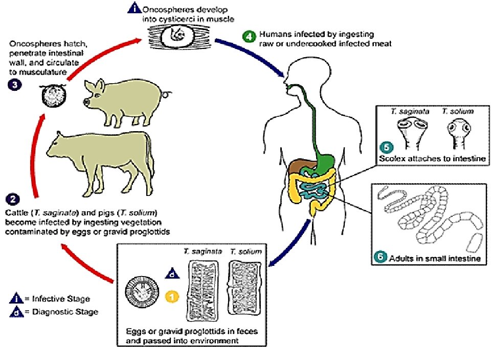
Life cycle • The evacuated gravid segments extrude the eggs while crawling on the ground, vegetation, or other surfaces. Cattle grazing on the infested ground pick up the eggs, which hatch in the duodenum. The emerging embryos penetrate the mesenteric venules or lymphatics and reach the skeletal muscles or the heart where in about 2 months they transform into a typical cysticercus stage (called cysticercus bovis) which measures roughly 5 -10 mm and has a head like that of the adult worm, invaginated into a fluid-filled bladder. • Thereafter, for a period of more than a year, a person who eats the raw infected beef is subject to infection, the prepatent period usually is 10 -12 weeks.
• The human host become infected when the larval cyst (cysticercus) is ingested with poorly cooked infected meat. Whenever the larva escapes the cyst and passes to the small intestine where it attaches to the mucosa by the scolex suckers. The proglottids start in development and the worm matures in 3 to 4 months. • The adult worm may live in the small intestine as long as 25 years. During this period, the gravid proglottids pass with the feces. Eggs extruded from the proglottid contaminate and persist on vegetation for several days and when consumed by cattle they hatch and form cysticerci.
Pathogenesis & Symptomatology • The infection is ordinarily asymptomatic. • Only mild abdominal symptoms like discomfort, inconvenience or embarrassment resulting from the gravid proglottids crawling out of the anus (in this respect the infection resembles that of Enterobius vermicularis). • Toward the end of the prepatent period, diarrhea and abdominal cramps may occur. • Rarely, a mass of tangled worms may cause acute intestinal obstruction. • Occasionally, appendicitis or cholangitis can result from migrating proglottids.
Diagnosis 1. The detection of the eggs in feces. 2. The detection of the gravid proglottids evacuated in feces. This is not possible during the first 3 months following infection, prior to the development of adult tapeworms. Repeated examination and concentration techniques will increase the likelihood of detecting light infections. 3. The detection of the gravid proglottids migrates from the rectum onto the skin or clothing. For this reason, the diagnosis may be made by using adhesive cellophane tape technique, as for Enterobiasis.
• The eggs of Taenia saginata and T. solium are indistinguishable morphologically. • Microscopic identification of gravid proglottids (or more rarely the examination of the scolex) allows species determination. • Gravid proglottids are longer than wide and the two species, T. solium and T. saginata, differ in the number of the lateral uterine branches: T. solium contains 7 -13 lateral branches and T. saginata 12 -30 lateral branches. Note: Take extreme care when processing the fresh samples! Ingestion of eggs can result in Cysticercosis!
• Other diagnostic techniques used in cysticercosis depends upon serology. • MRI scans may reveal the presence of lesions in the brain. • Occasionally, the diagnosis is made histologically on surgical specimens. • Calcification in muscles usually appears three to five years after initial infection. • Western Blots may also be used in the diagnosis. Treatment: Niclosamide (Yomesan). Praziquantel. Quinacrine hydrochloride.
2 Taenia solium This parasite causes pork tapeworm infection. In most respects, Taenia solium resembles T. saginata, but it is shorter, usually having a length of fewer than 3 meters due to a smaller number of proglottids (fewer than 1000) and smaller gravid proglottids. The scolex has rostellum with double circles of alternating large and small hooks and also has 4 suckers anteriorly.
• Gravid proglottids actively migrate from the anus or are passed in the feces. Eggs discharged by migrating proglottids or are become free when they disintegrate on the ground. • To develop, the eggs must be ingested by the pig or by the man himself. • The hexacanth embryo hatch in the duodenum, migrate through the intestinal wall and reach the blood and lymphatic channels which carry them to the skeletal muscle and myocardium. • At that time embryos transform into cysticerci (cysticercus cellulosae), glistening pearly white in 2 -3 months. • When people eat pork containing viable cysticerci, the larvae are digested out of the meat and the heads evaginate from the bladder, become attached to the wall of the intestine, and mature in 5 -12 weeks (direct infection).
Pathogenesis & Symptomatology • Taenia solium taeniasis is less frequently symptomatic than Taenia saginata taeniasis. • The main symptom is often the passage of proglottids. • The most important feature of Taenia solium taeniasis is the risk of development of Cysticercosis • The infection with the adult worm produces the same clinical manifestations as in the infection with T. saginata. However, because of its shorter length, there is less likelihood of developing intestinal obstruction. Diagnosis • Although eggs of T. solium may be found in the feces or on anal swabs, specific diagnosis is based on demonstration of the relatively small number of lateral arms of the uterus.
Treatment • Niclosamide and Praziquantel are the drugs of choice. • Niclosamide may causes disintegrate and release the eggs into the bowel lumen, possibly increasing the hazard of cysticercosis. 3 Hymenolepis nana (The dwarf tapeworm) • Dwarf tapeworm infection in humans is primarily limited to children in warm climates. • The adult worms found in the small intestine of the human and mice. It is characterized by its ability to complete its life cycle in one host. • H. nana is the smallest tapeworms of man. The entire worm has a length of only 15 -40 mm and 1 mm in breadth.
• The scolex is small, provided with 4 suckers and a rostellar crown of 20 -30 minute hooklets. • The neck is long and slender, followed by approximately 200 proglottids which are broader than they long. • The terminal gravid proglottids usually disintegrate before separating from the strobila, so that the eggs are randomly mixed with the feces. • The average infection consists of few to several worms, but thousands have been reported from some patients. • The eggs are nearly spherical; there are two membranous shells, the inner one has polar thickenings, each provided with 4 -8 long threadlike filaments extending into the space between the inner and outer shells.
Life cycle • This worm can complete its life cycle without a need to intermediate host, although it could grow in the fleas and the grain beetles as an intermediate host. • When eggs are swallowed, they hatch in the duodenum and the liberated embryos (Hexacanth embryo) penetrate into the stroma of the villi where in 5 -6 days they transform into cysticercoid larvae. • These cysticercoids then attach to the mucosa, and in about 2 weeks they develop into complete worms. Thus, both the larval and adult stages develop in the same individual. • In the heavy infections, it seems entirely probable that internal autoinfection may have occurred as a result of hatching of eggs in the upper levels of the small intestine following regurgitation
Pathology & Symptomatology The infection may produce no detectable symptoms or it may be responsible for diarrhea, anorexia, vomiting, insomnia, loss of appetite and weight, irritability, pruritus of the nose and anus, urticaria. Heavy infection invariably is pathogenic, causing diarrhea, abdominal pain, anorexia and nervous disorders. Diagnosis • It is based on demonstration of the characteristic eggs in the stools. Concentration techniques and repeated examinations will increase the possibility of detecting light infections. Treatment: Niclosamide in course of (5 -7) days. • also Praziquantel.