CHAPTER 6 ANATOMY OF FLOWERING PLANT Anatomy The


: Made up of sclerenchymatous cells. Absent in primary phloem")


 leaf: of leaf shows three parts: epidermis, mesophyll and vascular")


 leaf: anatomy of isobilateral leaf is similar to that of")


- Slides: 63
CHAPTER: 6 ANATOMY OF FLOWERING PLANT
Anatomy: The branch of biology that deals with the study of internal structure. Tissue: It is a group of cells having a common origin and usually performing a common function. Classification of tissue: Meristematic and permanent
PLANT TISSUE MERISTEMATIC TISSUE APICAL MERISTEM LATERAL MERISTEM PERMANENT TISSUE INTERCALARY MERISTEM SIMPLE PARENCHYMA COLLENCHYM A COMPLEX SCLERENCHYMA XYLEM PHLOEM
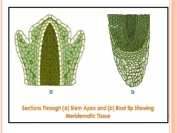
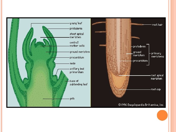
Meristematic tissue: Cells of meristematic tissues are in a state of continuous division. Cells are of thin cell wall and dense protoplasm. Present at the tips of roots and shoots. The meristems which occur at the tips of roots and shoots and produce primary tissues are called apical meristems.
Root apical meristem occupies the tip of a root while the shoot apical meristems occupy the distinct region of the stem axis. The meristems which occurs between mature tissues is called intercalary meristems. Both apical and intercalary meristems are primary meristems because they appear early in life of a plant and contribute to the formation of the primary plant body.
The meristems that occur in the mature regions of roots and shoots of the plants are called secondary or lateral meristems. They are cylindrical meristems. E. g. Fascicular vascular cambium, inter fascicular cambium and cork cambium. Lateral meristems are responsible for producing secondary tissues.
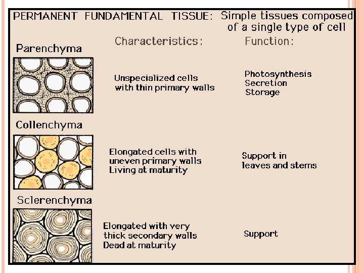
Permanent tissue: When primary and secondary meristems became structurally and functionally specialised and lose the ability to divide then permanent tissue is formed. Cells of permanent tissues do not divide further. Permanent tissues are of two types: Simple: All cells similar in structure and function. Complex: Having different types of cells.
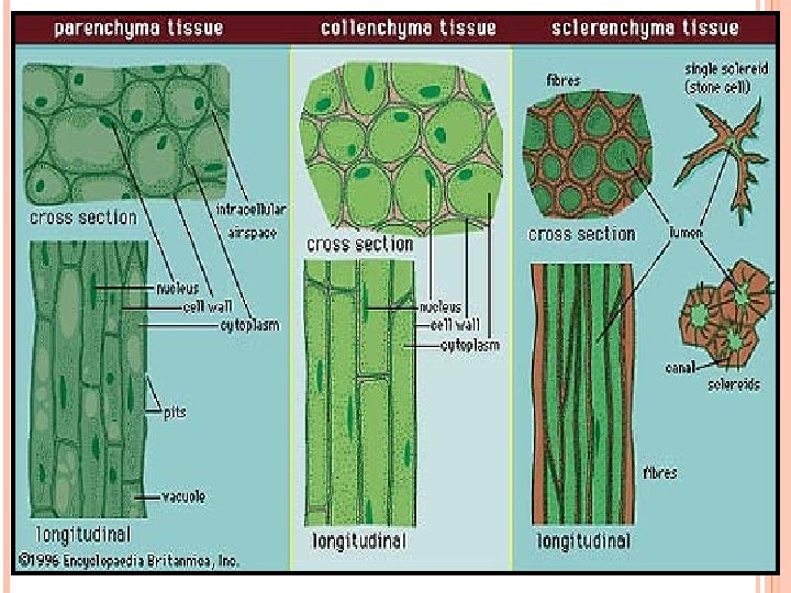
ØSimple tissues: Parenchyma: Forms the major component within organs of plant. Cells are generally isodiametric. Cells may be spherical, oval, round, polygonal or elongated in shape. Thin cell walls made up of cellulose. Closely packed cells or have small intercellular spaces. They perform various functions like photosynthesis, storage, secretion etc.
PARENCHYMA
PARENCHYMA
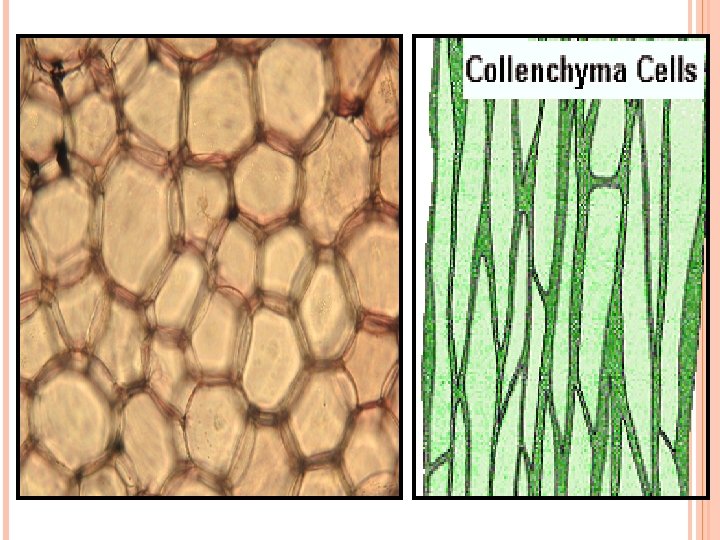
Collenchyma: In dicot it occurs below the epidermis. Found either as a homogenous layer or in patches. It consists of cells which are much thickened at the corner due to deposition of cellulose, hemicelluloses and pectin. Cells may be oval, spherical or polygonal. Sometimes cells contain chloroplasts and perform photosynthesis. Intercellular spaces are absent. Provide mechanical support to the growing parts of the plants (young stem, petiole of a leaf)
COLLENCHYMA
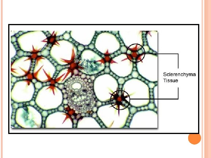
Sclerenchyma: Consists of long, narrow cells with thick and lignified cell walls having a few or numerous pits. Usually dead cells and without protoplasts. It may be either fibres or sclereids. Fibres are thick walled, elongated and pointed cells generally occurring in groups in various parts of the plant. The sclereids are spherical, oval or cylindrical, highly thickened dead cells with very narrow cavities (lumen). Commonly found in the fruit walls of nuts, pulp of fruits like guava, pear and sapota; seed coats of legumes and leaves of tea. Sclerenchyma provides mechanical strength to organs of plant.
SCLERENCHYMA
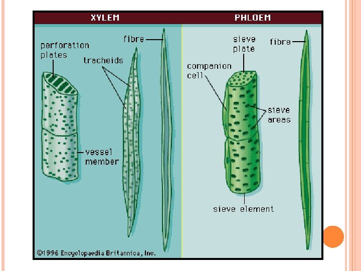
Complex Xylem: Helps tissues: Xylem and phloem. in conduction of water and minerals from roots to stem and leaves. It provides mechanical strength to the plant parts. Composed of four different kinds of elements: tracheids, vessels, and xylem fibre and xylem parenchyma. Gymnosperm lack vessels in their xylem.
Components Tracheids: Elongated of xylem: or tube like cells with thick and lignified wall and tapering cells. Dead cells without protoplasm. In angiosperm tracheids and vessels are the main water transporting elements.
Vessels: It is a long cylindrical tube – like structure made up many cells called vessel members. Presence of lignified walls and large central cavity. Dead cells and devoid of protoplasm. Vessels members are interconnected through perforations in their common walls.
Xylem fibres: Xylem parenchyma: Have highly thickened walls having central lumens. These may either be septate or aseptate. Cells are living and thin walled. Cell walls are made up of cellulose. They store food in the form of starch or fat or other substances like tannins.
Proto xylem: The first formed primary xylem elements are called proto xylem. Meta xylem: The later formed primary xylem is called meta xylem. Endarch: In stems, the proto xylem lies towards the centre (pith) and meta xylem lies towards periphery. Then the condition is called endarch. Exarch: In roots the proto xylem lies towards periphery and meta xylem lies towards the centre. Then the condition is called Exarch.
Phloem: Transports food material, usually from leaves to other parts of the plant. Composed of sieve tube, companion cell, and phloem parenchyma and phloem fibres. Gymnosperm lacks sieve tubes and companion cells. They have albuminous cells and sieve cells.
Components Sieve These tube: of phloem: are long tube like structures arranged longitudinally and are associated with the companion cells. There end walls are perforated in a sieve like manner to form the sieve plates. A mature sieve tube element possesses a peripheral cytoplasm and a large vacuole but lacks a nucleus. The function of sieve tube elements are controlled by the nucleus of companion cells
Companion cells: Specialised parenchymatous cells closely associated with sieve tube elements. Helps in maintaining the pressure gradient in the sieve tubes. Phloem Made parenchyma: up of elongated, tapering cylindrical cells with dense cytoplasm and nucleus. The cell wall is of cellulose and has pits. Stores food material and other substances like resins, latex and mucilage. Phloem parenchyma is absent in most of the monocots.
Phloem fibres: (Bast fibres): Made up of sclerenchymatous cells. Absent in primary phloem but found in the secondary phloem. Cells are elongated, unbranched and have pointed, needle like apices. Cell wall is quite thick. At maturity they lose their protoplasm and become dead. Phloem fibres of jute, flax and hemp are used commercially.
Proto phloem: The first formed primary phloem consists of narrow sieve tubes called as proto phloem. Meta phloem: The later formed phloem has bigger sieve tubes called meta phloem.
The tissue system Epidermal tissue system: Forms the outer most covering of the whole plant body. Consists of epidermal cell, stomata, epidermal appendages- the trichomes and hairs.
Epidermis: Outer most layer of the primary plant body. Made up of elongated, compactly arranged cells. It forms a continuous layer. Usually single layer. Cells are parenchymatous with small amount of cytoplasm lining the cell wall and a large vacuole. Epidermis often covered with thick waxy layer called cuticle that prevents the loss of water. Cuticle is absent in roots.
Stomata: Present in epidermis of leaves. It regulates the process of transpiration and gaseous exchange. Each stoma is composed of two bean shaped (in dicot) cells called guard cells. In monocot guard cells are dumb bell shaped. The outer walls of guard cells (away from the stomatal pore) are thin and inner walls (towards the stomatal pore) are highly thickened. The guard cells possess chloroplasts and regulate the opening and closing of stomata. Epidermal cells in the vicinity of guard cells are called subsidiary cells. The stomatal aperture, guard cells and subsidiary cells together called stomatal apparatus.
Root hair: Unicellular and it is elongations of the epidermal cells. Helps in absorption of water and minerals from the soil. Trichomes: Epidermal hairs on stem are called trichomes. It is usually multicellular. May be branched or unbranched, soft or stiff. Trichomes help in preventing water loss during transpiration. They may be secretory in function.
The ground tissue system: the ground tissue. All tissues except epidermis and vascular bundles constitute the ground tissue. It consists of simple tissues (Parenchyma, Collenchyma and Sclerenchyma) Parenchymatous cells are usually present in cortex, pericycle, pith and medullary rays, in the primary stems and roots. In leaves it is called as mesophyll. ( thinwalled chloroplast containing cells)
The vascular tissue system: Consists of xylem and phloem. Xylem and phloem together form vascular bundle. In dicot stem cambium is present between xylem and phloem. Cambium possesses the ability to form secondary xylem and secondary phloem. Such vascular bundle is called open vascular bundle.
In monocots vascular bundles have no cambium and they do not form secondary tissues are called closed vascular bundles. When Xylem and phloem are arranged in an alternate manner on different radii then it is called radial vascular bundles. E. g. in roots. Conjoint vascular bundles are common in stems and leaves.
Anatomy The of Dicot root: outer most layer is epidermis. Many of epidermal cells form unicellular root hairs. Cortex consists of several layers of thin walled parenchymatous cells with intercellular spaces. The innermost layer of cortex is endodermis. Endodermis is single layer of barrel shaped cells without intercellular spaces. Presence of casparian strips.
Presence of pericycle which is next to endodermis and consists of a few layers of thick walled parenchymatous cells. The pith is small or inconspicuous. Conjuctive tissues are present between xylem and phloem. 2 -4 xylem and phloem patches are present. Later a cambium ring develops between the xylem and phloem. Endodermis, pericycle, vascular bundle and pith constitute the stele.
Anatomy It of monocot root: has epidermis, cortex, endodermis, pericycle, vascular bundles and pith. More than 6 xylem bundles in the monocot root. Pith is large and well developed. Monocot roots do not undergo any secondary growth.
Anatomy Epidermis of dicot stem: is the outer most protective layer of the stem and is covered with thin layer of cuticle. Epidermis bears trichomes and a few stomata. Below epidermis there is cortex that consists of multilayer. Cortex consists of three subzones. The outer hypodermis consists of a few layers of collenchymatous cells just below the epidermis that provide mechanical strength to the young stem.
Below the hypodermis is cortical layer and it consists of rounded thin walled parenchymatous cells with intercellular spaces. The inner most layer of cortex is endodermis. The cells of endodermis are rich in starch grains and the layer is called starch sheath. Pericycle is present on the inner side of the endodermis.
Presence of medullary rays in between the vascular bundles. Vascular bundles are arranged in a ring. Each vascular bundle is conjoint, open with endarch proto xylem. Presence of pith that consists of large number of rounded parenchymatous cells with intercellular space.
Anatomy Monocot of monocot stem: stem has a sclerenchymatous hypodermis. Large numbers of vascular bundles are present that are scattered. Presence of parenchymatous ground tissue. Vascular bundles are conjoint and closed. Peripheral bundles are generally smaller than centrally located ones. The phloem parenchyma is absent. Water containing cavities are present within the vascular bundles.
Dorsiventral Anatomy (Dicot) leaf: of leaf shows three parts: epidermis, mesophyll and vascular system. Upper and lower epidermis covers both the surfaces (adaxial and abaxial). Presence of cuticle above epidermis.
Lower epidermis generally bears more stomata than upper epidermis. Sometimes upper epidermis lack stomata. Mesophyll is present between upper and lower epidermis. Cells of mesophyll possess chloroplasts and perform photosynthesis.
Cell of mesophyll are of two types: Spongy and palisade parenchyma. Palisade parenchyma is present adaxially and is made up of elongated cells, arranged vertically and parallel to each other. Spongy parenchyma is situated below palisade parenchyma and extends to the lower epidermis.
The cells of spongy parenchyma are oval or round and loosely arranged having large spaces and air cavities. Vascular bundles are seen in the veins and the midrib. The size of vascular bundles is dependent on the size of the veins. The vascular bundles are surrounded by a layer of thick walled bundle sheath cells.
Isobilateral The (Monocot) leaf: anatomy of isobilateral leaf is similar to that of the dorsiventral leaf in many ways. Still there are some differences. Stomata are present on the both surfaces of epidermis. Mesophyll not differentiated into palisade and spongy parenchyma. Presence of bulliform cells (modified adaxial epidermal cell along with veins) in grasses.
Secondary growth: The increase in girth is called secondary growth. The tissues involved in secondary growth are the two lateral meristems: vascular cambium and cork cambium. Vascular cambium: The meristematic layer that is responsible for cutting off vascular tissues (xylem and phloem) is called vascular cambium. In young stem it is present in patches as a single layer between xylem and phloem and later it forms a complete ring.
Formation of cambial ring: In dicot stems, the cells of cambium present between primary xylem and primary phloem is the intrafasicular cambium. The cells of medullary rays, adjoining this intrafasicular cambium become meristematic and form the Interfascicular cambium. Thus a continuous cambial ring is formed.
Activity The of the cambial ring: cambial ring becomes active and begins to cut off new cells, both towards the inner and the outer side. The cells cut off towards pith mature into secondary xylem and the cells cut off towards periphery mature into secondary phloem. The cambium is generally more active on the inner side than on the outer, so the amount of secondary xylem produced is more than the secondary phloem.
The primary and secondary phloem get gradually crushed due to continuous formation of more secondary xylem. The primary xylem however remains more or less intact, in or around the centre. At some places, the cambium forms a narrow band of parenchyma which passes through the secondary xylem and the secondary phloem in radial direction. These are the secondary medullary rays.
S. No. 1. Spring Wood Autumn wood In the spring season, cambium is very active and produces a large number of xylary elements having vessels with wider cavities. The wood formed during this season is called spring wood or early wood. In winter, the cambium is less active and forms fewer xylary elements that have narrow vessels this wood is called autumn wood or late wood.
2. Lighter in colour Darker in colour 3. Has a density lower Has a higher density. • Annual ring: Spring and autumn woods that appear as alternate concentric rings constitute an annual ring. It gives an estimate of the age of the tree.
Heart wood: In old trees, the greater part of secondary xylem is dark brown due to deposition of organic compounds like tannins, resins, oil, gums, aromatic substances and essential oils in the central or innermost layers of the stem. These substances make it hard, durable and resistant to attacks of microorganisms and insects. This type of wood is called heart wood. The heart wood does not conduct water but it gives mechanical support to the stem.
Sapwood: The peripheral region of the secondary xylem is lighter in colour and is known as the sapwood. It is involved in the conduction of water and minerals from root to leaf.
Cork As cambium: the stem continues to increase in girth due to the activity of vascular cambium, the outer cortical and epidermis layers get broken need to be replaced to provide new protective cell layers. This is done by another meristematic tissue called cork cambium or phellogen usually developed in cortical region. Phellogen is a couple of layers thick. It is made of narrow, thin walled and nearly rectangular cells.
Phellogen cuts off cells on both sides. The outer cells differentiate into cork or phellem and the inner cells differentiate into secondary cortex or phelloderm. The cork is impervious to water due to deposition of suberin in the cell wall. The cells of secondary cortex are parenchymatous. Phellogen, phellem and phelloderm are collectively called periderm.
Due to activity of cork cambium, pressure builds up on the remaining layers peripheral to phellogen and ultimately these layers die and slough off and bark is formed. Bark that is formed early in the season is called early or soft bark. Towards the end of the season late or hard bark is formed.
Lenticels: At certain regions, the phellogen cuts off closely arranged parenchymatous cells on the outer side instead of cork cells. These parenchymatous cells soon rupture the epidermis, forming lens- shaped openings called lenticels. It permits the exchange of gases between the outer atmosphere and the internal tissue of the stem. Lenticels occur in most woody trees.
Secondary growth in roots: In the dicot root, the vascular cambium is completely secondary in origin. It originates from the tissue located just below the phloem bundles, a portion of pericycle tissue, above the proto xylem forming a complete and continuous wavy ring, which later becomes circular.