ANATOMY OF FLOWERING PLANTS Plant Tissues Tissues are


: a)These are oval or spherical in shape and have very")


 together constitute")


- Slides: 70
ANATOMY OF FLOWERING PLANTS
Plant Tissues
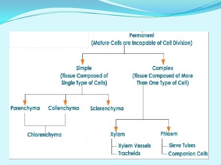
Tissues - are group of ce. IIs performing a specific function. - are structurally and/functionally similar. - Have a common origin and function. Types: A. Meristematic tissues (meristems) B. Permanent tissues
Plant tissues Meristematic tissues Permanent tissues Apical Intercalliary Lateral meristematic tissues Simple permanent tissues Parenchyma Collenchyma Sclerenchyma Complex permanent tissues Xylem Phloem
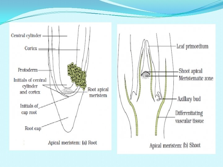
MERISTEMATIC TISSUES Made up of cells which have the capability to divide. Restricted to specialised regions and responsible for the growth of plants.
TYPES OF MERISTEM
CHARACTERISTICS OF MERISTEMATIC TISSUES Cells are thin walled. They have abundant cytoplasm and a prominent nucleus. They retain the power of division. Cells are arranged compactly without intercellular spaces.
TYPES OF MERISTEMS
CLASSIFICATION OF MERISTEMS APICAL MERISTEM Occurs at tips of shoot and root. LATERAL MERISTEM Arranged parallel to the sides of organs of plant. Cells formed Cells produced by differentiate into lateral meristems Primary tissues of differentiate into plant body secondary tissues. Causes an increase Causes increase in in length of the girth or width of plant body. the plant organ, Eg. Fascicular cambium and cork cambim. INTERCALARY MERISTEM Occurs in between mature/ permanent tissues. Produces cells which form primary tissues in the plant body. Causes increase in the length of plant organ.
CLASSIFICATION BASED ON ORIGIN • PRIMARY MERISTEM: Meristems that appear early in life of a plant and contribute to the formation of the primary plant body. Eg. Apical meristem , intercalary meristem • SECONDARY OR LATERAL MERISTEM: meristems that occur in the mature regions of roots and shoots of many plants. Eg. fascicular vascular cambium, interfascicular cambium and cork cambium.
Permanent tissues - formed from meristematic tissues - tissues that attained their mature form and perform specific functions - they stop dividing Types: Simple permanent tissues Complex permanent tissues
Simple permanent tissues - consist only of one kind of cells
1. Parenchyma - are the general purpose ce. IIs of plants - cells are rounded in shape & have uniformly thin walls found in all parts of the plants. - living at maturity, have large vacuoles - location Ieaf, stem (pith), roots, fruits Functions: *basic metabolic function (respiration, photosynthesis (ch. Iorenchyma in Leaf) & protein synthesis) *storage (potatoes, fruits, & seeds) *wound healing and regeneration
Parenchyma
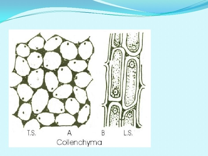
2. Co. IIenchyma - Greek word kolla which means “glue” - cells are elongated (up to 2 mm long) with unevenly thickened walls ( thin on the sides but thick at the angles where 2 or more cells meet) - differentiates from parenchyma cells & are alive at maturity Functions: Provides mechanical Support – to the growing parts of the plant
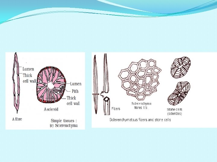
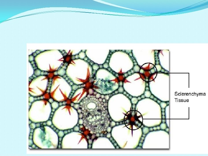
SCLERENCHYMA Long narrow cells with thick & lignified cell walls with few or more pits. Dead cells without protoplast. Cell may be either fibres or sclereids. Fibres occur in groups, thick walled, elongated and pointed cells. sclereids are spherical or oval highly thickened cells with very narrow lumen , found in nuts wall & fruit pulps. Function-mechanical support to organs.
SCLERENCHYMA CELLS SCLEREIDS(STONE CELLS): a)These are oval or spherical in shape and have very thick walls with a narrow lumen. b) Occurs in the shells of nuts and in the pulp of fruits of pear , guava and sapota, making them gritty. FIBERS: a)These are elongated and thick walled cells with tapering ends. b)Fibers are present in xylem, phloem and also in cortex and pericycle.
Complex tissues v Are made up of more than one type of cells. v There are two types of complex tissues. They are Xylem and Phloem. v They are called vascular or conducting tissues.
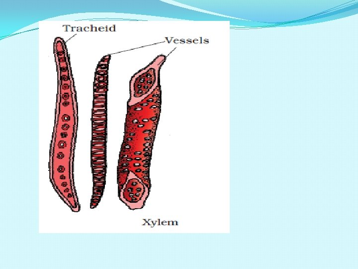
Xylem - Greek Word xy. Ios means “wood” - transports water and dissolved nutrients from the roots to a. II parts of the plant. TYPES: PRIMARY XYLEM – differentiates from procambium in the apical meristem & occurs throughout the primary plant body. SECONDARY XYLEM – differentiates from vascular cambium & is commonly called wood. PROTOXYLEM- The first formed primary xylem elements. METAXYLEM: The later formed primary xylem.
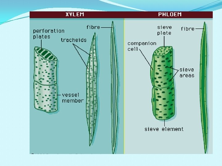
XYLEM- ELEMENTS Composed of four kinds of. TRACHIEDS : elongated tube like dead cells with thick lignified walls and tapering ends. VESSELS : long , cylindrical lignified dead cells with large central cavity. vessels are inter connected through perforations in their walls. XYLEM FIBRES: have highly thickened walls &obliterated central lumens. XYLEM PARENCHYMA: living & thin walled cells.
Xylem
longitudinal section Primary xylem cross section
FUNCTIONS OF XYLEM TRACHIEDS-main water conducting tissue of angiosperm and gymnosperm. VESSELS-main water transporting element of angiosperm. XYLEM PARENCHYMA-store food & other substances like tannins and conducts water Radially. XYLEM FIBRES -Provides mechanical strength to plants.
ENDARCH PRIMARY XYLEM OF STEM
EXARCH PRIMARY XYLEM OF ROOT
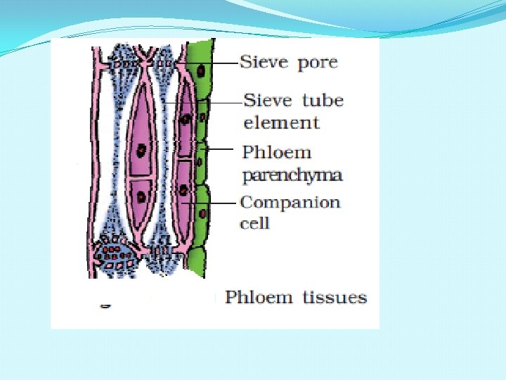
PHLOEM - Greek word phloios meaning, “bark” - transports dissolved organic / food materials from the Ieaves to the different parts of the plant - glucose in phloem moves in a. II directions Types 1. Primary phloem – differentiate from procambium and extends throughout the primary body of the plant. 2. Secondary ph. Ioem – differentiates from the vascular cambium and constitute the inner layer of the bark.
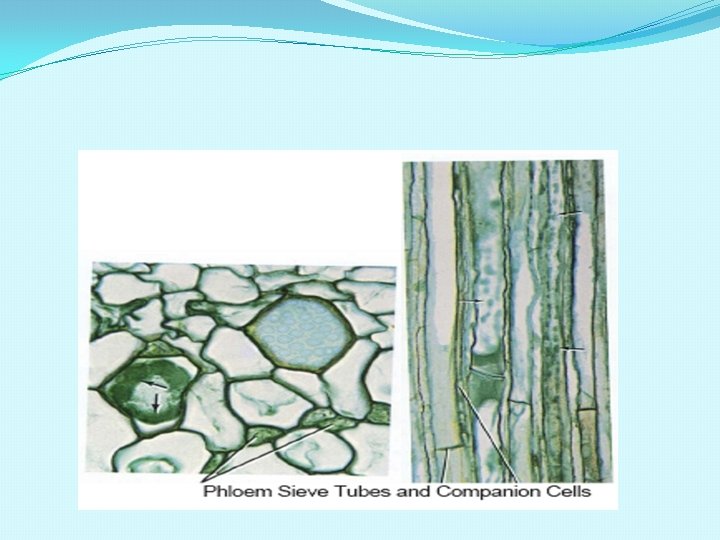
PHLOEM ELEMENTS SIEVE TUBE-long, tube like, associated with companion cells, lacks nucleus. COMPANION CELLS-specialised parenchyma, closely associated with sieve tube, nucleated. PHLOEM FIBRES(BAST FIBRES)sclerechymatous thick walled dead cells. e; g jute, flax, hemp. PHLOEM PARENCHYMA-elongated , cylindrical , nucleated cells having pits.
Phloem
FUNCTIONS OF PHLOEM üTransports food from leaves to other parts. üPhloem parenchyma stores food and other substance like resin, latex & mucilage. üPhloem fibres are used commercially. üCompanion cells maintains pressure gradient in sieve tube cells.
TISSUE SYSTEMS OF PLANT
PLANT TISSUES
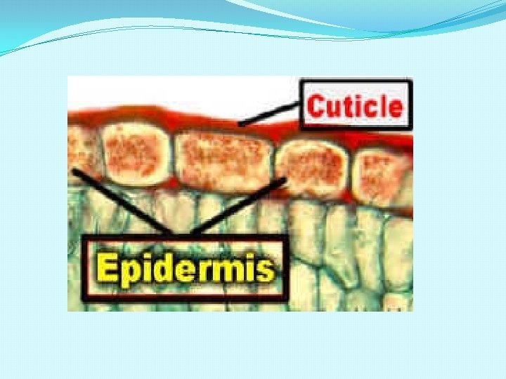
EPIDERMAL TISSUE SYSTEM v. Consists of epidermal cells, stomata & epidermal appendages such as trichomes and root hair. v. Epidermis-single layered, compactly arranged cells. v. Cuticle- the waxy layer sometimes cover epidermis. Cuticle checks water loss. v. Root hair-unicellular, absorb water & minerals. v. Trichomes-multicellular, branched or unbranched present in shoot system. prevent loss of water.
UNICELLULAR AND MULTICELLULAR HAIR
CUTICLE
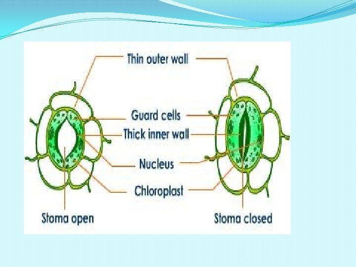
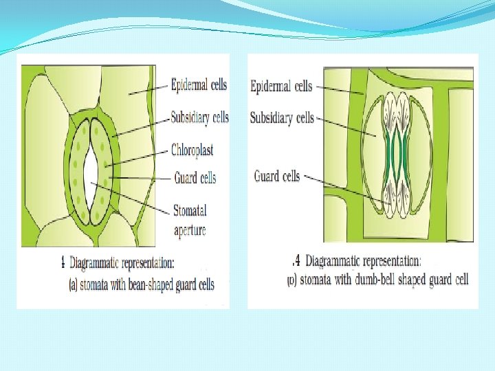
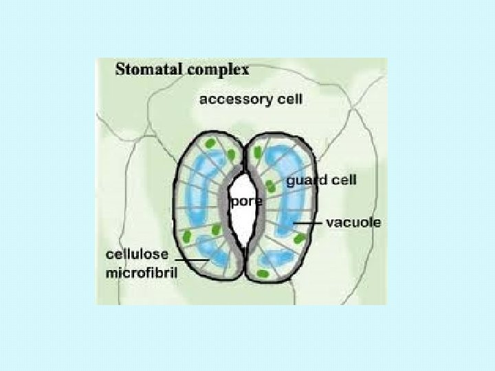
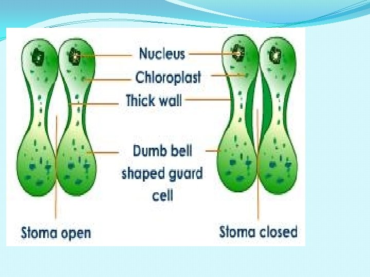
STOMATA �Each stoma surrounded by two guard cells; they regulate opening and closing of the stoma for transpiration and exchange of gases. �Guard cells are bean shaped in dicots and dumb bell shaped in monocots. �Outer wall of guard cell is thin and inner wall is thick. �Guard cell have chloroplast and carry out photosynthesis. �Epidermal cells in vicinity of guard cell become specialised in their size and shape and are called subsidiary cells.
STOMATAL APPARATUS Stomata aperture, guard cells and surrounding epidermal cells (subsidiary cells) together constitute the stomatal apparatus.
THE VASCULAR TISSUE SYSTEM Includes xylem and phloem. CONJOINT VASCULAR BUNDLE: when xylem and phloem occur along the same radius. OPEN VASCULAR BUNDLE: A strip of cambium present between xylem and phloem. CLOSED VASCULAR BUNDLE: There is no cambium in vascular bundle. RADIAL VASCULAR BUNDLE: When xylem and phloem occur along different radii, alternating with each other.
VASCULAR TISSUE SYSTEMS
TYPES OF VASCULAR BUNDLES
THE GROUND TISSUE SYSTEM �All tissues in a plant body except epidermis and vascular bundles, constitute ground tissue. �Consists of parenchyma, collenchyma & sclerenchyma. �Cortex, pericycle, pith & medullary rays contain ground tissue. �Mesophyll cells of leaf are ground tissues.
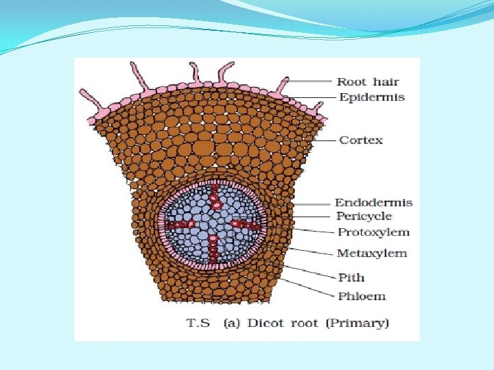
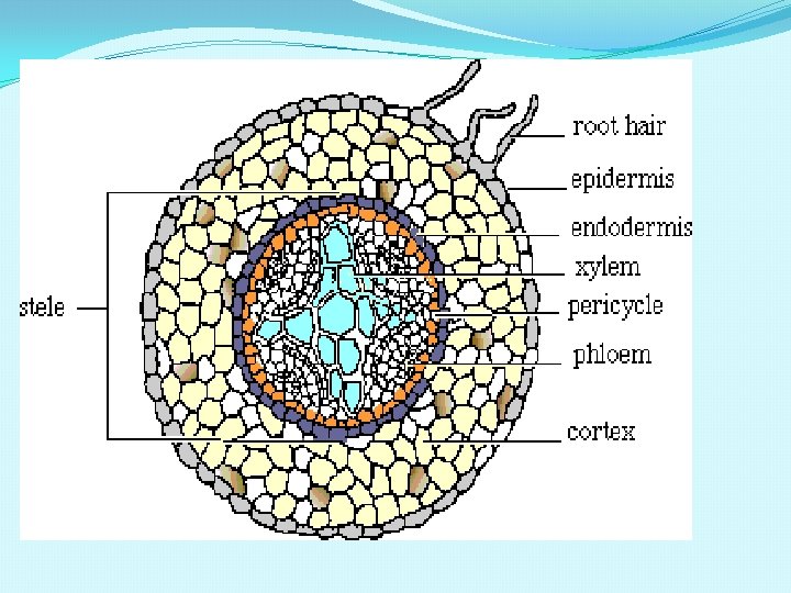
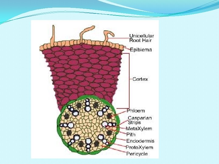
ANATOMY OF DICOT ROOT
T S OF DICOT ROOT epidermis cortex phloem Vascular cambium xylem pericycle endodermis
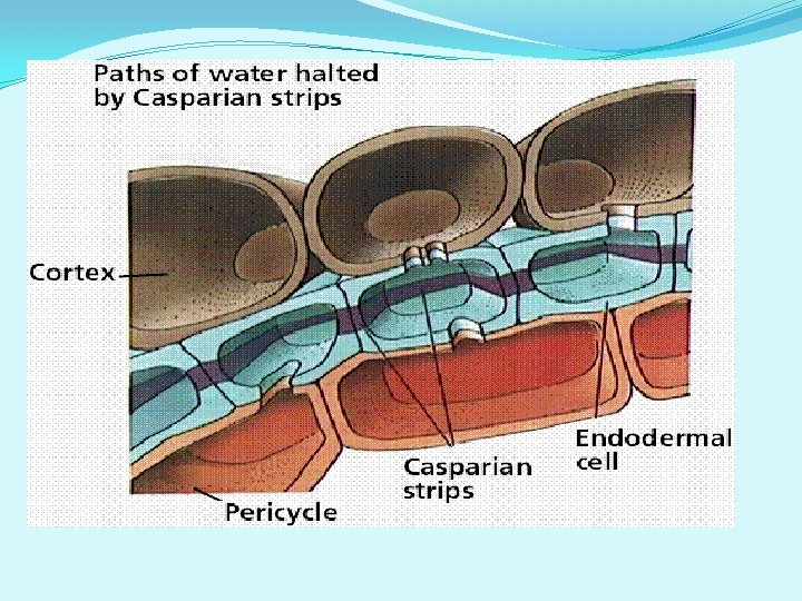
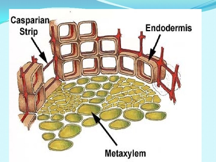
ANATOMY OF DICOT ROOT �EPIDERMIS: �Single layer of compact cells �Unicellular hairs �Cuticle absent. �CORTEX: �Made of several layers of parenchyma �Stores large amount of starch grains �Cells with intercellular spaces �ENDODERMIS: �Inner most layer of cortex. �Well developed casparian strips on their tangential and radial walls. �Endodermal cells opposite to the xylem groups do not have casparian strips and such cells are called passage cells.
�PERICYCLE: �Inner to endodermis �One or two layers of thin walled cells �Takes part in formation of cambium for secondary growth. �VASCULAR BUNDLES: �Radial arrangement �Number of xylem ranges from two (diarch) to six (hexarch), commonly tetarch are seen. �Xylem exarch. �Parenchyma cells that lie between xylem and phloem constitude the conjunctive tissue. �PITH: �Small or absent and made of parenchyma cells.
CONJUNCTIVE TISSUE: The parenchymatous cells which lie between the xylem and the phloem are called conjunctive tissues. Usually 2 - 4 xylem and phloem patches. STELE: All tissues on the inner side of the endodermis such as pericycle , vascular bundle and pith constitute the stele.
ANATOMY OF MONOCOT ROOT �EPIDERMIS: �Single layer of compact cells �Unicellular hairs �Cuticle absent. �CORTEX: �Made of several layers of parenchyma. �Stores large amount of starch grains. �Cells with intercellular spaces. �ENDODERMIS: �Inner most layer of cortex. �Well developed casparian strips on their tangential and radial walls. �Endodermal cells opposite to the xylem groups do not have casparian strips and such cells are called passage cells.
PERICYCLE: Inner to endodermis One or two layers of thin walled cells In old roots , the cells become lignified and thick walled. VASCULAR BUNDLES: Radial arrangement Polyarch. Xylem exarch. PITH: Large and well developed. Made of parenchyma cells.
DIFFERENCES BETWEEN DICOT AND MONOCOT ROOT DICOT ROOT MONOCOT ROOT 1. Cortex simple and homogenous. 1. Cortex well developed. 2. Endodermis is prominent with casparian strips. 2. Endodrmis more prominent. Thickening in radial and inner wall more prominent. 3. Number of xylem and phloem bundles is small; varies from 2 -5. 3. Xylem and phloem bundles are numerous; more than 6. 4. Conjunctive tissues are parenchymatous. 4. Conjunctive tissues are sclerenchymatous. 5. Pith reduced or absent. 5. Pith well developed. 6. Secondary growth takes place. 6. No secondary growth. .