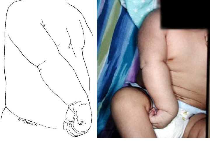
CASE 1 CASE 1 Dystocia Waiters tip sign


 excessive displacement of the head to")
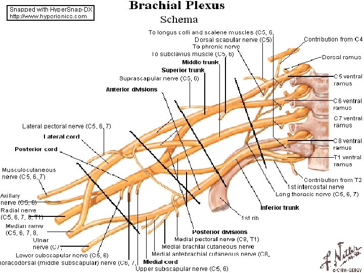
 C 5 and C 6 roots")
 and elevates scapula) Lateral pectoral nerve:")
 ulnar and median nerves all the")
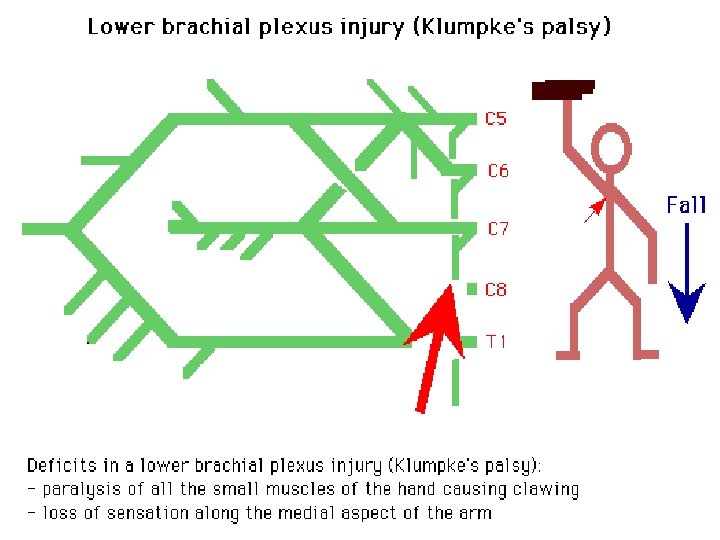
 C 8 and T 1 roots")


, ulnar nerve at")


 pressure")
 I.")

 groove")


 sudden and often repeated use of the forearm extensor muscles previously")


 Weak flexion @ shoulder joint Flexion of")


 is a frequent diagnosis considered in such patients and is")


- Slides: 59
CASE 1
CASE 1
Dystocia Waiter’s tip sign http: //www. skepticalob. com/2012/10/except-for-the-nerve-damage-the-baby-was-unscathed. html
Upper Lesions of the Brachial Plexus (Erb-Duchenne Palsy) excessive displacement of the head to the opposite side depression of the shoulder on the same side in infants during a difficult delivery in adults after a blow to or fall on the shoulder
Upper Lesions of the Brachial Plexus (Erb-Duchenne Palsy) C 5 and C 6 roots Medial rotation Pronation suprascapular nerve the nerve to the subclavius musculocutaneous nerve axillary nerve 1. s u p r a s p i n a t u s abductor of the shoulder 2. i n f r a s p i n a t u s lateral rotator of the shoulder 3. s u b c l a v i u s depresses the clavicle 4. b i c e p s b r a c h i i supinator of the forearm, flexor of the elbow, weak flexor of the shoulder 5. g r e a t e r p a r t o f t h e b r a c h i a l i s flexor of the elbow 6. c o r a c o b r a c h i a l i s flexor of the shoulder 7. d e l t o i d abductor of the shoulder 8. t e r e s m i n o r lateral rotator of the shoulder Sensatıon loss over the lateral sıde of the arm
Dorsal scapular nerve: Levator scapulae, rhomboids (Retracts (adducts) and elevates scapula) Lateral pectoral nerve: Pectoralis major (medial rotation, flexion and adduction of the arm) extension Lattisimus dorsi= thoracodorsal nerve
Lower Lesions of the Brachial Plexus (Klumpke Palsy) ulnar and median nerves all the small muscles of the hand Claw(ed) hand hyperextension of metacarpophalangeal joints flexion of the interphalangeal joints
Lower Lesions of the Brachial Plexus (Klumpke Palsy) C 8 and T 1 roots loss of sensation along the medial side of the arm 8 th cervical nerve damaged + medial side of the forearm, hand, and medial two fingers Foerster (1933)
MONONEUROPATHIES The pattern of distribution of peripheral nerve involvement is very helpful in reaching a diagnosis. Mononeuropathies, especially if an entrapment site, are often an isolated phenomenon, possibly related to pregnancy, DM, thyroid disease or occupation, but importantly may also occur as features of a more generalised disorder, such as hereditary neuropathy with liability to pressure palsies (HNPP) or amyloidosis.
Mononeuropathies occurring outside entrapment sites are more important to investigate fully, especially if vasculitis is suspected as this need careful evaluation for treatment. If the pattern suggests a single nerve or plexus lesion at an unusual site of compression or invasion, such as a radial nerve lesion compressed on a chair in a patient following an overnight binge, or invasion of the brachial plexus with breast malignancy, this is clearly important to detect. PALSY
Focal and multifocal neuropathies Entrapment neuropathy—for example, carpal tunnel syndrome (CTS), ulnar nerve at elbow Myxoedema, acromegaly Amyloidosis Diabetes Hereditary neuropathy with liability to pressure palsies (HNPP A) Vasculitis Multifocal motor neuropathy
Entrapment neuropathies occur when nerves chronically compressed or mechanically injured at specific locations. isolated peripheral nerve injuries occurring at specific locations where a nerve is mechanically constricted in a fibrous or fibro-osseous tunnel or deformed by a fibrous band. In some instances the nerve is injured by chronic direct compression, and in other instances angulation or stretching forces cause mechanical damage to the nerve.
Angulation and stretch injury are important mechanisms of nerve injury for ulnar neuropathies associated with gross deformity of the elbow joint (“tardy ulnar palsy”). Recurrent compression of nerves by external forces may also cause focal nerve injuries such as ulnar neuropathy at the elbow and deep branch lesions of the ulnar nerve in the hand. Although these latter neuropathies do not satisfy the strict definition of “entrapment neuropathies”, they are often considered in a discussion of the topic.
Long Thoracic Nerve Injuries serratus anterior C 5, C 6, C 7 blows to or pressure on the posterior triangle of the neck during the surgical procedure of radical mastectomy Difficulty in raising the arm above the head Inferior border of scapula not closely applied to the chest wall Protrude posteriorly Winged scapula
CASE 2 Here is the shoulder’s x-ray of the patient! SYMPTOMS loss of skin sensation over the lower half of the deltoid muscle. What is your possible diagnosis? 18
Axillary Nerve Injuries posterior cord of the brachial plexus (C 5 and 6) pressure of a badly adjusted crutch pressing upward into the armpit shoulder dislocations Quadrangular space fractures of the surgical neck of humerus deltoid and teres minor Loss of skin sensation over the lower half of deltoid region (lateral part of the arm) Upper lateral cutaneous nerve of the arm Impaired abduction of the shoulder (the other one: Supraspinatus only) Shoulder weakness Difficulty lifting the arm above the head
Axillary Nerve Injuries posterior cord of the brachial plexus (C 5 and 6) I. M. injections Operations around the shoulder runs transversely under cover of the deltoid at the level of the surgical neck of the humerus
CASE 3 A NIGHT AT E. R. You are taking the history from your patient. Here is what he says (his symptoms) He says he was painting the ceiling and fell. , suddenly. He has a terrible pain in his arm. Here is what you find (his clinical findings) Localized pain in his right forearm No sensory loss No wristdrop, the wrist can be extended. 21
Radial Nerve Injuries commonly damaged in the axilla & in the spiral (radial) groove
Radial Nerve Injuries @ Axilla pressure of the upper end of a badly fitting crutch drunk falling asleep with one arm over the back of a fractures and dislocations of the proximal end of the humerus Motor Triceps, anconeus, and long extensors of the wrist No extension of the elbow joint, wrist joint, and the fingers Wristdrop (flexion of the wrist) Supination good brachioradialis, supinator down, but biceps brachii
Radial Nerve Injuries @ Axilla Sensory A small loss of skin sensation down the posterior surface of the lower part of the arm down a narrow strip on the back of the forearm A variable area of sensory loss on the lateral part of the dorsum of the hand on the dorsal surface of the roots of the lateral 3 ½ fingers Trophic Changes Slight
Radial Nerve Injuries @ Spiral Groove of Humerus At the time of fracture of the shaft of the humerus Following the formation of the callus Pressure of the back of the arm on the edge of the operating table Prolonged application of a tourniquet to the arm in a person with a slender triceps temporary radial palsy
Radial Nerve Injuries @ Spiral Groove of Humerus most commonly @ distal part of the groove Motor Inability to extend the wrist &fingers Wrist drop Sensory A variable small area of anesthesia dorsal surface of the hand dorsal surface of roots of lateral 3 ½ fingers Trophic changes Very slight or absent
potential space located anterior to the proximal radius starting from the level of the humeroradial joint extending past the proximal edge of the supinator posterior interosseus nerve The radial nerve bifurcates into deep and superficial branches anterior to the lateral epicondyle of the humerus, between the brachialis and the brachioradialis, in the lateral border of the cubital fossa. After passing through the two heads of the supinator muscle, the deep branch becomes the posterior interosseous nerve.
Radial tunnel syndrome DIAGNOSIS ANATOMY Radial nerve Lateral part of the elbow, radial tunnel below the supinator Tenderness and pain @ lateral side of the elbow
Tennis Elbow(Lateral epicondylitis) sudden and often repeated use of the forearm extensor muscles previously been much used extensor carpi radialis brevis Tenderness and pain @ lateral side of the elbow Pain on wrist extension, pain when shaking hands, and frequently a weakened grip. In tennis elbow, the tenderness is mostly right where the tendon attaches to the lateral epicondyle of the elbow. In radial tunnel syndrome, the place that is most tender is about two inches further down the arm, right over where the radial nerve goes into the supinator muscle.
Deep branch of the Radial Nerve Injuries Motor nerve Extensor muscles @ posterior compartment of the forearm fractures of the proximal end of the radius dislocation of the radial head Supinator Intact Extensor carpi radialis longus No wrist drop No sensory loss
Superficial Radial Nerve Injuries a variable small area of anesthesia over the dorsum of the hand dorsal surface of the roots of the lateral 3 ½ fingers
Musculocutaneous Nerve Injuries Rarely injured (protected position) Weak flexion @ shoulder joint Flexion of the forearm @ elbow by remainder of brachialis + flexors of forearm Weak supination supinator radial nerve Sensory loss along the lateral side of the forearm lateral cutaneous nerve of the forearm
Median Nerve Injuries occasionally in the elbow region supracondylar fractures of the humerus most commonly by stab wounds or broken glass just proximal to the flexor retinaculum
Injuries to the Median Nerve @ the Elbow Motor pronator & flexor muscles of forearm (EXCEPT? ) thenar muscles Forearm in supine position- Weak wrist flexion-accompanied by adduction No flexion @ interphalangeal joints of index & middle fingers Weak flexion @ metacarpophalangeal joints –interossei- FIST Middle & index fingers remain straight (extended) POPE’S BLESSING
Injuries to the Median Nerve @ the Elbow Motor No Flexion of the terminal phalanx of the thumb Thenar eminence flattened Thumb laterally rotated & adducted APE HAND DEFORMITY
Injuries to the Median Nerve @ the Elbow Sensory Lost skin sensation @lateral half or less of the palm of the hand palmar aspect of lateral 3 ½ fingers distal part of dorsal surfaces of lateral 3 ½ fingers
Injuries to the Median Nerve @ the Elbow Vasomotor changes Skin area affected warmer & drier Arteriolar dilatation and absence of sweating / loss of sympathetic control Trophic changes Chronic cases dry and scaly skin nails crack easily atrophy of the pulp of the fingers
Injuries to the Median Nerve @ the Motor Wrist Thenar muscles & first two lumbricals Thenar eminence flattened Thumb laterally rotated and adducted Ape-like hand No opposition of the thumb MAKE A FIST, SLOWLY Index & middle fingers lag behind the ring & little fingers
CASE 4 Dr. Eda, a gynecologist sends her patient , 25 years old pregnant female , a cashier at a mall in downtown @ the end of her first trimester to Dr. Süleyman who is an internist An expert from Dr. Eda’s request of consultation letter for Dr. Süleyman Free T 4 0. 4 ng/dl 0. 8 – 2. 4 ng/dl Hypothyroidism?
CASE 4 Dr. Süleyman examines the patient as a consulting internist, Finds the following during his examination: Symptoms of depression Increased sensitivity to cold Poor muscle tone Burning sensations in the thumb & index finger The anxious patient asks: What is wrong with me, doctor? Dr. Süleyman replies with a tone of affection in his voice: «You are pregnant and your thyroid gland does not work efficiently and plus you have …………………. . X………………» What is X?
Carpal tunnel syndrome (CTS) is a frequent diagnosis considered in such patients and is the most frequent entrapment neuropathy. Approximately 1. 6% of adults describe symptoms consistent with CTS. Clinically, the syndrome consists of a burning pain or “pins and needles” along the distribution of the median nerve to the lateral three and a half fingers and weakness of thenar muscles. It is produced by compression of the median nerve within the tunnel. The syndrome can often be treated effectively with splinting and avoidance of repetitive motions and awkward wrist positions; however, carpal tunnel release is ultimately performed in 25% to 50% of these patients. Solomon DH, Katz JN, Bohn R, Mogun H, Avorn J. Nonoccupational risk factors for carpal tunnel syndrome. J Gen Intern Med. 1999 May; 14(5): 310 -4. Pregnancy experience CTS due to hormonal changes (high progesterone levels) and water retention, which is common during pregnancy During hypothyroidism and pregnancy fluid is retained in tissues, which swells the tenosynovium. Obesity also increases the risk of CTS: individuals classified as obese (BMI > 29) are 2. 5 times more likely than slender individuals (BMI < 20) to be diagnosed with CTS is also work-related, occupational disease and associated with overuse. Bonfiglioli R, Venturi S, Graziosi F, Fiorentini C, Mattioli S. [Carpal tunnel syndrome among supermarket cashiers]. G Ital Med Lav Ergon. 2005 Jan-Mar; 27(1): 106 -11. [Article in Italian]
Carpal Tunnel Syndrome MOST COMMON PERIPHERAL NERVE INJURY IN THE UPPER LIMB
Carpal Tunnel Syndrome Burning pain or “pins and needles” along the distribution of the median nerve to the lateral 3 ½ fingers Weakness of thenar muscles No paresthesia over thenar eminence palmar cutaneous branch of the median nerve
Ulnar Nerve Injuries most commonly injured @ @ elbow where it lies behind the medial epicondyle usually associated with fracture @ wrist where it lies with the ulnar artery in front of the flexor retinaculum.
Injuries to the Ulnar Nerve @ the Elbow CUBITAL TUNNEL SYNDROME (2 ND most common) Motor Flexor carpi ulnaris & medial half of flexor digitorum profundus ring & little fingers No flexion of the terminal phalanges of the ring & little fingers Flexion of wrist = abduction paralysis of flexor carpi ulnaris medial border of the front of the forearm flattned/wasted All the small muscles of the hand paralyzed EXCEPT ? fibro-osseus tunnel between the medial epicondyle and flexor carpi ulnaris
Injuries to the Ulnar Nerve @ the Elbow Motor Extensor digitorum can abduct the fingers to a small extent when metacarpophalangeal joints are hyperextended Impossible to adduct the thumb adductor pollicis paralyzed Froment’s sign Grip a piece of paper between the thumb and index fingers Froment sign: The patient is asked to hold the paper between the thumb and index finger. (A) With the intact ulnar nerve, the patient is able to make use of the adductor pollicis. ( B) When the ulnar nerve is deficient, the patient compensates for the denervated adductor by using the flexor pollicis longus (median nerve innervated).
Injuries to the Ulnar Nerve @ the Elbow Motor 2 medial lumbricals & interossei Hyperextended metacarpophalangeal joints Flexed interphalangeal joints fourth & fifth fingers “claw” deformity main en griffe
Injuries to the Ulnar Nerve @ the Elbow Motor Flattening of hypothenar eminence Loss of the convex curve to the medial border of the hand Hollowing between metacarpal bones @ dorsum of the hand wasting of dorsal interossei
Injuries to the Ulnar Nerve @ the Elbow Sensory Loss of skin sensation anterior & posterior surfaces of medial 1/3 of the hand medial 1 ½ fingers Vasomotor Changes warmer and drier skin area arteriolar dilatation and absence of sweating /loss of sympathetic control
Injuries to the Ulnar Nerve @ the Wrist Motor Small hand muscles paralyzed, wasted – EXCEPT 3 thenar @ first 2 lumbricals Claw hand More obvious Flexor digitorum profundus intact Marked flexion of the terminal phalanges Ulnar paradox Higher lesion Less obvious claw deformity More proximal injury Less claw
ULNAR NERVE ENTRAPMENT SYNDROMES PROXIMAL/ @ ELBOW CUBITAL TUNNEL SYNDROME BETWEEN MEDIAL EPICONDYLE & FLEXOR CARPI ULNARIS Q: Medial half of Flexor digitorum profundus affected in which one most? DISTAL/ @ WRIST GUYON’S CANAL Roof: Palmaris brevis, hamate, pisiforme bones & Flexor carpi ulnaris
Injuries to the Ulnar Nerve @ the Wrist Sensory Main ulnar nerve Palmar cutaneous branch Posterior cutaneous branch 6. 25 cm, 2 inch above the pisiform bone palmar surface of the medial 1/3 hand medial 1 ½ fingers the dorsal aspects of the middle and distal phalanges of the same fingers
Rotator Cuff Tendinitis Subscapularis stabilizing the shoulder joint Supra-infra spinatus Teres MINOR Lesions of the cuff common cause of pain in the shoulder region Subacromial bursitis Supraspinatus tendinitis Pericapsulitis spasm of pain in the middle range of abduction when the diseased area impinges on the acromion
Rupture of the Supraspinatus Tendon advanced cases of rotator cuff tendinitis necrotic supraspinatus tendon calcified or rupture Hold humeral head @ glenoid fossa at the beginning of abduction No initation of abduction of the arm, unless passively assited for the first 15°
Anterior interosseous nerve syndrome flexor pollicis longus flexor digitorum profundus pronator quadratus Pinch deformity Pronation to index, sometimes middle fingers
Venipuncture • obtaining blood for laboratory testing • administering fluid and intravenous drugs Cephalic vein
OTHER
Auscultatory Triangle latissimus dorsi trapezius medial border of scapula
Levator scapulae levator scapula which connects the neck and shoulder. most usual complaint pain when trying to turn the head to the side where it hurts, often turning the body instead of the neck to look behind. It is often associated with a headache but not always. The most common causes for developing this kind of stiff neck are; turning the head to one side while typing, long phone calls without a headset, sleeping without proper pillow support with the neck tilted or rotated, sitting in a chair with armrests too high and exposure of the neck to a cold draft. There are other causes basically from shortening of this muscle as illustrated with using a cane that is too long. Activities such as vigorous tennis, swimming the crawl stroke and watching a tennis match rotating the head back and forth can also cause a stiff neck.