Cardiovascular SystemHeart SALADIN CHAPTER 19 Overview of Cardiovascular


![Gross Anatomy of the Heart • Right coronary artery [R. atrium, pacemaker] posterior interventricular](https://slidetodoc.com/presentation_image_h2/04c43ee8627c11f2fa8cc4139b2f1e10/image-27.jpg "Gross Anatomy of the Heart • Right coronary artery [R. atrium, pacemaker] posterior interventricular")


 node")


")


")


 • CO is the amount of blood pumped by each ventricle")
 • HR is the number of heart beats per minute •")
 • Cardiac reserve is the difference between resting and maximal CO")
 • Heart Rate – Tachycardia >100 beats/min – Bradycardia < 60")
 • Factors affecting HR – ANS – Medulla oblongata - cardiac")
 – Chemical Regulation • Hormones – Epinephrine stimulates SA node –")
 • Ion related: – Hypercalcemia – really slow rate; hypo -->")
")
 • Stroke Volume – Preload, or degree of stretch, of cardiac")

 • Homeostatic Imbalances – Congestive heart failure (CHF) • Pumping efficiency")
 • Developmental Aspects of the Heart – Fetal heart structures that")
 • Atherosclerosis - fatty deposits occur in vessels walls leading to")
 – Treatments: • Balloon angioplasty, laser angioplasty, coronary bypass surgery, stents")

- Slides: 78
Cardiovascular System/Heart SALADIN CHAPTER 19
Overview of Cardiovascular System/Heart • Pulmonary & Systemic Circuits - heart is 2 pumps – Pulmonary - from right V --> Pulmonary trunk --> pulmonary arteries - lungs --> pulmonary veins --> left A
Overview of Cardiovascular System/Heart • Pulmonary & Systemic Circuits - heart is 2 pumps – Systemic - left V --> aorta --> other arteries --> veins --> superior & inferior vena cavas --> right A
Overview of Cardiovascular System/Heart • Position & Size of Heart – Approximately the size of your fist - ~ 9 cm wide, 13 cm from base to apex, 6 cm from anterior to posterior; weighs about 300 g.
Overview of Cardiovascular System/Heart • Position & Size of Heart – Location • Superior surface of diaphragm • In mediastinum of thoracic cavity • Left of the midline
Overview of Cardiovascular System/Heart • Position & Size of Heart – Location • Anterior to the vertebral column, posterior to the sternum
• Position & Size of Heart
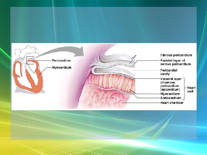
Overview of Cardiovascular System/Heart • Pericardium – a double-walled sac around the heart. – Protects and anchors the heart [to diaphragm & sternum] • Prevents overfilling of the heart with blood
Overview of Cardiovascular System/Heart • Pericardium – a double-walled sac around the heart. – Protects and anchors the heart [to diaphragm & sternum] • Allows for the heart to work in a relatively friction-free environment
Overview of Cardiovascular System/Heart • Pericardium – a double-walled sac around the heart. – Composed of: • A superficial fibrous pericardium
Overview of Cardiovascular System/Heart • Pericardium – a double-walled sac around the heart. – Composed of: • A deep two-layer serous pericardium – Parietal layer - internal surface of fibrous pericardium – Visceral layer covers surface of the heart – Separated by fluid-filled pericardial cavity
Gross Anatomy of the Heart • Heart Wall – Epicardium – visceral layer of the serous pericardium – Myocardium – cardiac muscle layer for contraction of the heart
Gross Anatomy of the Heart • Heart Wall – Endocardium – endothelial on inner surface – Fibrous skeleton of the heart – crisscrossing, interlacing layer of connective tissue in septa, around valves & in tissue between - insulates, reinforces.
Gross Anatomy of the Heart • Chambers of the Heart – Consist of: • 2 atria plus auricles - thin walls • 2 ventricles - L thickest wall; trabeculae carne on surfaces
Gross Anatomy of the Heart • Chambers of the Heart – Consists of: • Chamber boundaries marked by sulci – Coronary sulcus – Interventricular sulcus
• Chambers of the Heart
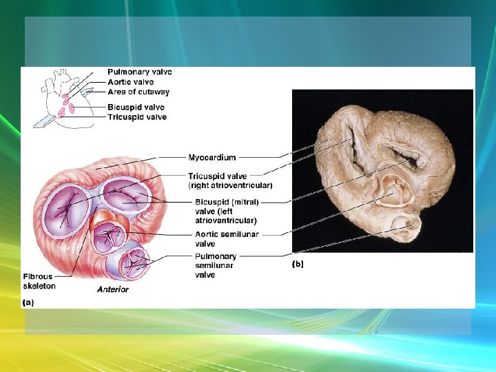
Gross Anatomy of the Heart • Heart Valves - ensure unidirectional blood flow through the heart. – Atrioventricular (AV) valves lie between the atria and the ventricles • AV valves prevent backflow into the atria when ventricles contract
Gross Anatomy of the Heart • Tricuspid valve – Between right atrium and right ventricle • Bicuspid valve (mitral valve) – Between left atrium and left ventricle – Chordae tendenae anchor flaps to the papillary muscles.
Gross Anatomy of the Heart
Gross Anatomy of the Heart
Gross Anatomy of the Heart – Semilunar Valves • Semilunar valves prevent backflow of blood into the ventricles – Aortic semilunar valve » lies between left ventricle and aorta – Pulmonary semilunar valve » lies between right ventricle and pulmonary trunk
Gross Anatomy of the Heart • Blood flow through heart chambers – RA --> RV--> lungs --> LA--> systemic circ --> RA
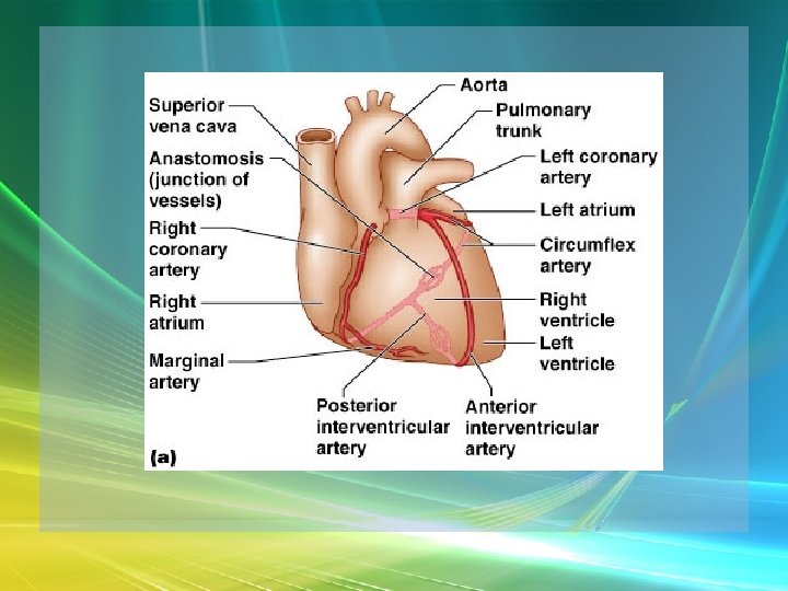
Gross Anatomy of the Heart • Coronary Circulation – Coronary circulation is functional blood supply to the heart muscle itself – Supplies 250 m. L/min = 5% of circulating blood
Gross Anatomy of the Heart – Arterial Supply • Ascending aorta left and right coronary arteries • Left coronary artery interventricular artery [interventricular septum, ant. both ventricles] (anastamoses with posterior interventricular) [both ventricles, 2/3 of interventricular septum], and the circumflex artery [L atrium, posterior L ventricle]
Gross Anatomy of the Heart • Right coronary artery [R. atrium, pacemaker] posterior interventricular [post. both ventricles]& marginal arteries • MI = necrosis of heart tissue – usually due to artery blockage
Gross Anatomy of the Heart • Collateral routes ensure some blood delivery to heart even if major vessels are occluded. • Diastolic flow > systolic [opposite other tissues]
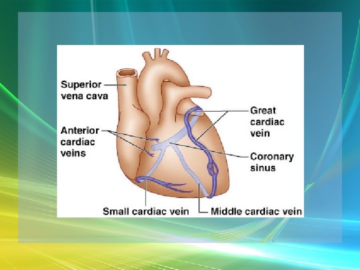
Gross Anatomy of the Heart • Coronary Circulation – Venous Supply • Coronary arteries cardiac veins coronary sinus right atrium
Cardiac Conduction System & Cardiac Muscle • Nerve Supply – ANS – Sympathetic speeds up, PS slows down. – Medulla – cardioaccelerator center –>sympathetic nerves – T 1 -T 5 C ganglia Cardiac nerves ventricular myocardium ↑ force of contraction. Also ↑coronary blood flow in sympathetic mode
Cardiac Conduction System & Cardiac Muscle – Cardioinhibitory center – sends to vagus [to SA & AV nodes] ↓HR – Steady firing of Vagus nerves to control HR = vagal tone
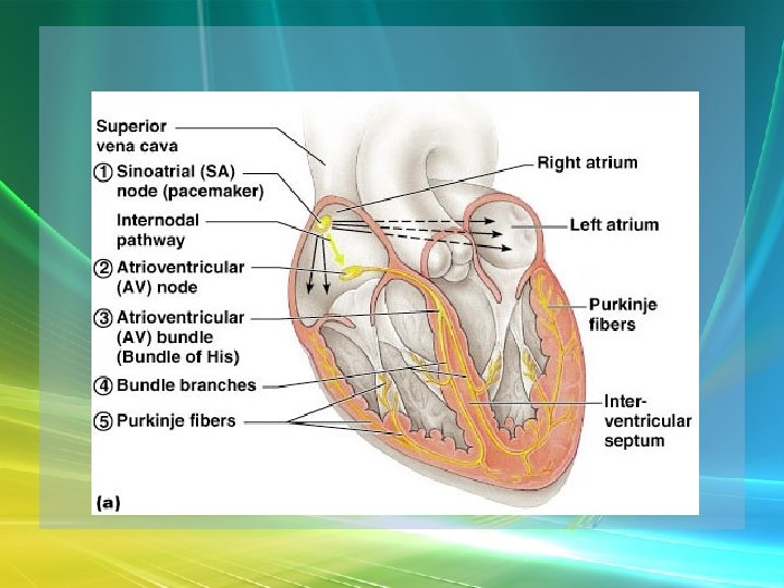
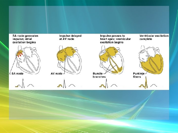
Cardiac Conduction System & Cardiac Muscle • Conduction System – Autorhythmic cells: • Self excitable - Initiate action potentials • Found in SA node, AV bundle, R & L bundle branches, and Purkinjee fibers [same order as signal passage].
Cardiac Conduction System & Cardiac Muscle – Sequence of Excitation • Sinoatrial (SA) node impulses about 75 times/minute • Action potentials gap junctions through intercalated discs across atria=atria contract
Cardiac Conduction System & Cardiac Muscle • AV node delays impulse ~ 0. 1 second • Impulse passes from atria to ventricles via the AV bundle [bundle of His – superior interventricular septum].
Cardiac Conduction System & Cardiac Muscle • AV bundle splits into 2 paths in interventricular septum (= bundle branches) – Bundle branches carry impulse toward apex – Purkinje fibers carry impulse to apex & ventricular walls
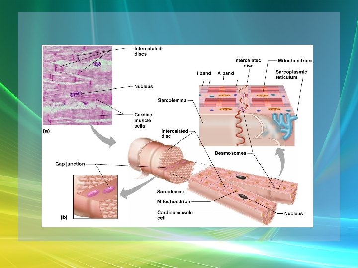
Cardiac Conduction System & Cardiac Muscle • Properties of Cardiac Muscle Fibers – Striated, short, fat, branched, and interconnected – Intercellular spaces filled with loose connective tissue with capillaries
Cardiac Conduction System & Cardiac Muscle – Intercalated discs anchor cardiac cells together and allow passage of ions – Ca 2+delivery – wider & fewer T-tubules; no triads
Cardiac Conduction System & Cardiac Muscle • Metabolism of Cardiac Muscle – Almost exclusively aerobic – Myoglobin stores O 2, glycogen stores glucose – Extra large mitochondria [25% of cell]
Electrical & Contractile Activity • Normal pattern triggered by SA node = sinus rhythm [70 -80/min] • Outside stimuli can cause firing from ectopic focus – usually AV node
Electrical & Contractile Activity • Arrhthymias = uncoordinated contractions – Blocks - action potential propagation problem [damage to AV node]. – Fibrillation - asynchronous contraction [control taken away from SA node].
Electrical & Contractile Activity • Pacemaker Physiology – Upon stimulation, Na+ enters and depolarization begins opening of fast Ca channels action potential K channels open K leaves
Electrical & Contractile Activity • Impulse Conduction – Delay at AV node of 100 msec [to enhance ventricular filling] – SA signal --> AV node in 0. 05 sec – Ventricular myocardium conducts at 0. 5 m/sec, but Purkinjee, etc. are much faster • keeps the ventricles synchronous
Electrical & Contractile Activity • Electrical Behavior of Myocardium – Cardiac Muscle Contraction • Heart muscle: differences from skeletal muscle – Stimulated by nerves and self-excitable (automaticity) resting potential is -90 m. V – Contracts as a unit
Electrical & Contractile Activity – Sequence • Upon stimulation, Na+ enters and depolarization begins opening of fast Ca channels action potential • Depolarization stimulates release of Ca 2+ from SR • Calcium binds to troponin & opens site for myosin to attach to actin
Electrical & Contractile Activity • Ca 2+ - 10 -20% enters from extracellular space stimulates sarcoplasmic reticulum to release the other 90%. Fast Ca 2+ channels only open when Slow Na+ channels are open. • Has a long (250 ms) absolute refractory period [prevents tetanus]
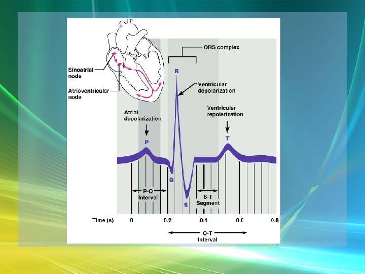
Electrical & Contractile Activity • Electrocardiogram – Electrical activity is recorded by electrocardiogram (ECG) – Typically attaches to wrists, ankles and 6 chest positions
Electrical & Contractile Activity – P wave corresponds to atrial depolarization – QRS complex corresponds to ventricular depolarization – T wave corresponds to ventricular repolarization – Arial repolarization record is masked by the larger QRS complex
Electrical & Contractile Activity – Intervals-time spans between waves • P-Q- time from beginning of atrial contraction to beginning of ventricular contraction • S-T-plateau phase of ventricular contraction • Q-T-from first of ventricular depolarization to end of ventricular repolarization
Electrical & Contractile Activity – Interpretations: • Enlarged P --> atrial hypertrophy • Missing/inverted p --> SA damage • Enlarged Q --> MI • Enlarged R --> ventricular hypertrophy • See figures 19. 1 & 19. 8 for examples
Blood Flow, Heart Sounds & Cardiac Cycle • Pressure & Flow – Fluid dynamics depend on pressure & resistance – Pressure is measured here in mm. Hg = Torr by the sphygmomanometer
Blood Flow, Heart Sounds & Cardiac Cycle • Heart Sounds – Heart sounds (lubb-dupp) are associated with closing of heart valves – Two sounds in normal heart • 1 st sound (lubb) occurs as AV valves close • 2 nd sound (dupp) occurs when SL valves close • 3 rd & 4 th heard with rapid vent. filling & atrial contraction
Blood Flow, Heart Sounds & Cardiac Cycle • Phases of Cardiac Cycle – Ventricular filling – mid-to-late diastole • Heart blood pressure is low as blood enters atria and about 80% flows into ventricles • AV valves are open; aortic and pulmonary are closed.
Blood Flow, Heart Sounds & Cardiac Cycle • AV node fires atrial depolarization p wave and the atria contract sends the remaining 20% into ventricles = atrial systole • Atria then relax
Blood Flow, Heart Sounds & Cardiac Cycle – Ventricular systole • ↑ ventricular pressure results in closing of AV valves • Isovolumetric contraction phase; ventricular pressure continues to ↑.
Blood Flow, Heart Sounds & Cardiac Cycle – Isovolumetric relaxation – early diastole • Ventricles relax • Backflow of blood in aorta and pulmonary trunk closes semilunar valves • Continuing atrial filling and further relaxation opens AV valves
Cardiac Output (CO) • CO is the amount of blood pumped by each ventricle in one minute • CO = heart rate (HR) X stroke volume (SV)
Cardiac Output (CO) • HR is the number of heart beats per minute • SV is the amount of blood pumped out by a ventricle with each beat
Cardiac Output (CO) • Cardiac reserve is the difference between resting and maximal CO • CO (ml/min) = HR (75 beats/min) x SV (70 ml/beat) = 5250 ml/min (5. 25 L/min)
Cardiac Output (CO) • Heart Rate – Tachycardia >100 beats/min – Bradycardia < 60 – Maximum CO usually around 160
Cardiac Output (CO) • Factors affecting HR – ANS – Medulla oblongata - cardiac center with cardioacceleratory center [sympathetic] & cardioinhibitory center [parasympathetic] • Stimulation – increasing HR-sympathetic • Inhibition-parasympathetic [vagus]
Cardiac Output (CO) – Chemical Regulation • Hormones – Epinephrine stimulates SA node – Thyroid hormones increase HR • Other – Caffeine inhibits c. AMP clearance in 2 nd messenger system – Nicotine stimulates catecholamine secretion
Cardiac Output (CO) • Ion related: – Hypercalcemia – really slow rate; hypo --> rapid rate – Hypernatremia-blocks contractions – Hyperkalemia-can cause cardiac arrest; hypo makes cells harder to stimulate
Cardiac Output (CO)
Cardiac Output (CO) • Stroke Volume – Preload, or degree of stretch, of cardiac muscle cells before they contract is the critical factor controlling stroke volume – The greater the stretching before systole, the greater the force of the contraction (Frank. Starling Law)
• Stroke Volume
Cardiac Output (CO) • Homeostatic Imbalances – Congestive heart failure (CHF) • Pumping efficiency too low for body needs – is caused by: Coronary atherosclerosis, persistent high blood pressure, multiple myocardial infarcts, dilated cardiomyopathy (DCM)
Cardiac Output (CO) • Developmental Aspects of the Heart – Fetal heart structures that bypass pulmonary circulation • Foramen ovale connects the two atria • Ductus arteriosus connects pulmonary trunk and the aorta
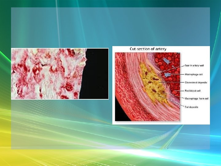
Cardiac Output (CO) • Atherosclerosis - fatty deposits occur in vessels walls leading to occlusion. – Possible causes: Damage to vessel lining -- > infiltration by phagocytic cells [absorb cholesterol & fats] – Platelets adhere to damaged lining --> clots – Related to too much LDL.
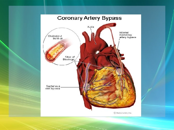
Cardiac Output (CO) – Treatments: • Balloon angioplasty, laser angioplasty, coronary bypass surgery, stents
Balloon Cath + Stent