Anatomy Phys EOC Review SC 912 L 14


 OSTEOBLASTS = Build bone (reforming) Bone must")


 Inhale Exhale Diaphragm ____ Diaphragm_______intercostal muscles contract ______intercostal")
 Inhale J Exhale L Diaphragm contracts Diaphragm relaxes")


 Normal rate = ____? _____ breaths per")


")


")


")

 and cerebrospinal fluid Don’t get mixed")


 broke his first 2 cervical vertebra in a horse riding")


 (Rest & Digest)")


 Oh Once One Takes The Anatomy Final Very Good Vacations")
")
 = surrounds brain & spinal cord")


 The #1 Killer in the USA! Risk Factors: high BP")
 (some of which is preventable with")


")
 High BP = “hypertension”")


")


")

-Potassium (K) Pump = returns neuron to its resting stage by pumping Na")
 junction = synapse between motor neuron & muscle Nerve impulse goes down")
")


- Slides: 108
Anatomy & Phys EOC Review!
SC. 912. L. 14. 12 Describe the anatomy and histology of bone tissue. 2 questions (1 low, 1 med complexity) Students will know the parts of the bone. May include: anatomical long bone structures Microscopic bone tissue Cartilage Joints Bone marrow (red & yellow) Bone repair Bone remodeling It will NOT include bone diseases.
Long Bone structure
Microscopic Bone Structure
Joints & cartilage
Red vs Yellow marrow Location Function Red Yellow * flat bones (pelvis, sternum, ribs, skull, shaft (diaphysis) vertebra, scapula) of long bones * in spongy ends of (femur, humerus) long bones produce blood cells store fat
Bone Repair
Bone Remodeling OSTEOCLASTS = Kill bone (resorption) OSTEOBLASTS = Build bone (reforming) Bone must be broken down to release calcium and phosphorus. Balance of resorption & reforming results in healthy bone. Overactive resorption by osteoclasts results in osteoporosis. We have a brand new skeleton every 10 years!
SC. 912. L. 14 Identify the major bones of the axial and appendicular skeleton. 4 Questions (2 easy, 2 medium, 2 high-level) Students will know all 206 bones of the adult body. It will NOT include a diagram of an adult skeleton with specific bony landmarks (example lateral epicondyle of the right femur).
Axial vs Appendicular skeleton
Identifying major bones www. anatomyarcade. com
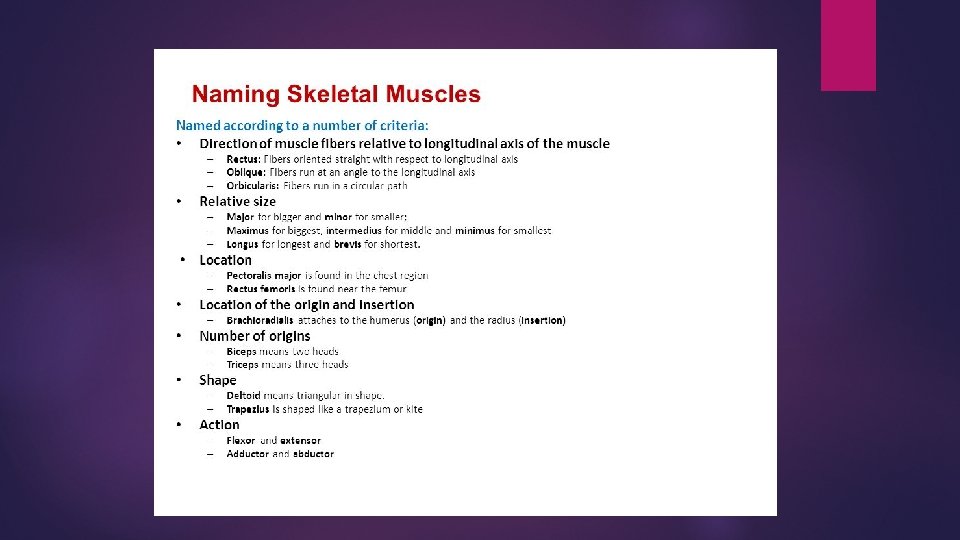
SC. 912. L. 14. 20 Identify the major muscles of the human on a model or diagram. 6 Questions (2 easy, 2 medium, 2 high-level) Students will know major muscle groupings It will NOT include origin, insertion, or specific action.
Identifying major muscles www. anatomyarcade. com
Face muscles
SC. 912. L. 14. 44 Describe the physiology of the respiratory system including the mechanisms of ventilation, gas exchange, gas transport and the mechanisms that control the rate of ventilation. 4 questions (2 easy, 2 medium) Students will know mechanisms for ventilation including gas exchange. It may include information on asthma and bronchitis.
Respiratory System diagram
Muscles involved in breathing
Diaphragm during breathing
Boyle’s Law In a closed system, Volume and Pressure are inversely proportional. When V increases, P decreases. When V decreases, P increases. https: //www. youtube. com/watch? v=e. R 49 g 3 ub. TBg https: //www. youtube. com/watch? v=N 5 xft 2 f. Iq. QU https: //www. youtube. com/watch? v=27 yq. J 9 v. J 5 k. Q
Mechanism of Breathing (Negative Pressure breathing) Inhale Exhale Diaphragm ____ Diaphragm_______intercostal muscles contract ______intercostal & ab muscles contract Volume _______ Pressure ____ Pressure____ Air rushes ______ Air rushes _____
Mechanism of Breathing (Negative Pressure breathing) Inhale J Exhale L Diaphragm contracts Diaphragm relaxes External intercostal muscles contract Internal intercostal & ab muscles contract Volume of thorax increases decreases Pressure increases Air rushes IN Air rushes OUT
https: //www. youtube. com/watch? v=Mf 8 x. Tqfspp 4
Lung model
Gas Exchange Occurs in 2 places: between capillaries & alveoli between capillaries & cells Occurs by simple diffusion Diffusion = moving from area of high concentration to area of low conc.
Gas Transport
How is O 2 & CO 2 carried in blood?
Control of ventilation rate (ventilation = breathing) Normal rate = ____? _____ breaths per min 12 -20 breaths per min Controlled by medulla oblongata of brain stem Inhaling Sends out nerve impulses to diaphragm & external intercostals, causing them to contract Exhaling Stops sending nerve impulses to muscles, causing them to relax
Asthma
Bronchitis
Triggers of asthma & bronchitis
SC. 912. L. 14. 32 Describe the anatomy & phys of the endocrine system. 4 questions (2 easy, 2 medium) Students will know all endocrine organs and main types of hormones. Items may include endocrine diseases, however students will NOT be held accountable for specific differences related to this benchmark.
Endocrine System Endocrine GLANDS release HORMONES into the blood. Hormones = chemical messengers that tell another part of body to do something (kind of like what nerve impulses do!) E. g. Pancreas (gland) releases Insulin (hormone) that travels in blood to all the cells of the body to cause them to uptake sugar from the blood. Not enough Insulin, cells don’t take it in, too much sugar remains in blood… High blood sugar = “Diabetes”!
Endocrine system maintains homeostasis by NEGATIVE feedback (hormones REVERSE a change)
Pituitary
Thyroid & Parathyroid
Thymus
Adrenal glands
An easy way to remember adrenal cortex hormones!
Adrenal glands help you cope with STRESS!
Pancreas
Type I & Type II Diabetes
Ovaries (female)
Estrogen!
Monthly cycle of Female hormones! On what days of a 28 -day cycle can a woman most likely get pregnant, if Day 1 is the first day of her period? around ovulation (days 11 – 18) How many weeks is full-term pregnancy? 38 weeks What question does an OB/GYN ask you to determine baby’s due date? When was Day 1 of your last period? (then They add 40 weeks to that!)
TESTES!
Testosterone
SC. 912. L. 14. 30 Compare endocrine and neural controls of physiology. 2 questions (1 easy, 1 medium) Students will know the physiological differences between neural control and endocrine control. Items may include neurological and endocrine diseases, however students will not be held accountable for specific differences related to this benchmark.
Comparison of Nervous & Endocrine
SC. 912. L. 14. 28 Identify the major functions of the spinal cord. 4 questions (2 easy, 2 medium) Students will know the structure & functions of the spinal cord. Items may include functions of the spinal cord. (Really? )
Structure of spinal cord (cross-section)
Reflex arc http: //ib. bioninja. com. au/_Media/reflex-arc. mp 4
Location of spinal cord Protected by vertebrae (spine) and cerebrospinal fluid Don’t get mixed up between “spine” & “spinal cord”! Spine is hard bone Spinal cord is soft nerve tissue
Where spinal cord ends & cauda equina begins!
Spinal Cord anatomy and spinal cord injuries
An easy way to remember… Number of & functions of spinal nerves
Christopher Reeve (original “Superman”) broke his first 2 cervical vertebra in a horse riding competition when he was 42.
SC. 912. L. 14. 21 Describe the anatomy, histology & phys of the CNS & PNS & name the major divisions of the nervous system. 4 Questions (2 easy, 2 medium) Students will know all the structures of the PNS and CNS, including lobes of the brain and cranial nerves. Students will understand what the somatic, autonomic, parasympathetic, and sympathetic nervous systems control. Items will NOT include the function of specific peripheral nerves. Items will NOT include specific neurotransmitters.
Divisions of nervous system
CNS vs PNS
Sympathetic vs Parasympathetic (Fight or Flight) (Rest & Digest)
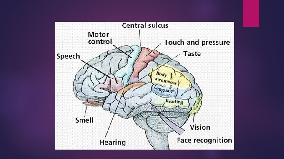
External BRAIN Internal
Corpus callosum = millions of myelinated axons connecting left & right brain so they can communicate
meninges
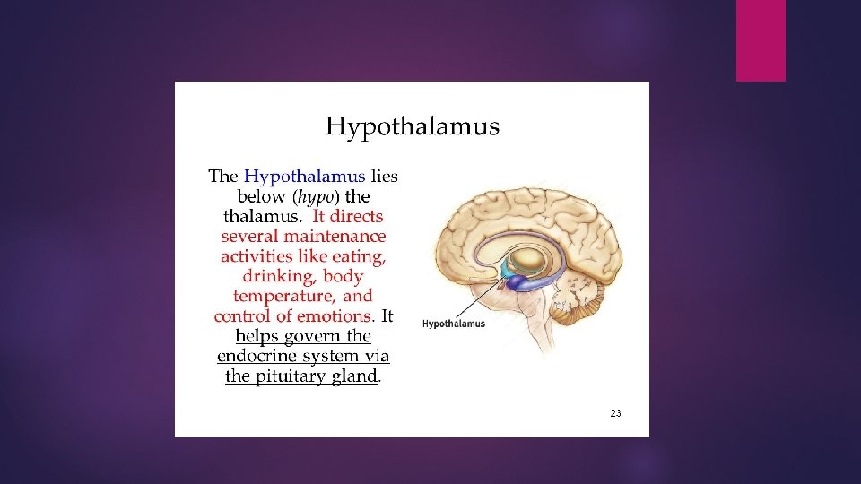
Internal structures of brain
Cranial Nerves (12 pair) Oh Once One Takes The Anatomy Final Very Good Vacations Are Heavenly
Cranial Nerve Functions (probably don’t have to know all these functions)
Cerebrospinal fluid (CSF) = surrounds brain & spinal cord
Functions of CSF Protection – It acts as a cushion for the brain, limiting neural damage in cranial injuries. Buoyancy – By being immersed in CSF, the net weight of the brain is reduced to approximately 25 grams. This prevents excessive pressure on the base of the brain. Chemical stability – The CSF creates an environment to allow for proper functioning of the brain. E. g. Maintaining low extracellular K+ for synaptic transmission.
CSF is produced by ventricles of brain
Concussion = mild brain injury
SC. 912. L. 14. 36 Describe the factors affecting blood flow through the cardiovascular system SC. 912. L. 14. 39 Describe hypertension and some of the factors that produce it. 6 Questions (3 easy, 3 medium) Students will know how to diagnose different cardiovascular diseases based on given signs and symptoms. Items may include varying values of blood pressure and cholesterol in order to draw conclusions based on presented data (includes sex, height, weight, life style).
http: //www. heart. org/idc/groups/heartpublic/@wcm/@global/documents/downloadable/u cm_305699. pdf
Coronary Heart Disease (CHD) The #1 Killer in the USA! Risk Factors: high BP High fat/cholesterol Overweight Inactivity Smoking diabetes genetics https: //www. youtube. com/watch? v=CIv. A 71 c. QJm. Q
Leading causes of death in USA (CDC, 2011) (some of which is preventable with diet & exercise & non-smoking!)
Coronary Heart Disease https: //www. youtube. com/watch? v=T 8 zkvdkz. U 7 A Caused by: Atherosclerosis = narrowing of arteries caused by fat & calcium, reduces blood flow, & increases blood pressure Arteriosclerosis = hardening of arteries Can lead to: Angina = chest pain Arrhythmia = abnormal heart rhythm Aneurism = bulge in artery Thrombus = blood clot Embolus = moving blood clot Hypertension = high blood pressure Heart Attack (Coronary Thrombosis) = blood clot in blood vessel going to HEART Stroke = blood clot in blood vessel going to BRAIN
Heart Murmur = leaky heart valve causing back flow of blood Abnormal Heart Sounds Baby born with heart outside chest!
Cholesterol = fat-like substance used to protect nerves, make part of cell membranes & make sex hormones We need it, just not in excess!
The Good, the Bad and the Ugly
Cholesterol (keep it under 200)
Blood Pressure (Keep it under 140/90) High BP = “hypertension”
SC. 912. L. 14. 18 Describe signal transmission across a myoneural junction. SC. 912. L. 14. 24 Identify general parts of a synapse & describe the physiology of signal transmission across a synapse. 2 questions (1 easy, 1 medium) Students will know all parts of neuron & sodium/potassium pump. Students will know order of action potential along a neuron to the myoneural junction. Items may include major neurotransmitters, including acetylcholine.
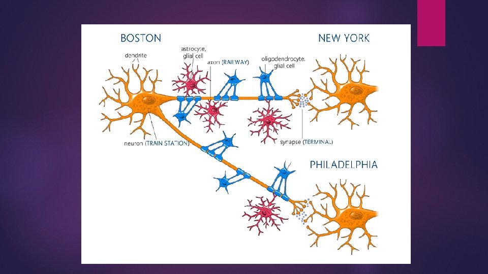
Structure of Neuron
Glial cells = supporting cells (Schwann cells, astrocyte, oligodendrocyte, satellite cell, microglia)
Satellite cells = surround and protect cells bodies Schwann cells = made of myelin to insulate axon
Oligodendrocytes = produce myelin
Microglia = macrophages that ingest bacteria (10 -15% of all cells in brain)
Action Potential = nerve impulse
Sodium (Na)-Potassium (K) Pump = returns neuron to its resting stage by pumping Na to outside & K to inside of axon
Myoneural (neuromuscular) junction = synapse between motor neuron & muscle Nerve impulse goes down axon to synapse to myofibrils of muscle.
Signal transmission across a synapse (synapse = gap between 2 neurons)
Neurotransmitters = chemicals that transmit the nerve impulse across a synapse acetylcholine = controls muscle contraction, heart rate, digestion & memory. norepinephrine = arousal, as well as in learning & mood regulation. serotonin = regulation of sleep, mood and eating. Dopamine = regulating movement and experiencing pleasure. GABA = inhibits the firing of neurons glutamate = strengthens synaptic connections between neurons. Endorphin = binds to opiate receptors and moderates pain
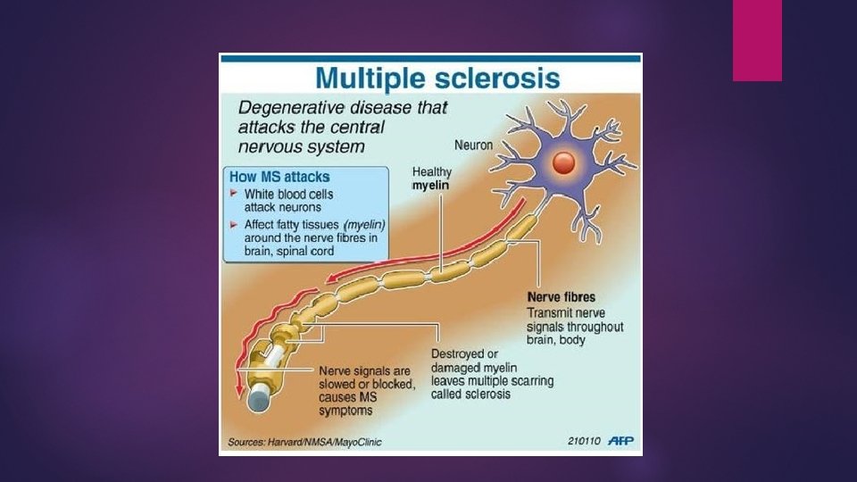
Multiple sclerosis early symptoms
SC. 912. L. 14. 50 Describe the structure of vertebrate sensory organs. Relate structure to function in vertebrate sensory systems. 4 questions (2 easy, 2 medium) Students should know all structures and functions of vision, hearing, balance, olfactory, and tasting. Items may include focus on injuries to certain structures and its effects on the body.
VISION
How the EYE is like a CAMERA
HEARING Semicircular canals = “Organs of Balance” they are filled with fluid, which moves when you move cochlea = body's microphone, converts sound waves into electrical impulses which are passed on to the temporal lobe of brain via the auditory nerve.
BALANCE Cerebellum Aids in coordination By receiving impulses from frontal lobe of brain Maintains balance By receiving impulses from semicircular canals of inner ear Aids in muscle tone So all muscle fibers are slightly tensed
OLFACTORY
TASTING
SC. 912. L. 14. 46 Describe the physiology of the digestive system, including mechanical digestion, chemical digestion, absorption and the neural and hormonal mechanisms of control. 4 QUESTIONS (2 easy, 2 medium) Students will know the mechanisms of digestion, including absorption areas of macromolecules. Items will not include digestive disorders or diseases.
Digestive System alimentary canal accessory organs
Source, substrate, product & optimal p. H for digesting carbs, proteins, fats
Villi & microvilli increase the surface area of small intestine for maximum ABSORPTION of nutrients (glucose, amino acids, fatty acids)