Adrenal Glands Ayman Mismar Associate Prof of Endocrine


")


 Crescentic (semilunar)")


 �B- Abnormal")


 � 2 - Hypercortisolism (")
 1. 2. 3. 4. Solitary adrenal adenomas (80 -90%) Bilateral")


 B. Adrenal Hyperplasia")
 �Ectopic ACTH syndrome (small")


 Pediatric Malignant Normotensive Extra-adrenal Multiple")


 �Anterior �Posterior retroperitoneal")


- Slides: 67
Adrenal Glands Ayman Mismar Associate Prof. of Endocrine Surgery The University of Jordan
Anatomy
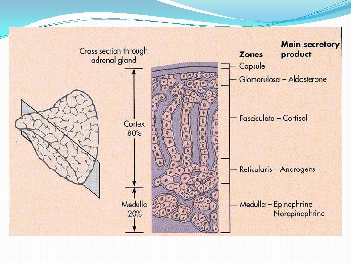
Adrenal Gland Anatomy was first described in 1563. Is located above (or attached to) the upper pole of the kidney. Is pyramidal in structure and weighs ~ 4 g. Consists of the adrenal cortex and adrenal medulla Activities are regulation of fluid volume and stress response
Adrenal Gland • Blood supply Arterial - receives 7 cc/gram minute • arterial sources of flow: 1. Inferior phrenic artery 2. Aorta 3. Renal artery Venous – single main vein most important surgical structure - right → post IVC - left → renal vein
Adrenal Gland �Cortex - mesoderm �Medulla - neuroectoderm �Renal agenesis, found in normal anatomic position �Size- 5 x 3 x 1 x 5 cm �Retroperitoneal structure, contained in its own subcompartment w/in Gerota’s fascia
Comparison between Rt. & Lt. Suprarenals Right Suprarenal Left Suprarenal Triangular (pyramidal) Crescentic (semilunar) Does NOT reach the hilum of the right kidney Reaches the hilum of the left kidney The hilum is directed upwards The hilum is directed downwards Its vein is short and drains to the IVC Its vein is long and drains to the left renal vein. 6
Physiology
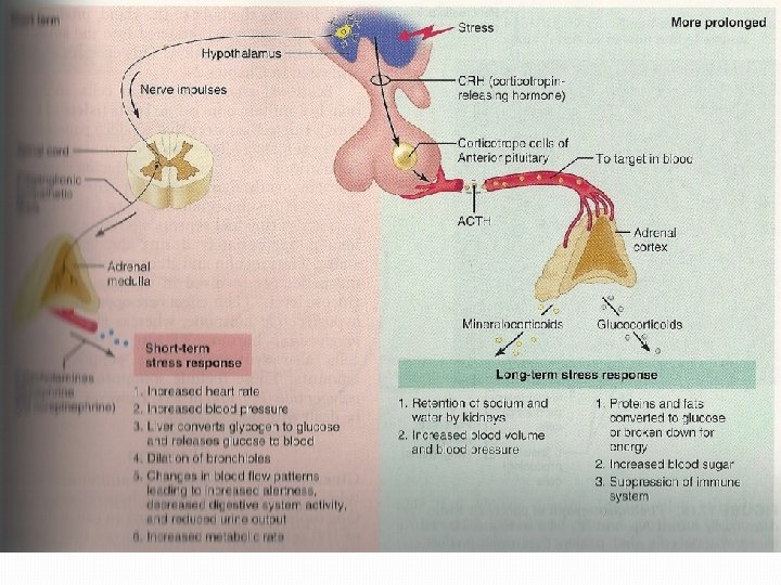
The General Adaptation Syndrome
The General Adaptation Syndrome
Disosrders of the adrenal gland
Disorders of Adrenal glands: �A- Abnormal anatomy: ( diffuse or localized swelling) �B- Abnormal physiology: ( hyper or hypo secretion of hormones)
Neoplasms arising from cortex and metastases �Adenoma �Carcinoma �Myelolipoma �Metastatic carcinoma
Cortical Adenoma �Solitary nodule �Usually encapsulated by rim of connective tissue �Usually small: <50 gm, < 5 cm diameter (on the average, much smaller than cortical carcinomas) �Yellow color (majority, because of lipid-laden cells) �Black color (minority, due to excess lipofuscin pigment) �Cured by surgical excision �Endocrine function is unpredictable; 3 syndromes are �no hyperfunction (“silent”) �hypercortisolism �hyperaldosteronism
CT Adenoma Characteristics �Sharp margins �Smooth, homogenous, lipid rich �Most <10 Hu on noncontrast images �Washout >50% @ 15 min
�Secreting tumors: � 1 - Hyperaldosteronism ( Conn`s syndrome) � 2 - Hypercortisolism ( Cushing syndrome) � 3 - Hyperandrogenism ( Precocious puberty) � 4 - Pheochromocytoma
Primary Hyperaldosteronism (Conn's syndrome) 1. 2. 3. 4. Solitary adrenal adenomas (80 -90%) Bilateral adrenal hyperplasia (10 -20%) Adrenal Carcinoma (rare) Unilateral Adrenal Hyperplasia (very rare)
Clinical features of Conn's syndrome �Often asymptomatic �Frontal headache �Muscle weakness to flaccid paralysis decreased muscle strength (because of low potassium level) �Polyuria and Polydipsia �Hypertension
Investigation of Conn's syndrome A. 1. 2. 3. Serum Electrolytes Serum Potassium decreased Serum Sodium increased (Mild) Metabolic Alkalosis B. Increased serum level of aldosterone C. Greatly decreased serum level of renin D. Sodium loading test
Localization �Ultrasound. �CT Scan. �MRI �Angiographic Blood Sampling.
Treatment of Conn's syndrome A. Adrenal Adenoma Surgical excision (unilateral adrenalectomy) B. Adrenal Hyperplasia Spironolactone (Aldactone)
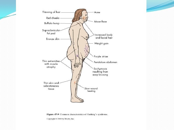
Cushing's syndrome Endogenous : ACTH dependent 85% �Cushing's disease (pituitary) �Ectopic ACTH syndrome (small cell lung carcinoma) �Ectopic CRH syndrome (bronchial carcinoid tumours) ACTH independent 15% �Common: Adrenal adenoma, adrenal carcinoma �Less common: Micronodular hyperplasia, macronodular hyperplasia
Exogenous �Glucocorticoid treatment Pseudo-Cushing's syndrome �Major depressive disorder �Alcoholism �Obesity
Investigation of Cushing's syndrome Screening tests �Urinary free cortisol The measurement of 24 hour excretion of cortisol in urine integrates the variations in plasma free cortisol concentrations seen during the entire day; three consecutive 24 hour urine specimens. � There is a false negative rate of 5. 6%, with false positives of 1% in normal people and 5% in obese individuals.
�Dexamethasone suppression tests In normal subjects, administration of a supraphysiological dose of glucocorticoid results in suppression of ACTH and hence of cortisol secretion (cortisol <50 nmol/l). This is the basis of dexamethasone suppression tests, of which there are several types.
Treatment of Cushing's syndrome �Transsphenoidal surgery �Pituitary radio-therapy � Bilateral or unilateral adrenalectomy � Medical therapy
Pheochromocytoma Rule’s of 10 • • Bilateral Familial (non-sporadic) Pediatric Malignant Normotensive Extra-adrenal Multiple *Childhood presentation breaks the rules- 25% bilateral, multiple, extra-adrenal
Pheo �Triad: headache, tachycardia, diaphoresis �Other symptoms: Pallor, flushing, palpitations, abd/chest pain, weakness, N/V, psychosis �Small tumors more likely symptomatic
Pheo- The Diagnosis �Plasma free metanephrines : most sensitive test seen 99% of patients � 24 h urinary catecholamines (2 x normal is diagnostic) �VMA �Clonidine suppression test (0. 3 mg oral, test 3 hrs later) >50% reduction catecholamines NO pheo
Pre-op Management �Early alpha blockad �Goal to control hypertension- phenoxybenzamine �Do NOT use b-blocker before alpha �IV hydration �Prevent cardiac arrhythmias
Adrenal Cortical Carcinoma �Mean age 50 years �Larger: typically > 100 gm; mean weight 900 grams (BIG!) �Gross clues for malignancy (some apparent by imaging) �Large size, hemorrhage, necrosis, invasion of adrenal vein �Absolute criteria for malignancy: invasion & metastasis � 5 year survival: 20 -30%; most tumor-related deaths occur within first two years after diagnosis
4. The adrenal “Incidentaloma” �Definition: “an adrenal mass, generally more than 1 cm. diameter, that is discovered accidentally during a radiologic examination performed for indications other than an evaluation for adrenal disease. ” *
Nature of Incidentally Found Adrenal Masses �Review of 2, 005 incidentally-discovered adrenal masses: � Nonfunctioning adenoma 82% � Functioning: Cushing’s 5% � Pheo 5% � Aldosteronoma 1% � Malignancy: Metastasis 3% � ACC 4% � Young WF, et al. Endocrinol Clin N Am. 2000
Incidental Adrenal Mass Management �Hormonally active → surgical removal �> 5 cm → removal (with a scalpel) �< 3 cm observe �Surveillance Recommendations: Old: CT at 6 months then Annual endocrine eval for 4 years New: If mass stable on scans @ 3 m and 1 yr then no further workup
Young, WF; New England Journal of Medicine, 356: 601 -610, 2007, fig. 1
Surgical approach
Surgical Management of Adrenal Neoplasms �Before the era of laparoscopic procedures, conventional open adrenalectomy was the only surgical approach to adrenal neoplasms �Since the introduction of laparoscopic adrenalectomy by Gagner et al. in 1992, the majority of benign adrenal lesions have been removed by various laparoscopic techniques.
Indications �The choice of approach will depend on size, location, possibility of malignancy, and surgeon experience with the different techniques. �The open approach to adrenalectomy is considered the gold standard for masses suspected of being adrenal carcinoma
Indications �Conn syndrome �Cushing syndrome �pheochromocytoma �large myelolipoma �metastatic tumors �adrenocortical carcinoma � neuroblastoma
Conventional Open Methods �The flank or posterior �The anterior or transabdominal �The transthoracic or thoracoabdominal
�The standard approach has the patient in the prone position with arms extended over the head �The incision is made over the 11 th or 12 th rib, with or without a “hockey stick” extension cephalad
Posterior Approach �Also referred to as the lumbodorsal approach �The advantage of this approach is allowing the surgeon to both remain in an extraperitoneal dissection plane and offers access to both adrenal glands �The incision is made through the bed of the twelfth rib
Posterior Approach Disadvantages : �Since exposure with this approach is limited, it should only be used to remove tumors up to 5 cm in size �Intra-abdominal exploration for other pathology or metastases is impossible �Two separate incisions are required for bilateral adrenalectomy
Flank Approach
Flank Approach �Incision is made over the 11 th rib and carried down through the muscle and fascial layers over the 11 th rib �The rib is mobilized posteriorly taking care to avoid injuring the neurovascular bundle �The rib is then resected and entry is made into the retroperitoneal space through the lumbodorsal fascia.
Flank Approach �This approach offers excellent exposure in obese patients �there is a risk of injury to the pleural reflection at the level of the 11 th rib in the midaxillary line and inadvertent entry into the pleural cavity or lung injury.
Anterior Subcostal Approach
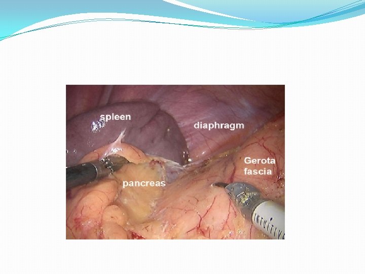
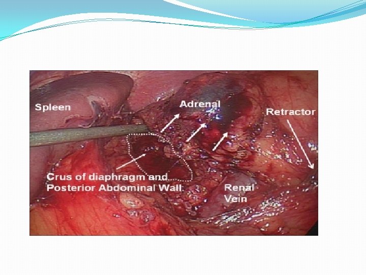
Anterior Subcostal Approach �The subcostal approach is initiated with the patient supine and the incision is made two fingerbreadths below the costal margin. �The adrenal is identified by its characteristic yellowish color, lobulated appearance, and clearly definable blunt lateral edge. �The vascular attachments are usually on or near the medial and superior edges of the gland rather than on its broad surfaces.
Anterior Subcostal Approach �This approach offers the advantage of being well known to all surgeons and allows for bilateral exploration. �However, it caries a higher rate of ileus, and may be difficult with obese patients
Thoracoabdominal Approach
Thoracoabdominal Approach �The incision is then made through the 8 th or 9 th intercostal space through the muscle layers and carried medially through the costal cartilage �The incision is carried through the anterior and posterior rectus sheaths and the pleura is entered and the lung packed out of the field �Care is taken during division of the diaphragm to not be too central as the phrenic nerve can be damaged with careless division
Thoracoabdominal Approach �This approach offers excellent exposure to the retroperitoneum and the great vessels. �However, it has the highest morbidity of all the approaches with risk of prolonged ileus, pulmonary complications, and necessary chest tube �Most authors reserve this approach for large tumors and adrenal carcinoma
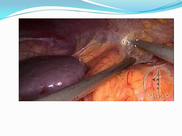
Laparoscopic approaches �Lateral transperitoneal (flank) �Anterior �Posterior retroperitoneal
The lateral transperitoneal laparoscopic approach �Four trocars are inserted in the flank area, one under the subcostal area at the anterior axillary line and three under the twelfth rib along a line lateral to the first one �This method allows one to inspect much of the abdominal cavity and provides wider exposure for removing relatively larger glands than can be removed with the retroperitoneal approach.
The lateral transperitoneal laparoscopic approach
The Anterior Laparoscopic Approach �four to five trocars are inserted, one above the umbilicus and two subcostally, one at the anterior axillary line and another at the midclavicular line. �The advantages and disadvantages of the anterior approach are similar to the lateral transperitoneal or flank approach �more dissection and retraction is required which may lead to more bleeding and other injuries
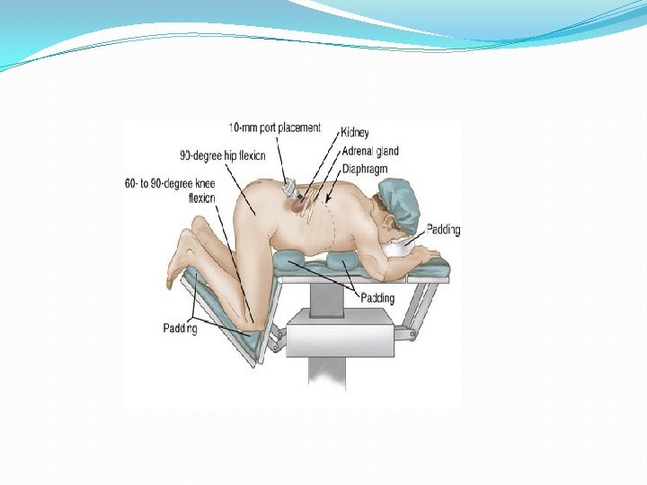
The posterior or retroperitoneal laparoscopic approach �The patient is placed in a semi-jackknife prone position. �A balloon trocar is inserted 2. 5 cm lateral to the twelfth rib into the retroperitoneum to create a working space. �Three more trocars are introduced into the created retroperitoneal space under direct vision
The posterior or retroperitoneal laparoscopic approach Advantages �the retroperitoneal approach allows one to perform a bilateral adrenalectomy without repositioning the patient �It also avoids adhesions from previous operations and other abdominal conditions that might interfere with the operation
The posterior or retroperitoneal laparoscopic approach Disadvantages � However, the retroperitoneal approach precludes intra-abdominal exploration. � Due to its limited working space, large tumors cannot be removed (up to 5 cm).
Thank You