Histology of the Endocrine System Adrenal glands Dr

 gland, human US Federal Government")

 gland Source Undetermined")

. – General function: Maintain")


, LM Fasciculata Humio Mizoguti, Kobe Univ Sch Med, slide")
 of the adrenal cortex")
, LM Humio Mizoguti, Kobe Univ Sch Med, slide 549")
 It consists of small rounded cells that are arranged")


 and norepinephrine (noradrenalin), both catecholamines. Two cell types,")


- Slides: 23
Histology of the Endocrine System Adrenal glands Dr. Ahmed Abdulhussein AL-Huchami
Location of the adrenal (suprarenal) gland, human US Federal Government
Suprarenal gland has two parts that are of different embryonic origin, these are: 1. Cortex (90%) – derived from the genital ridge of the mesoderm. 2. Medulla (10%) – derived from neural crest cells.
Adrenal (suprarenal) gland Source Undetermined
Human adrenal, low power LM Bailey’s Histology
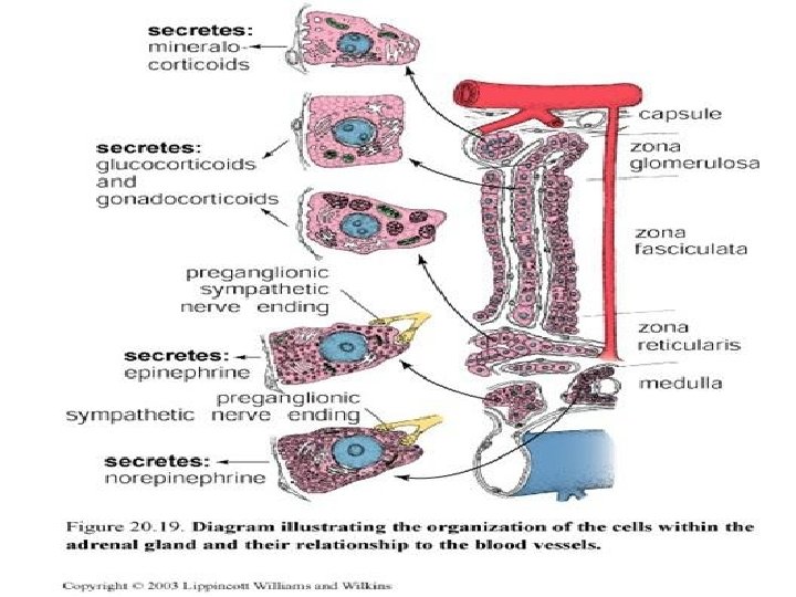
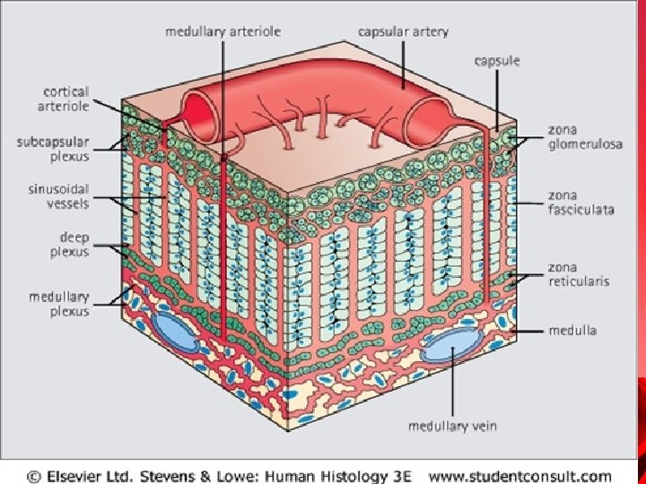
Adrenal cortex Zona glomerulosa – Main hormone: Aldosterone (a mineralocorticoid). – General function: Maintain blood electrolyte balance. – Main control: Angiotensin II. Zona fasciculata – Main hormone: Cortisol (a glucocorticoid). – General function: Includes regulating glucose and fatty acid metabolism, and response to stress. – Main control: Pituitary ACTH. Zona reticularis – Hormones: Some cortisol androgens. – Function and control: Similar to zona fasciculata.
Adrenal cortex, human, LM Hadley Kirkman slide collection, slide K 285
glomeruloza Outer layer forming 15% pyramidal and arranged in rounded or arched groups that are surrounded by capillaries. � Their nuclei are rounded and central while cytoplasm is lightly basophilic
Adrenal cortex, human, H&E, LM Humio Mizoguti, Kobe Univ Sch Med, slide 547
Zona glomerulosa (source of aldosterone), LM Fasciculata Humio Mizoguti, Kobe Univ Sch Med, slide 548
Zona fasciculata: It is the thickest layer (65 – 80%) of the adrenal cortex Cells are large polyhedral with rounded central nucleus and pale foamy cytoplasm. These cells are arranged in parallel columns of 1 or 2 cells thickness. The cytoplasm of zona fasciculata cells contains numerous lipid droplets � During slide preparation (by typical histological procedures) lipid is extracted, thus gives vacuolated or spongy appearance to these cells and that's why they are called Spongyocytes.
Zona fasciculata (source of cortisol), LM Humio Mizoguti, Kobe Univ Sch Med, slide 549
Zona reticularis: Thin layer (10%) It consists of small rounded cells that are arranged in branching and anatomizing cords. Cells have rounded central nucleus and deeply basophilic cytoplasm (because they contain fewer lipid droplets and more lipofuscin pigment). This layer secrets the weak androgen (dehydroepiandrosterone - DHEA) which is converted to testosterone in several other tissues
Zona reticularis, LM Medulla Zona fasciculata Humio Mizoguti, Kobe Univ Sch Med, slide 550
Adrenal medulla The adrenal medulla is composed of large, pale-staining polyhedral cells arranged in cords or clumps and supported by a reticular fiber network. When medullary cells are exposed to an oxidizing agent such as potassium bichromate caticholamines (adrenalin and noradrenalin) will be oxidized giving a brown coloration to their cytoplasm, therefore these cells are called chromaffin cells and the reaction is called Chromaffin reaction. Unlike cells of the cortex , contain many granules, contain one or more of the catecholamines, epinephrine or norepinephrine. About 80% of the catecholamine secreted from the adrenal is epinephrine.
Adrenal medulla Hormones – Epinephrine (adrenalin) and norepinephrine (noradrenalin), both catecholamines. Two cell types, one for E and one for N. – General function: Acute response to stress. – Main control: Preganglionic sympathetic innervation. . Also called "chromaffin cells" – Cells of the adrenal medulla are examples of "chromaffin cells, " containing catecholamine granules that stain brown with potassium dichromate. Neurons of sympathetic ganglia are also chromaffin cells. The term is used in pathology.
Adrenal medulla, LM Humio Mizoguti, Kobe Univ Sch Med, slide 565
EM of adrenal medulla: norepinephrine and epinephrine cells Nucleus Norepinephrine Epinephrine Nucleus Stan Erlandsen Medical Histology Slide Collection, slide MH 9/G/2 -P
THANK YOU