Embryology organogenesis Development and teratology of nervous system

Embryology /organogenesis/ Development and teratology of nervous system.

Neuroectoderm NOTOCHORD DEVELOPMENT Neural plate NOTOCHORD - induces neural plate development 2

Neural plate – thickened area of embryonic ectoderm neuroectoderm pseudostratif. columnar ep. Pharyngeal membrane Primitive streak and node Notochord Cloacal membrane 3
 - neural folds -")
NEURULATION – invagination of neural plate (day 16 - 24) - neural folds - neural groove - neural tube - neural crest 4 notochord

Day 20 Neural folds 5

Day 22, 23 Neuroporus anterior closes on D 25 closes on D 27 Neuroporus posterior 6

NEURAL CREST 7

Odontoblasts Leptomeningeal cells 8

EKTOMESENCHYME 9
")
Histogenesis of neural tube The wall of neural tube: (simple → pseudostratified neural epithelium) Cell proliferation 3 zones: Ependymal Intermediate Marginal zone Ependyma Gray matter White matter 10 (in medulla spinalis)
 Intermediate zone (gray matter) (mantle zone)")
HISTOGENESIS of NEURAL TUBE Marginal zone (white matter) Intermediate zone (gray matter) (mantle zone) Ependymal zone (germinal) 11

Histogenesis of neural tissue In spinal cord white matter gray matter ependyme Three zones line neural tube (the spinal cord and brain stem). Marginal zone (white matter) – without neurons, but with axons of neurons and glial cells Mantle zone (gray matter) – neuroblasts + spongioblasts give rise to bodies of neurons and glial cells 12 Ependymal zone (germinal) – lining of central canal

In brain and cerebellum gray matter white matter ependyme In brain and cerbellum: mantle zone cells migrate through marginal layer and the gray matter coveres white matter. Some neurons stay in white matter nuclei. 13

Spinal cord development Dorsal horns future white matter sensory zone future gray matter motor zone Ventral horns 14
 2. Mantle layer (gray matter) 3. Marginal layer")
SPINAL CORD: 1. Ependymal layer (germinal) 2. Mantle layer (gray matter) 3. Marginal layer (white matter) 15

Positional changes of spinal cord the end fo the 2 nd month the 5 th month Vertebrate canal grows more rapidly than spinal cord and caudal end of spinal cord doesn‘t extend the entire length of canal in adult; it terminates at L 2 in adults #. new-born child # pia mater Cauda equina 16

Brain development • Brain develops from cranial part of neural tube • Week 4 – three primary brain vesicles: - prosencephalon (forebrain) - mesencephalon (midbrain) - rhombencephalon (hindbrain) 17

3 primary → 5 secondary vesicles: week 5 Lamina terminalis week 4 Telencephalon 1 Prosencephalon Diencephalon Optic vesicle Neurohypophysis Epiphysis Mesencephalon 2 3 4 Rhombencephalon Metencephalon Cerebellum Pons Myelencephalon 18 1 – ventriculi lat. , 2 – ventriculus tertius, 3 – aqueductus cerebri, 4 – ventriculus quartus

Midbrain flexure Cervical flexure 19

20

Myelination of nerve fibers from the 4 th prenatal month to the end of 2 nd postnatal year 21

CNS malformations • failure neurulation (absence of notochord inductive influence or teratogen influence on neuroectodermal cells) • defects of spinal cord • defects of brain • difficult malformations of CNS are usually connected with skull or spinal column (vertebral) defects. Etiology: usually multifactorial (fever, drugs during gravidity, hypervit. A etc. ) or hereditary disposition. Folic acid use influence normal development of CNS. Sonography detects anomalies. 22
 of vertebral arches • Menigocele • Menigomyelocele • Menigohydromyelocele")
Spinal cord malformations Defects (clefts) of vertebral arches • Menigocele • Menigomyelocele • Menigohydromyelocele spina bifida cystica • Myeloschisis – complete cleft of spinal column in the whole length 23

24

25
 hairy patch 2) hemangioma Urodynamics 3)")
Examples of external signs of spina bifida: 1) hairy patch 2) hemangioma Urodynamics 3) skin appendage 4) lipomatous mass 26
 (with myeloschisis) 27")
Brain malformations • Anencephalia (†) (with myeloschisis) 27

28

Brain malformations MICROCEPHALIA ANENCEPHALIA 29

30

Hydrocephalus - accumulation of abundant cerebrospinal fluid in brain ventricular system, - etiology: stenosis or obliteration of aqueductus cerebri between 3 rd and 4 th ventricles fluid is accumulated in lateral ventricles pushes on the brain tissue (is thinned); internal pressure complicates drenage of fluid insubarachnoid space; - until skull suture don‘t ossify – skull can grows extremely. 31

HYDROCEPHALUS ventriculoperitoneal shunt 32

33
 34")
Brain and meninges hernia(tion) 34

35

36

Neurohypophyseal diverticle of diencephalon floor + Rathke‘s pouch of stomodeum roof

 – diverticulum of the roof of diencephalon the floor of diencephalon")
Pineal gland (epiphysis) – diverticulum of the roof of diencephalon the floor of diencephalon

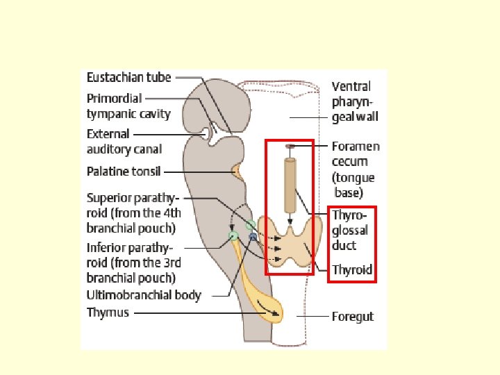
Thyroid gland

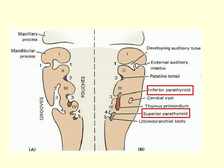
Descensus of thyroid gland Ductus thyroglossus Ultimobranchial body – the 4 th endodermal pouch – parafollicular cells
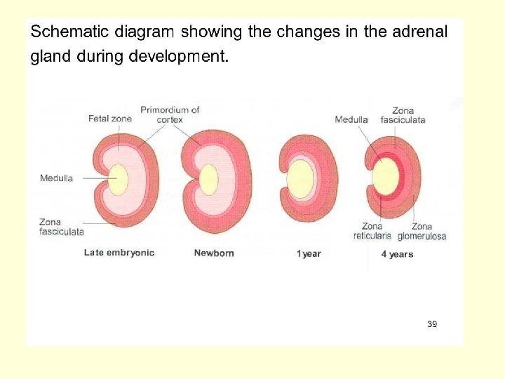
 MEDULLA - neuroectoderm (neural crest)")
CORTEX - mesoderm (coelomic epithelium) MEDULLA - neuroectoderm (neural crest)

46
 – dendrite(s) Nissl")
Terms • • • Neuron – perikaryon – axon (= neurite) – dendrite(s) Nissl bodies = rough ER Axon hillock Myeline sheath Schwann sheath Mesaxon Internodium Node of Ranvier Neuron – classification Synapse (presynaptic knobe, synaptic cleft, postsynaptic memrane) • Neurotransmitters 47
 Ependyma - tanycytes")
Terms • • Neuroglia - classification Oligodendroglia Astrocytes Microglia (of Horteg) Ependyma - tanycytes Schwann cells Satelite cells in CNS in PNS 48

Special histology - questions • • • Structure of the brain cortex. Cyto- and myeloarchitecture. Structure of the cerebellum. Synapses of the cerebellum. Microscopic structure of the spinal cord. Microscopic structure of ganglia and peripheral nerves. Ependyma, plexus chorioideus and meninges. 49
 • Cajal cells, Martinotti cells, granular")
Terms • Brain cortex – 6 layers (lamina) • Cajal cells, Martinotti cells, granular and pyramidal cells • Membrana limitans gliae superficialis et profunda (seu perivascularis) • Brain barrier • Cerebellum – 3 layers of cortex (stratum) • Purkinje cells, basket cells, granular cells • Glomeruli cerebellares • Mossy and climbing fibers 50

Terms • Dura mater – arachnoidea – pia mater • Endoneurium – perineurium – epineurium • Plexus chorioideus 51

52

53
 A myelinated axon in the peripheral nervous system and (b) its")
Fig. 1 (a) A myelinated axon in the peripheral nervous system and (b) its development. Each Schwann cell myelinates a single axon, to which it is directly apposed. During development (anticlockwise) Schwann cells loosely ensheath axons and the myelin sheath grows around the axon to form concentric layers, which become tightly apposed 54

Fig. 3 Myelination in the central nervous system. A single oligodendrocyte myelinates numerous axons (a) and, in section, concentric layers of myelin are seen to spiral around the axon (b). Myelin sheaths are arranged along axons in segments 1 mm long separated by short nodes, and would appear as large sheets if they were unwrapped from around the axon 55

56

57

58

roof plate central canal ependymal layer mantle layer marginal layer floor plate 59

60

61

Invagination of neural plate neural folds + neural groove 62

Neural tube and neural crest Neuroporus ant. , post. Neural crest Neural tube 63

future brain future spinal and autonomic ganglia future spinal cord 64

65

66

67
- Slides: 67