The Autonomic Nervous System and the Adrenal Medulla


 2) 3) q synapse with")


 2) cholinergic fibers – secrete acetylcholine adrenergic fibers –")
 are cholinergic (sympathetic and parasympathetic) q all or almost")


")


 q")

 five subclasses (M 1 -M")


 under the influence of")


 1) 2) 3) 4) 5) preventing")

 1) neostigmine, pyridostigmine,")


- Slides: 73
The Autonomic Nervous System and the Adrenal Medulla Prof. dr. Zoran Valić Department of Physiology University of Split School of Medicine
q q q portion of the nervous system that controls most visceral functions of the body arterial pressure, gastrointestinal motility, gastrointestinal secretion, urinary bladder emptying, sweating, body temperature rapidity (seconds) and intensity of the change (100%) – most striking characteristics
General Organization of the ANS centers located in: q q q spinal cord brain stem hypothalamus q + limbic cortex operates through visceral reflexes (subconscious) sympathetic and parasympathetic nervous system
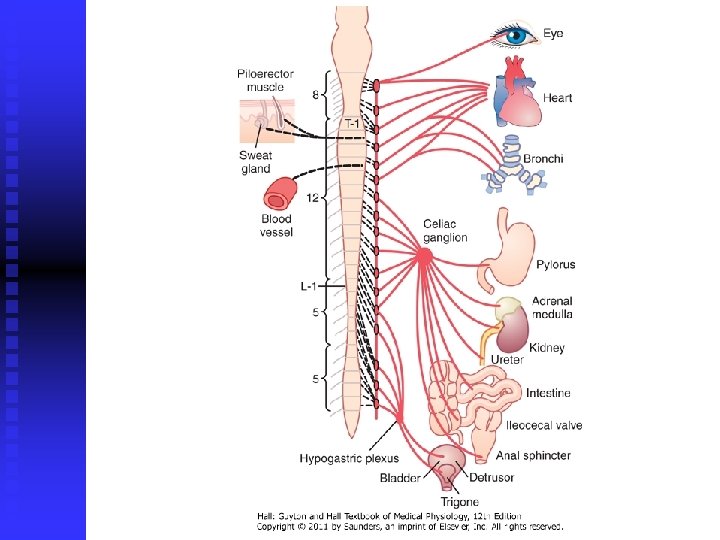
Physiologic Anatomy of the Sympathetic Nervous System two paravertebral sympathetic chains of ganglia two prevertebral ganglia q q q celiac hypogastric postganglionic nerves sympathetic nerve fibers originate in the spinal cord along with spinal nerves between cord segments T-1 and L-2
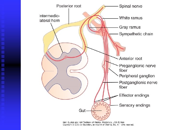
Preganglionic and Postganglionic Sympathetic Neurons q q q difference from skeletal motor nerves cell body of each preganglionic neuron lies in the intermediolateral horn of the spinal cord its fiber passes through an anterior root of the cord into the corresponding spinal nerve
q q after the spinal nerve leaves the spinal canal, the preganglionic sympathetic fibers leave the spinal nerve pass through a white ramus into one of the ganglia of the sympathetic chain
course of the fibers can be following: q 1) 2) 3) q synapse with postganglionic sympathetic neurons in the ganglion that it enters pass upward or downward in the chain and synapse in one of the other ganglia of the chain pass for variable distances through the chain and then through one of the sympathetic nerves radiating outward from the chain, finally synapsing in a peripheral sympathetic ganglion postganglionic fibers then travel to their destinations in the various organs
q q some of the postganglionic fibers pass back from sympathetic chain into spinal nerves through gray rami at all levels of the cord these are all very small type C fibers, and they extend to all parts of the body by way of the skeletal nerves they control: blood vessels, sweat glands, and piloerector muscles of the hairs they make 8% of fibers in average nerve
Segmental Distribution sympathetic pathways are not necessarily distributed to the same part of the body as the somatic spinal nerve fibers from the same segments: q q q T 1: T 2: T 3 -T 6: T 7 -T 11: T 12 -L 2: head neck thorax abdomen legs
q q distribution is only approximate and overlaps greatly it is determined partly by the locus in the embryo from which the organ originated
Adrenal Medullae q preganglionic sympathetic nerve fibers pass, without synapsing, from the intermediolateral horn cells of the spinal cord, through the sympathetic chains, then through the splanchnic nerves, and finally into the two adrenal medullae
q q these secretory cells embryologically are derived from nervous tissue and are actually themselves postganglionic neurons (they even have rudimentary nerve fibers) the endings of these fibers that secrete the adrenal hormones epinephrine and norepinephrine
Physiologic Anatomy of the Parasympathetic Nervous System q q q parasympathetic fibers leave the CNS through cranial nerves III, VII, IX, and X, additionally by 2 nd and 3 rd sacral spinal nerves (sometimes 1 st and 4 th) 75 percent of all parasympathetic nerve fibers are in the vagus nerves supply: heart, lungs, esophagus, stomach, entire small intestine, proximal half of the colon, liver, gallbladder, pancreas, kidneys, and upper portions of the ureters
+ external genitalia - erection
Preganglionic and Postganglionic Parasympathetic Neurons q q q preganglionic fibers pass uninterrupted all the way to the organ that is to be controlled (except in the case of a few cranial nerves) postganglionic neurons are located in the wall of the organ postganglionic fibers, a fraction of a millimeter to several centimeters in length, leave the neurons to innervate the tissues of the organ
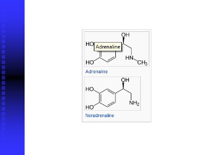
Cholinergic and Adrenergic Fibers 1) 2) cholinergic fibers – secrete acetylcholine adrenergic fibers – secrete norepinephrine (noradrenalin)
q all preganglionic neurons (fibers) are cholinergic (sympathetic and parasympathetic) q all or almost all of the postganglionic neurons (fibers) of the parasympathetic system are also cholinergic q most of the postganglionic sympathetic neurons (fibers) are adrenergic
q exemption: postganglionic sympathetic nerve fibers to the sweat glands, to the piloerector muscles of the hairs, and to a very few blood vessels are cholinergic
q q acetylcholine – parasympathetic neurotransmitter norepinephrine – sympathetic neurotransmitter
Secretion of ACh and NE q q few parasympathetic nerve endings are similar to, but much smaller, than those of the skeletal neuromuscular junction majority of fibers merely touch the effector cells of the organs that they innervate varicosities – bulbous enlargements – ACh and NE, mitochondria action potential entrance of Ca secretion from terminals or varicosities
ACh q q q synthesized in the terminal endings and varicosities stored in vesicles acetyl-Co. A + choline acetylcholinesterase from local connective tissue splits it to acetate ion and choline secreted is then transported back into the terminal nerve ending (reuptake)
NE synthesis in nerve endings, but is completed inside the secretory vesicles tyrosine (hydroxylation) DOPA (decarboxylation) dopamine (transport into vesicles, hydroxylation) NE (in AM, 80%, methylation) epinephrine removal: q q q reuptake (50 -80%) diffusion into surrounding body fluids (most of the remaining NE) enzymatic degradation (MAO, COMT, small amounts)
q q q NE secreted directly into a tissue remains active for only a few seconds NE and epinephrine secreted into the blood by the AM remain active until they diffuse into some tissue (COMT, liver) NE and epinephrine remain active for 10 to 30 seconds; but their activity declines to extinction over 1 to several minutes
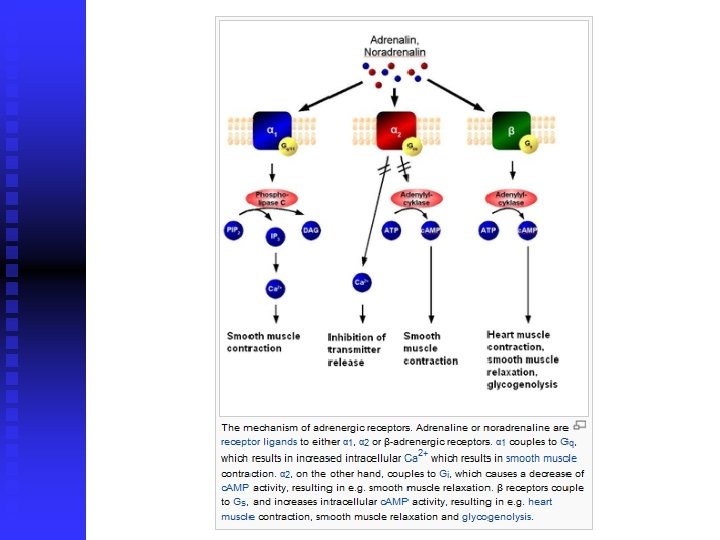
Receptors ACh and NE must first bind with specific receptors on the effector cells receptor is on the outside of the cell membrane binding of transmitter substance causes a conformational change in the structure of the protein molecule q q q 1) 2) change in cell membrane permeability activating or inactivating an enzyme
Change in Membrane Permeability q q opening or closing an ion channel most frequently sodium and/or calcium ion channels entrance of ions usually depolarizes the cell membrane and excites the cell exit of K ions from the cell leads to hyperpolarization (inhibition) of the cell
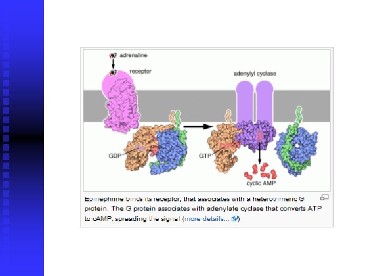
Altering Intracellular "Second Messenger" Enzymes q q binding NE adenylyl cyclase c. AMP exact effect depends on the chemical machinery of the effector cell
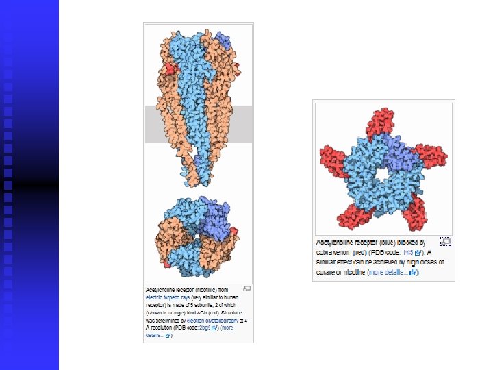
Acetylcholine Receptors nicotinic receptors 1. q muscarinic receptors 2. q q activated by nicotine activated by muscarine (a poison from toadstools) ACh activates both nicotinic and muscarinic receptors
1. Nicotinic receptors q q found at the synapses between the preganglionic and postganglionic neurons of both the sympathetic and parasympathetic systems also present at many nonautonomic nerve endings; at the neuromuscular junctions in skeletal muscle
ionotropic receptor (directly connected with the ion channel, does not utilize second messengers) q two subclasses: 1) nicotinic receptor of the muscle type 2) nicotinic receptor of the neuronal type q
2. Muscarinic receptors q q found on all effector cells that are stimulated by the postganglionic cholinergic neurons either the parasympathetic nervous system or the sympathetic system
q q metabotropic receptor (receptor coupled with G protein) five subclasses (M 1 -M 5)
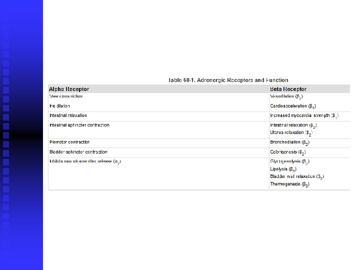
Adrenergic Receptors α-receptors 1. q β-receptors 2. q q q in turn divided into α 1 - i α 2 -receptors in turn divided into β 1 -, β 2 - i β 3 -receptors NE excites mainly α-receptors, β-receptors to a lesser extent epinephrine excites both types of receptors approximately equally
q q q metabotropic receptor (receptor coupled with G protein) under the influence of catecholamins isopropyl norepinephrine (isoprenaline or isoproterenol) acts on β-receptor
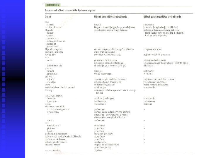
Excitatory and Inhibitory Actions q q excitatory effects in some organs but inhibitory effects in others – there is no generalization, one must learn all the separate functions of these two nervous systems on each organ when sympathetic stimulation excites a particular organ, parasympathetic stimulation sometimes inhibits it
q most organs are dominantly controlled by one or the other of the two systems
Function of the Adrenal Medullae q q stimulation of the sympathetic nerves to the AM causes large quantities of epinephrine (80%) and NE (20%) to be released, although proportions can differ almost the same effects as direct sympathetic stimulation, except that the effects last 5 to 10 times as long (2 -4 min)
q q epinephrine has a greater effect on cardiac stimulation, less vasoconstrictive effect (especially in muscle, represent a major segment of the vessels of the body) NE greatly increases the total peripheral resistance and elevates arterial pressure; epinephrine raises the arterial pressure to a lesser extent but increases the cardiac output more
q epinephrine has 5 to 10 times as great a metabolic effect as NE
Value of the Adrenal Medullae to the Function of the SNS q q stimulation of organs in two ways: directly by the sympathetic nerves and indirectly by the adrenal medullary hormones two means of stimulation support each other, sometimes substitution for the other – safety factor
Value of the Adrenal Medullae to the Function of the SNS q capability of epinephrine and NE to stimulate structures of the body that are not innervated by direct sympathetic fibers (all the cells in the body)
Relation of Stimulus Rate to Degree of Effect q only a low frequency of stimulation is required for full activation of autonomic effectors (difference to skeletal nervous system)
Relation of Stimulus Rate to Degree of Effect q q only one nerve impulse every few seconds suffices to maintain normal effect, and full activation occurs when the nerve fibers discharge 10 to 20 times per second full activation in the skeletal nervous system requires 50 to 500 or more impulses per second
Sympathetic and Parasympathetic "Tone" q q autonomic nervous system is continually active, and the basal rates of activity are known as sympathetic and parasympathetic tone value of tone is that it allows a single nervous system both to increase and decrease the activity of a stimulated organ
q q sympathetic tone normally keeps almost all the systemic arterioles constricted to about one-half their maximum diameter cutting the vagus nerves can cause serious constipation
Basal Secretion of AM q q q epinephrine – 0. 2 μg/kg/min NE – 0. 05 μg/kg/min quantities are considerable, enough to maintain the blood pressure even if all direct sympathetic pathways to the cardiovascular system are removed
Loss of Tone q q in the case of transection of the nerves blood vessels – within 5 to 30 seconds almost maximal vasodilation over weeks intrinsic tone in the smooth muscle of the vessels increases in the parasympathetic system, the compensation sometimes requires many months
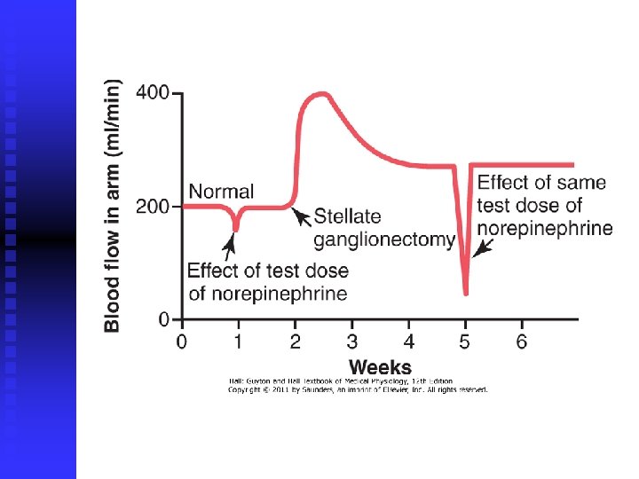
Denervation Supersensitivity q q first week after nerve is destroyed, the innervated organ becomes more sensitive to injected NE or acetylcholine denervation supersensitivity sympathetic and parasympathetic organs; increasing the response more than 10 -fold number of receptors in the postsynaptic membranes of the effector cells increases
Autonomic Reflexes regulating many visceral functions cardiovascular autonomic reflexes baroreceptor q q reflex – arterial pressure and HR gastrointestinal autonomic reflexes q q q smell of appetizing food or presence of food in mouth stretching of rectum other autonomic reflexes q q q emptying of the urinary bladder sexual reflexes
Mass Discharge by Sympathetic System q q q in some instances, almost all portions of the sympathetic nervous system discharge simultaneously as a complete unit fear or severe pain alarm or stress response
Activation in isolated portions of the sympathetic nervous system q q q process of heat regulation control sweating and blood flow in the skin local reflexes – heating a skin area causes local vasodilation and enhanced local sweating (cooling causes opposite effects) reflexes that control gastrointestinal functions; motor or secretory activity
Specific Localized Responses caused by Parasympathetic q q q control functions by the parasympathetic system are often highly specific parasympathetic cardiovascular reflexes usually act only on the heart salivary secretion, gastric secretion and pancreatic secretion frequently occurs at the same time; rectal emptying reflex often initiates a urinary bladder emptying reflex
"Alarm" or "Stress" Response of the Sympathetic Nervous System increases in many ways the ability of the body to perform vigorous muscle activity q 1) 2) 3) 4) 5) 6) 7) 8) arterial pressure blood flow to active muscles, inactive tissues rates of cellular metabolism throughout the body blood glucose concentration glycolysis in the liver and in muscle strength mental activity rate of blood coagulation
q q sympathetic stress response – extra activation of the body in states of stress state of rage – sympathetic alarm reaction, fight or flight reaction
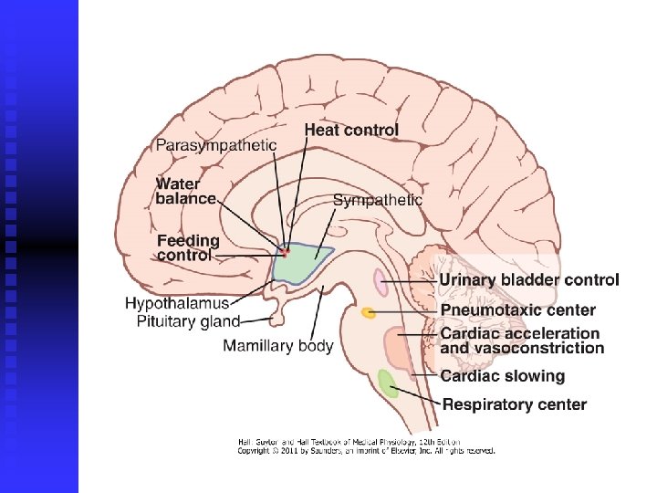
Medullary, Pontine, and Mesencephalic Control of the ANS q q q control of arterial pressure, HR, respiratory rate, glandular secretion, peristalsis, and degree of contraction of the urinary bladder transection below medulla causes arterial pressure to fall to less than one-half normal higher areas can also play a role (pressure, temperature) – diseases (peptic ulcer, constipation, heart palpitation, heart attack)
Pharmacology of ANS – Sympathetic Nervous System drugs that act on adrenergic receptors – sympathomimetic drugs 1) 1) 2) 3) α-receptors – phenylephrine β-receptors – isoproterenol β 2 -receptors – albuterol drugs that release NE from nerve endings 2) 1) ephedrine, tyramine, amphetamine
drugs that block adrenergic activity – sympahtolytics 3) 1) 2) 3) 4) 5) preventing synthesis and storage of NE – reserpine blocking release of NE – guanethidine blocking α-receptors – phentolamine blocking β-receptors – propranolol blocking β 1 -receptors – metoprolol blocking transmission of nerve impulses through the autonomic ganglia – hexamethonium
Pharmacology of ANS – Parasympathetic Nervous System drugs that act on cholinergic receptors – parasympathomimetics (cholinergic drugs) 1) 1) 2) ACh – various effects due to cholinesterase destruction in the blood and body fluids muscarinic receptors – pilocarpine and methacholine
drugs that have a parasympathetic potentiating effect – anticholinesterase drugs 2) 1) neostigmine, pyridostigmine, and ambenonium drugs that block cholinergic activity at effector organs – antimuscarinic drugs 3) 1) - atropine, homatropine, scopolamine; do not affect the nicotinic action of acetylcholine on the postganglionic neurons or on skeletal muscle
Drugs That Stimulate Postganglionic Neurons q q q injected ACh nicotine (drugs that can stimulate postganglionic neurons – nicotinic drugs) excites sympathetic and parasympathetic postganglionic neurons at the same time
Ganglionic Blocking Drugs q q q block impulse transmission from the preganglionic to the postganglionic neurons tetraethyl ammonium ion, hexamethonium ion, pentolinium block sympathetic and the parasympathetic systems simultaneously used for blocking sympathetic activity reducing arterial pressure, effects are difficult to control