
Chapter 11 Efferent Division Autonomic and Somatic Motor
- Slides: 40
Chapter 11 Efferent Division: Autonomic and Somatic Motor Control
About this Chapter • Autonomic division • Autonomic reflexes • Antagonistic controls • Control of cardiac and smooth muscle, and glands in homeostasis • Agonists and antagonists in research and medicine • Somatic motor division • CNS control of skeletal muscles through neuromuscular junctions
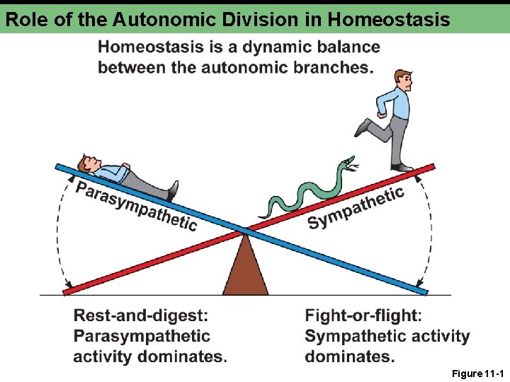
Role of the Autonomic Division in Homeostasis • Antagonistic branches • Parasympathetic • “Rest and digest” • Restore body function • Sympathetic • “Fight or flight” • Energetic action
Role of the Autonomic Division in Homeostasis Figure 11 -1
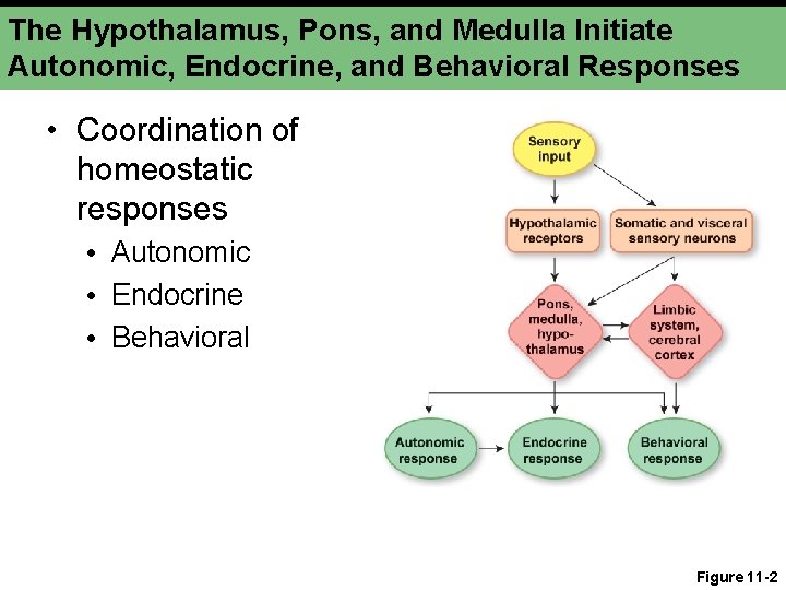
The Hypothalamus, Pons, and Medulla Initiate Autonomic, Endocrine, and Behavioral Responses • Coordination of homeostatic responses • Autonomic • Endocrine • Behavioral Figure 11 -2
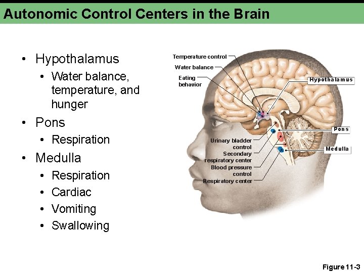
Autonomic Control Centers in the Brain • Hypothalamus • Water balance, temperature, and hunger Temperature control Water balance Eating behavior Hypothalamus • Pons • Respiration • Medulla • • Respiration Cardiac Vomiting Swallowing Pons Urinary bladder control Secondary respiratory center Blood pressure control Respiratory center Medulla Figure 11 -3
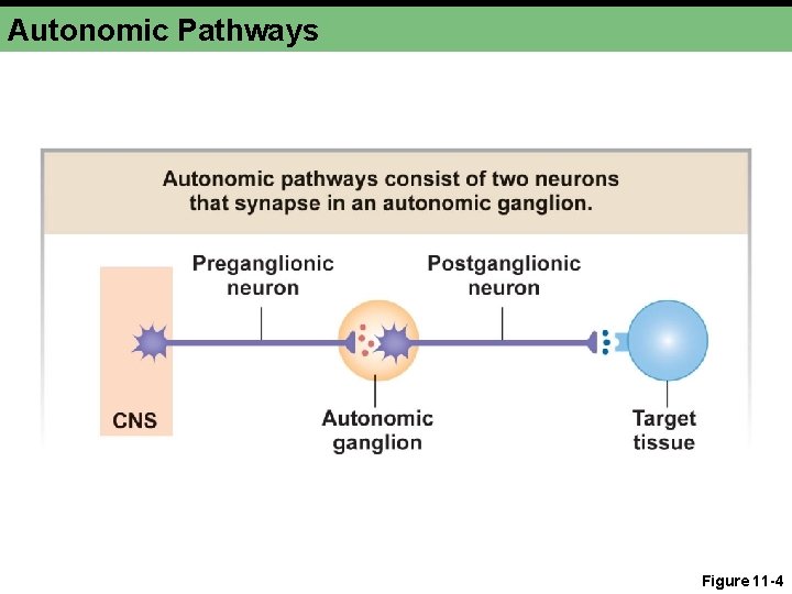
Autonomic Pathways Figure 11 -4
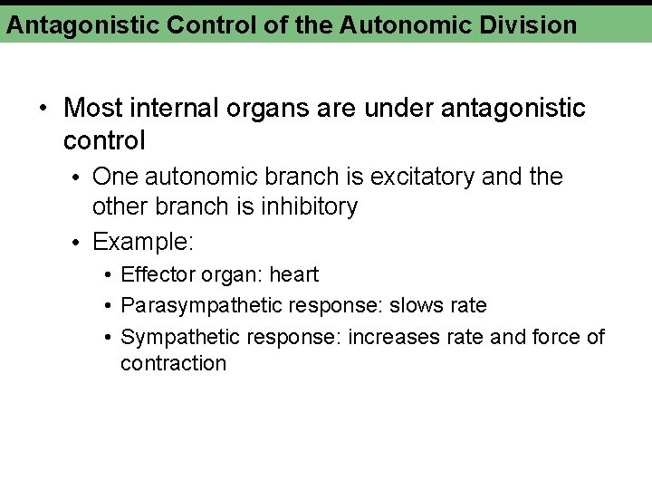
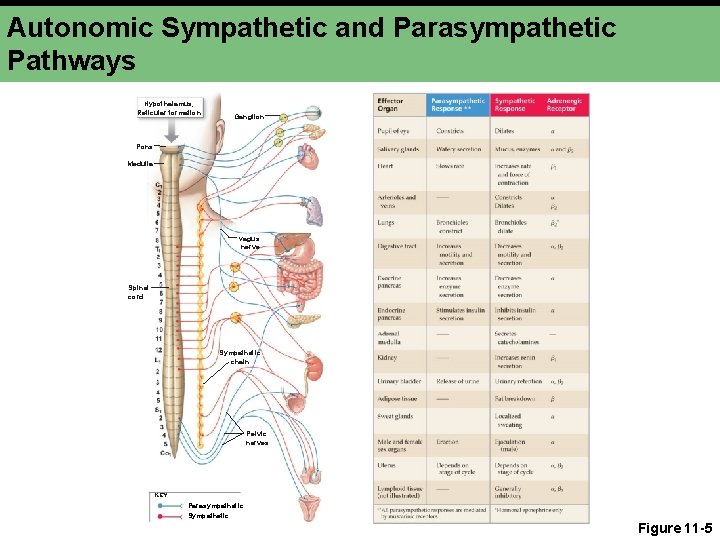
Antagonistic Control of the Autonomic Division • Most internal organs are under antagonistic control • One autonomic branch is excitatory and the other branch is inhibitory • Example: • Effector organ: heart • Parasympathetic response: slows rate • Sympathetic response: increases rate and force of contraction
Autonomic Sympathetic and Parasympathetic Pathways Hypothalamus, Reticular formation Ganglion Pons Medulla Vagus nerve Spinal cord Sympathetic chain Pelvic nerves KEY Parasympathetic Sympathetic Figure 11 -5
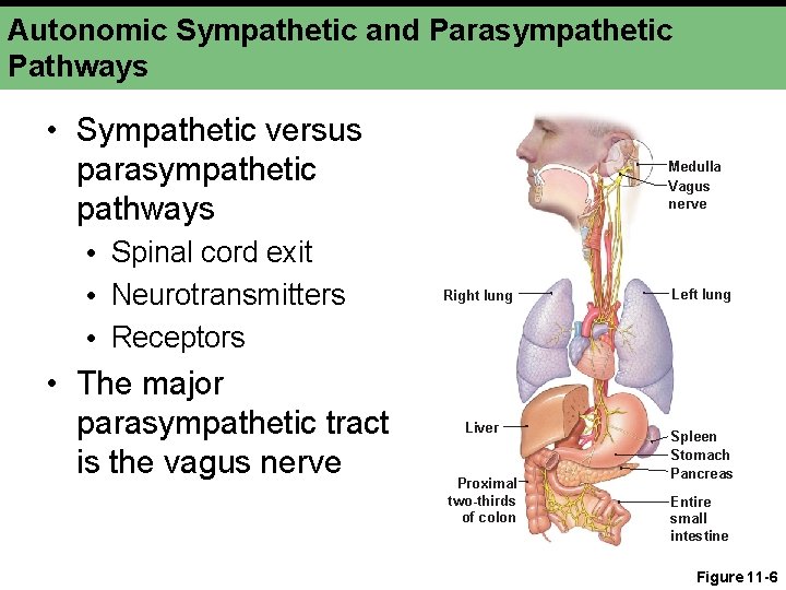
Autonomic Sympathetic and Parasympathetic Pathways • Sympathetic versus parasympathetic pathways Medulla Vagus nerve • Spinal cord exit • Neurotransmitters • Receptors Right lung • The major parasympathetic tract is the vagus nerve Liver Proximal two-thirds of colon Left lung Spleen Stomach Pancreas Entire small intestine Figure 11 -6
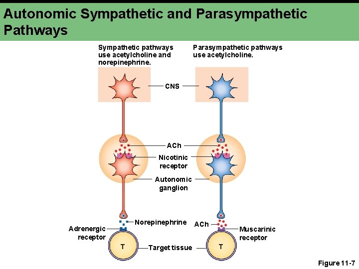
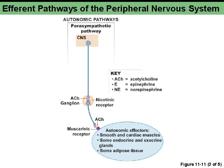
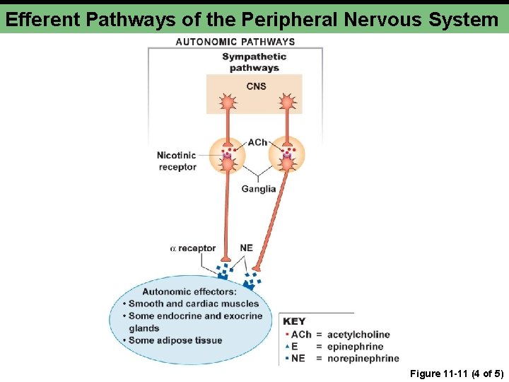
Autonomic Sympathetic and Parasympathetic Pathways Sympathetic pathways use acetylcholine and norepinephrine. Parasympathetic pathways use acetylcholine. CNS ACh Nicotinic receptor Autonomic ganglion Norepinephrine Adrenergic receptor T Target tissue ACh Muscarinic receptor T Figure 11 -7
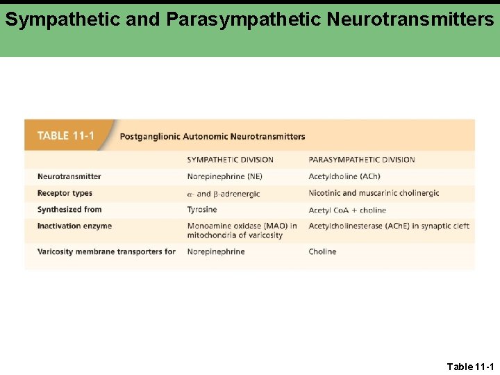
Sympathetic and Parasympathetic Neurotransmitters Table 11 -1
Autonomic Targets • • Autonomic pathways control: Smooth muscle Cardiac muscle Exocrine glands (select) Endocrine glands (select) Lymphoid tissue Adipose tissue
Autonomic Neuron Structure • Neuroeffector junction • Postganglionic axon • Varicosities • Axon • Neurotransmitter synthesis
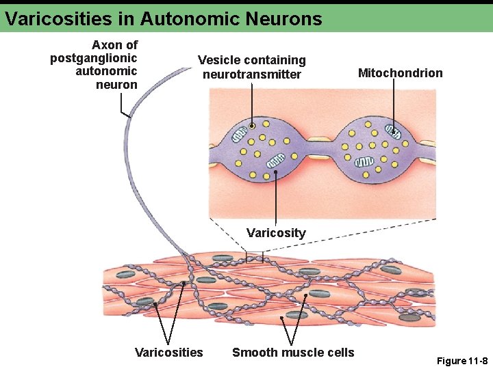
Varicosities in Autonomic Neurons Axon of postganglionic autonomic neuron Vesicle containing neurotransmitter Mitochondrion Varicosity Figure 11 -8 Varicosities Smooth muscle cells Figure 11 -8
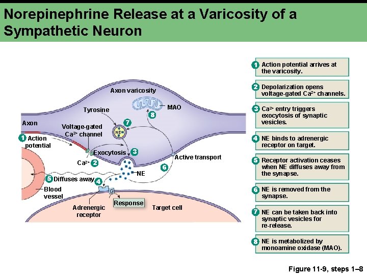
Norepinephrine Release at a Varicosity of a Sympathetic Neuron 1 Action potential arrives at the varicosity. 2 Depolarization opens voltage-gated Ca 2+ channels. Axon varicosity Tyrosine Axon 1 Action potential 8 7 Voltage-gated Ca 2+ channel NE Exocytosis 3 Active transport NE 5 Diffuses away 4 6 5 Receptor activation ceases when NE diffuses away from the synapse. 6 NE is removed from the synapse. G Adrenergic receptor 3 Ca 2+ entry triggers exocytosis of synaptic vesicles. 4 NE binds to adrenergic receptor on target. Ca 2+ 2 Blood vessel MAO Response Target cell 7 NE can be taken back into synaptic vesicles for re-release. 8 NE is metabolized by monoamine oxidase (MAO). Figure 11 -9, steps 1– 8
Sympathetic Branch: Stimulation • • • Pupil dilation Salivation Heart beat and volume Blood vessel and bronchiole dilation Fat breakdown Ejaculation
Sympathetic Branch: Inhibition • Digestion • Pancreas secretion • Urination
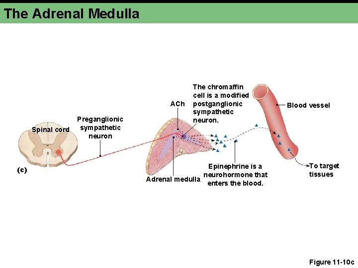
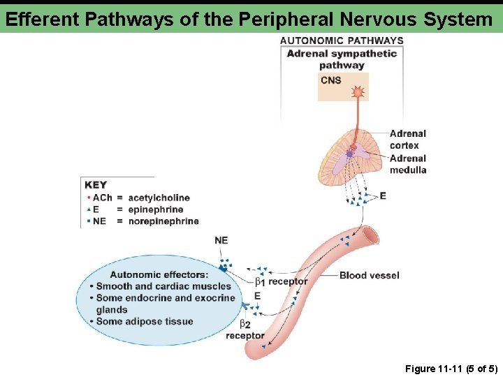
Adrenal Medulla • Primary neurohormone • Epinephrine • Multiple and distant targets
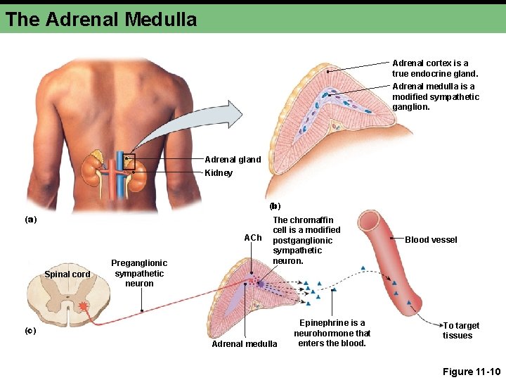
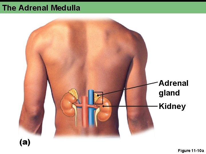
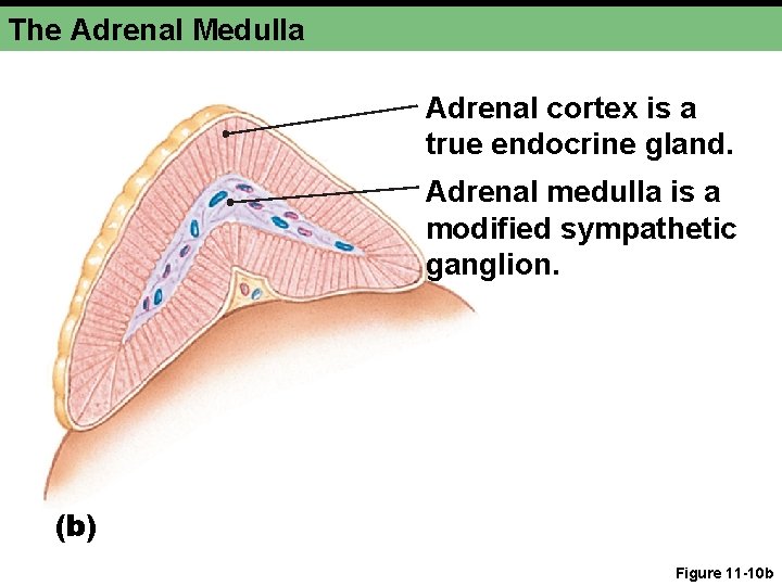
The Adrenal Medulla Adrenal cortex is a true endocrine gland. Adrenal medulla is a modified sympathetic ganglion. Adrenal gland Kidney (b) (a) ACh Spinal cord Preganglionic sympathetic neuron The chromaffin cell is a modified postganglionic sympathetic neuron. (c) Adrenal medulla Epinephrine is a neurohormone that enters the blood. Blood vessel To target tissues Figure 11 -10
The Adrenal Medulla Adrenal gland Kidney (a) Figure 11 -10 a
The Adrenal Medulla Adrenal cortex is a true endocrine gland. Adrenal medulla is a modified sympathetic ganglion. (b) Figure 11 -10 b
The Adrenal Medulla ACh Spinal cord (c) Preganglionic sympathetic neuron The chromaffin cell is a modified postganglionic sympathetic neuron. Epinephrine is a neurohormone that Adrenal medulla enters the blood. Blood vessel To target tissues Figure 11 -10 c
Parasympathetic Branch • Acetylcholine • Muscarinic receptors • G protein-coupled • Second messenger pathways • At least five subtypes
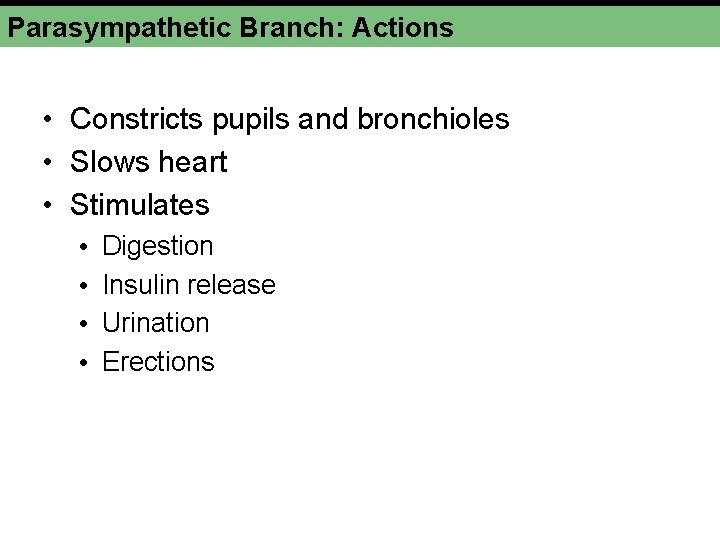
Parasympathetic Branch: Actions • Constricts pupils and bronchioles • Slows heart • Stimulates • • Digestion Insulin release Urination Erections
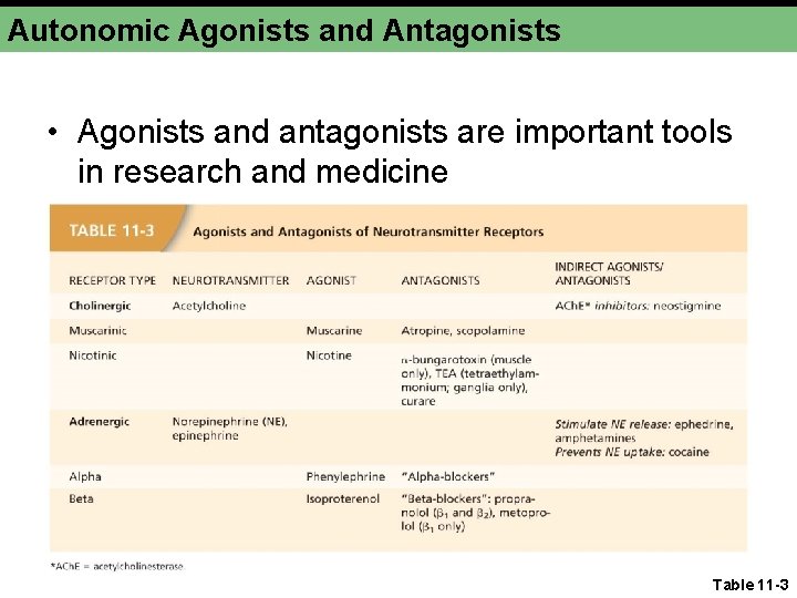
Autonomic Agonists and Antagonists • Agonists and antagonists are important tools in research and medicine Table 11 -3
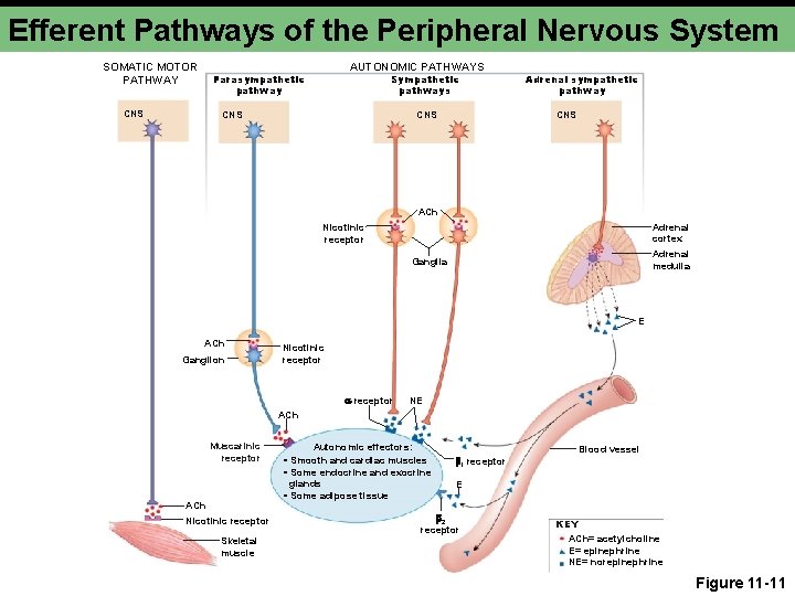
Efferent Pathways of the Peripheral Nervous System SOMATIC MOTOR PATHWAY CNS AUTONOMIC PATHWAYS Parasympathetic pathway Sympathetic pathways CNS Adrenal sympathetic pathway CNS ACh Adrenal cortex Nicotinic receptor Adrenal medulla Ganglia E ACh Ganglion Nicotinic receptor NE ACh Muscarinic receptor ACh Nicotinic receptor Skeletal muscle Autonomic effectors: • Smooth and cardiac muscles • Some endocrine and exocrine glands • Some adipose tissue Blood vessel 1 receptor E 2 receptor KEY ACh= acetylcholine E= epinephrine NE= norepinephrine Figure 11 -11
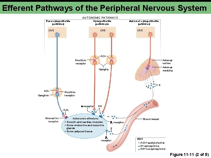
Efferent Pathways of the Peripheral Nervous System AUTONOMIC PATHWAYS Parasympathetic pathway Sympathetic pathways CNS Adrenal sympathetic pathway CNS ACh Adrenal cortex Nicotinic receptor Adrenal medulla Ganglia E ACh Ganglion Nicotinic receptor NE ACh Muscarinic receptor Autonomic effectors: • Smooth and cardiac muscles • Some endocrine and exocrine glands • Some adipose tissue Blood vessel 1 receptor E 2 receptor KEY ACh= acetylcholine E= epinephrine NE= norepinephrine Figure 11 -11 (2 of 5)
Efferent Pathways of the Peripheral Nervous System Figure 11 -11 (3 of 5)
Efferent Pathways of the Peripheral Nervous System Figure 11 -11 (4 of 5)
Efferent Pathways of the Peripheral Nervous System Figure 11 -11 (5 of 5)
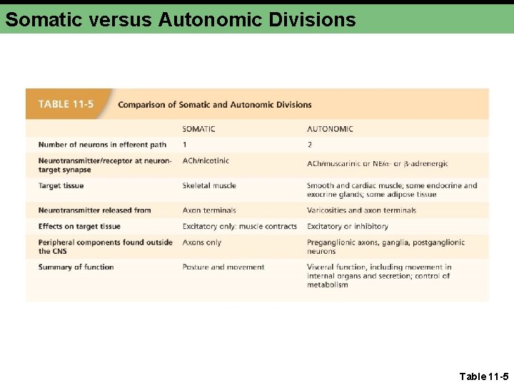
Somatic versus Autonomic Divisions Table 11 -5
Somatic Motor Division • Single neuron • CNS origin • Myelinated • Terminus • Branches • Neuromuscular junction
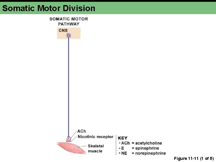
Somatic Motor Division Figure 11 -11 (1 of 5)
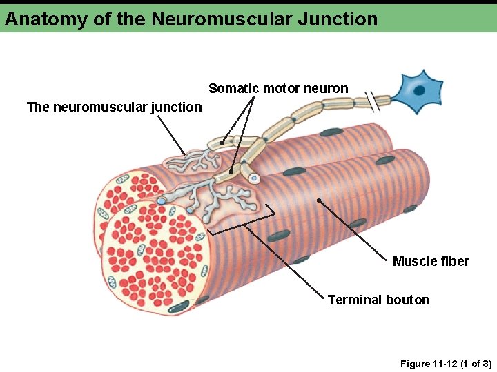
Anatomy of the Neuromuscular Junction Somatic motor neuron The neuromuscular junction Muscle fiber Terminal bouton Figure 11 -12 (1 of 3)
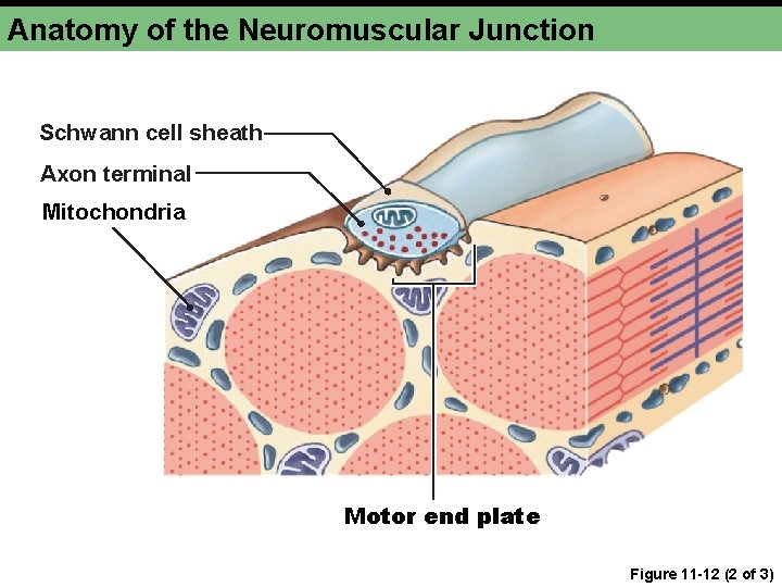
Anatomy of the Neuromuscular Junction Schwann cell sheath Axon terminal Mitochondria Motor end plate Figure 11 -12 (2 of 3)
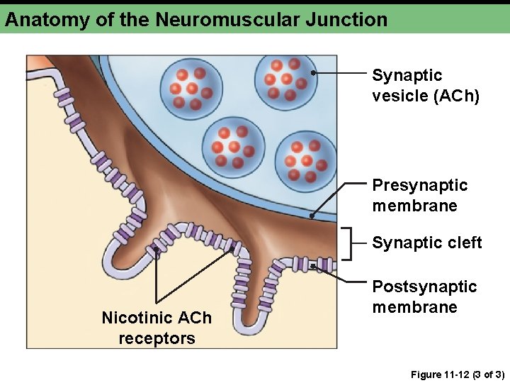
Anatomy of the Neuromuscular Junction Synaptic vesicle (ACh) Presynaptic membrane Synaptic cleft Nicotinic ACh receptors Postsynaptic membrane Figure 11 -12 (3 of 3)
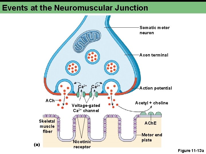
Events at the Neuromuscular Junction Somatic motor neuron Axon terminal Ca 2+ ACh Voltage-gated Ca 2+ channel Skeletal muscle fiber (a) Action potential Acetyl + choline ACh. E Nicotinic receptor Motor end plate Figure 11 -13 a
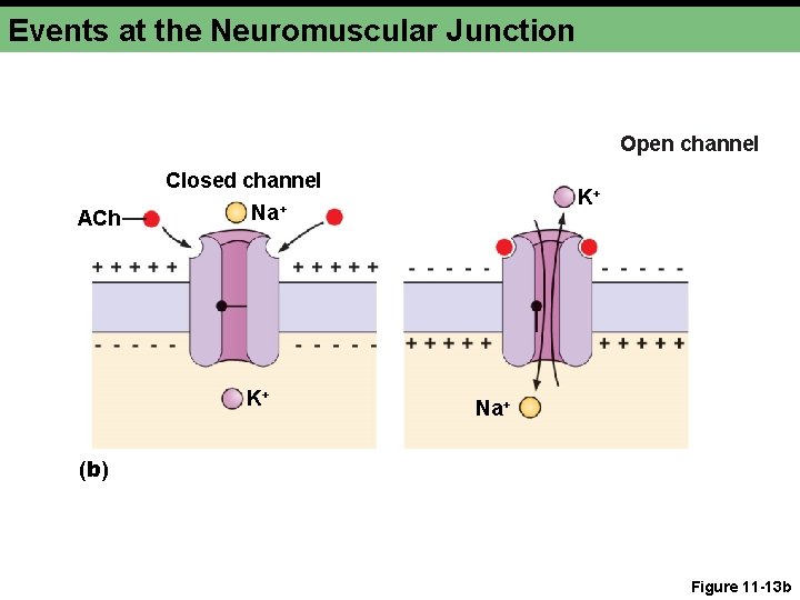
Events at the Neuromuscular Junction Open channel Closed channel ACh K+ Na+ (b) Figure 11 -13 b
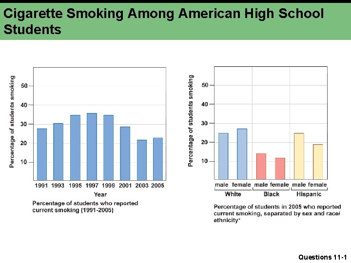
Cigarette Smoking Among American High School Students Questions 11 -1